Surgical Hip Dislocation (SHD) for FAI: Anatomy, Biomechanics & Advanced Management
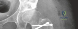
Key Takeaway
Surgical Hip Dislocation (SHD) offers unparalleled 360-degree visualization for complex femoroacetabular impingement (FAI) cases. It enables precise correction of severe osseous deformities, extensive chondrolabral damage, and management of challenging intra-articular pathologies, especially when arthroscopy is insufficient. This open approach allows meticulous protection of critical structures like the Medial Femoral Circumflex Artery (MFCA), mitigating AVN risk.
A 28-year-old active male presents with chronic anterior groin pain and mechanical locking. Imaging reveals a significant CAM lesion with an alpha angle of 85 degrees and a secondary labral tear. You are considering a surgical hip dislocation. Describe the anatomical landmark that must be identified and protected during this approach to prevent the most devastating complication, and explain exactly where it is found.
Candidate: You need to protect the medial femoral circumflex artery (MFCA) to avoid avascular necrosis of the femoral head. It is located near the piriformis tendon and the quadratus femoris muscle.
The candidate identifies the artery but fails to be anatomically precise regarding the "deep branch" or its specific relationship to the obturator externus/quadratus femoris interval. Examiners are looking for knowledge of the "retinacular vessels" and the exact path the surgeon must respect during the trochanteric flip.
The structure at risk is the deep branch of the Medial Femoral Circumflex Artery (MFCA). It is the primary blood supply to the femoral head. It runs posteriorly, typically between the piriformis and the superior gemellus, then courses along the obturator externus tendon to reach the posterior-superior capsule where it gives off the retinacular vessels. During a Ganz trochanteric flip, the osteotomy is performed anteriorly to move the greater trochanter, but the posterior soft-tissue envelope—containing the MFCA—must remain undisturbed. Protecting the quadratus femoris and the underlying obturator externus interval is mandatory to avoid iatrogenic AVN.
Examine this image. What is the clinical significance of the deformity shown, and how does this impact the biomechanics of the hip joint in FAI?
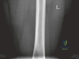
Candidate: This shows a Pincer-type deformity where there is acetabular overcoverage. This causes the labrum to get pinched between the femoral neck and the acetabular rim, which causes labral damage.
Simply stating "overcoverage" is too generic. A top-tier candidate must mention the "suction seal" of the labrum, the concept of "contre-coup" injury to the posterior-inferior cartilage, and the difference in cartilage damage patterns (central vs. peripheral) compared to CAM impingement.
This image depicts a Pincer impingement. Biomechanically, excessive acetabular coverage (e.g., coxa profunda or retroversion) leads to a loss of the normal hip "suction seal" function provided by the labrum. The labrum is crushed between the acetabular rim and the femoral neck, leading to hypertrophy and ossification of the labrum. Unlike CAM, which causes peripheral shear and delamination, Pincer impingement creates central chondral wear and often results in a 'contre-coup' injury on the postero-inferior aspect of the femoral head due to the levering effect created by the anterior rim contact.
You have performed the osteotomy and now need to fix the greater trochanter. Look at the post-operative imaging below. What complications are you trying to mitigate with this specific fixation method, and what post-operative monitoring is required?
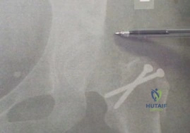
Candidate: You are using large screws to fix the trochanter to ensure it doesn't move. You are trying to avoid a nonunion of the trochanteric osteotomy, which would cause pain and a limp. You need to check X-rays to make sure it's healing.
Missing the clinical consequences of a nonunion (abductor weakness/Trendelenburg gait) and failing to discuss the rehabilitation implications—specifically how this fixation dictates the weight-bearing status.
The primary complication being mitigated is trochanteric nonunion. Because the gluteus medius and minimus exert significant tension on the osteotomy fragment, rigid fixation (typically 2-3 cortical screws) is essential to neutralize these forces. If nonunion occurs, the patient will suffer from persistent pain and a significant abductor lurch (Trendelenburg gait). Post-operative management involves strict weight-bearing restrictions (non-weight bearing or touch-down for 6-8 weeks) to allow for biological union. Additionally, I would monitor for hardware-related irritation, which may necessitate secondary removal once the osteotomy is consolidated (typically at 6-12 months).
