Clinical Diagnosis and Management of Peripheral Nerve Injuries and Complex Regional Pain Syndrome
Key Takeaway
The immediate clinical diagnosis of peripheral nerve injuries in the traumatized extremity requires rapid, targeted sensory and motor testing. Accurate assessment dictates subsequent surgical decision-making. Furthermore, recognizing and treating sympathetically mediated pain disorders, such as Complex Regional Pain Syndrome (CRPS), within the first six months is critical to optimizing functional outcomes and preventing irreversible dystrophic changes in the affected limb.
INTRODUCTION TO PERIPHERAL NERVE INJURIES IN TRAUMA
Immediately following a severe injury to an extremity, the clinical recognition of a peripheral nerve injury presents a formidable diagnostic challenge. In the acute trauma setting, the preservation of life and limb strictly adheres to Advanced Trauma Life Support (ATLS) protocols, rendering neurological assessment a secondary, albeit critical, priority. Furthermore, the patient's pain is often so severe that cooperation is severely limited, and altered sensorium due to concomitant head trauma or systemic shock may further obscure the clinical picture.
Despite these challenges, the early identification of peripheral nerve deficits is paramount. A missed nerve injury can lead to devastating functional consequences, irreversible motor end-plate degeneration, and severe medicolegal repercussions. Therefore, orthopedic surgeons must employ rapid, highly specific screening tests to evaluate the major nerves of the extremities. These tests rely on the assessment of "autonomous zones"—specific areas of skin innervated exclusively by a single nerve—and the evaluation of terminal motor branches.
CLINICAL PEARL:
The initial neurological examination must be documented meticulously before the administration of local anesthetics, regional blocks, or surgical intervention. A documented preoperative nerve deficit dictates the surgical approach and manages patient expectations regarding postoperative recovery.
THE ANATOMICAL IMPERATIVE IN NERVE EVALUATION
In evaluating peripheral nerve lesions, a precise, encyclopedic knowledge of neuroanatomy is non-negotiable. The surgeon must understand the exact anatomical course of the nerve, the precise level of origin of its motor branches, and the specific muscles that these branches innervate. This knowledge allows the clinician to localize the exact level of the lesion based on the pattern of motor and sensory deficits.
For instance, a high radial nerve palsy (proximal to the spiral groove) will result in the loss of elbow extension (triceps), wrist extension, and digit extension. Conversely, a lesion at the level of the arcade of Frohse (Posterior Interosseous Nerve syndrome) will spare the extensor carpi radialis longus (ECRL) and brevis (ECRB), resulting in preserved radial wrist extension but a loss of digit and thumb extension.
RAPID SCREENING PROTOCOLS FOR THE UPPER EXTREMITY
In the chaotic environment of the emergency department, comprehensive neurological testing is often impractical. Instead, the surgeon should rely on rapid screening procedures targeting the autonomous sensory zones and isolated motor functions of the median, ulnar, and radial nerves.
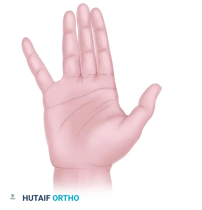
The Ulnar Nerve
The ulnar nerve is the primary motor nerve of the intrinsic muscles of the hand.
* Sensory Evaluation: Loss of pain perception or two-point discrimination in the volar tip of the little finger (fifth digit) is the hallmark of ulnar nerve injury. This is the absolute autonomous zone for the ulnar nerve.
* Motor Evaluation: Ask the patient to cross their index and middle fingers or forcefully abduct the digits against resistance (testing the dorsal interossei and abductor digiti minimi).
The Median Nerve
The median nerve provides critical sensory feedback to the working surface of the hand and motor innervation to the flexor-pronator mass and thenar musculature.
* Sensory Evaluation: Loss of pain perception in the volar tip of the index finger indicates median nerve injury.
* Motor Evaluation: Test the abductor pollicis brevis (APB) by asking the patient to palmar abduct the thumb against resistance. Alternatively, testing the flexor pollicis longus (FPL) by asking the patient to flex the interphalangeal joint of the thumb (the "OK" sign) evaluates the anterior interosseous nerve (AIN) branch.
The Radial Nerve
The radial nerve is responsible for the extension of the elbow, wrist, and digits.
* Sensory Evaluation: The autonomous sensory zone is located in the dorsal first web space.
* Motor Evaluation: The inability to extend the thumb (the "hitchhiker’s sign") usually indicates a radial nerve injury, specifically involving the extensor pollicis longus (EPL).
SURGICAL WARNING:
The Tendon Laceration Pitfall: In cases of penetrating trauma, the extensor or flexor tendons may be severed, rendering motor tests invalid. A severed EPL tendon will result in an absent hitchhiker's sign, mimicking a radial nerve palsy. Always utilize the tenodesis effect (passive wrist flexion to observe passive digit extension) to differentiate between a tendon laceration and a primary nerve palsy.
RAPID SCREENING PROTOCOLS FOR THE LOWER EXTREMITY
Similar rapid screening principles apply to the lower extremity, where high-energy trauma, such as knee dislocations or posterior hip dislocations, frequently compromises the sciatic nerve and its terminal branches.
The Sciatic and Tibial Nerves
The sciatic nerve divides into the tibial and common peroneal nerves at the apex of the popliteal fossa. The tibial nerve innervates the posterior compartment of the leg and the plantar aspect of the foot.
* Sensory Evaluation: Loss of pain perception in the sole of the foot usually indicates a sciatic or isolated tibial nerve injury.
* Motor Evaluation: Inability to actively plantarflex the ankle or toes indicates compromise of the gastrocnemius-soleus complex and deep toe flexors.
The Common Peroneal Nerve
The common peroneal nerve is highly susceptible to injury as it wraps around the fibular neck. It divides into the deep and superficial peroneal nerves.
* Sensory Evaluation: The autonomous zone for the deep peroneal nerve is the dorsal first web space. The superficial peroneal nerve supplies the dorsum of the foot.
* Motor Evaluation: Inability to extend the great toe (extensor hallucis longus) or dorsiflex the foot (tibialis anterior) indicates a peroneal or high sciatic nerve injury. This presents clinically as a "drop foot."
PITFALL:
As with the upper extremity, direct injury to the muscle bellies or tendons (e.g., a lacerated tibialis anterior tendon) may render these tests useless. Furthermore, an acute compartment syndrome of the anterior leg can rapidly cause ischemic neuropraxia of the deep peroneal nerve, presenting identically to a primary traumatic nerve laceration.
COMPLEX REGIONAL PAIN SYNDROME (CRPS) FOLLOWING NERVE INJURY
A critical, often debilitating sequela of peripheral nerve injury—even minor crush injuries or traction neuropraxias—is the development of Complex Regional Pain Syndrome (CRPS), historically referred to as Reflex Sympathetic Dystrophy (RSD) or causalgia.
CRPS is a neuropathic pain disorder characterized by autonomic dysfunction, severe hyperalgesia, allodynia, and trophic changes in the affected extremity. If left unrecognized and untreated, CRPS can lead to irreversible joint contractures, severe osteopenia, and permanent disability, completely negating the results of an otherwise successful orthopedic reconstruction.
Pathophysiology and Clinical Presentation
The exact pathophysiology of CRPS remains incompletely understood but involves a maladaptive inflammatory response, sympathetic nervous system dysfunction, and neuroplastic changes within the central nervous system. Patients typically present with pain that is disproportionate to the inciting event.
Clinical signs include:
* Sensory: Hyperesthesia and allodynia (pain from a non-painful stimulus, such as light touch).
* Vasomotor: Temperature asymmetry and skin color changes (mottled, cyanotic, or erythematous).
* Sudomotor/Edema: Asymmetrical sweating and non-pitting edema.
* Motor/Trophic: Decreased range of motion, motor weakness, tremor, and trophic changes to the hair, nails, and skin (shiny, thin skin).
Evidence-Based Management of CRPS
The successful management of CRPS requires a multidisciplinary approach, combining aggressive physical therapy with interventional pain management. The overarching goal is to break the cycle of sympathetically mediated pain to allow the patient to participate in functional rehabilitation.
Sympathetic Ganglion Blocks
Kleinert et al. and Lankford have extensively reported highly favorable results utilizing sequential stellate ganglion blocks combined with rigorous physical therapy in patients with CRPS involving the upper extremity. For lower extremity CRPS, lumbar sympathetic blocks are utilized.
- Efficacy: Pain relief and significantly improved range of motion have been reported in 80% to 93% of patients with CRPS following a series of sequential sympathetic blocks.
- Mechanism: The block temporarily halts the efferent sympathetic outflow to the extremity, causing vasodilation, warming of the limb, and a profound reduction in sympathetically maintained pain. This "window of analgesia" is immediately capitalized upon by physical therapists to mobilize stiff joints.
Intravenous Regional Blocks (Bier Blocks)
Poplawski, Wiley, and Murray reported on a cohort of 27 patients treated with intravenous regional blocks utilizing a combination of lidocaine and corticosteroids, followed immediately by standard physical therapy. This technique provides both regional anesthesia and localized anti-inflammatory effects, facilitating aggressive joint mobilization.
Surgical Sympathectomy
While the majority of patients respond to conservative measures and temporary blocks, a subset remains refractory. Studies indicate that approximately 19% of patients may experience only a temporary response to chemical sympathetic blocks. In these severe, recalcitrant cases, surgical sympathectomy (e.g., endoscopic thoracic sympathectomy for the upper extremity) may be required to provide permanent disruption of the sympathetic chain.
CLINICAL PEARL:
The Critical Time Window: Poplawski and colleagues found that the single most important prognostic factor in predicting a favorable outcome in CRPS is the time elapsed between the onset of symptoms and the initiation of treatment. An interval of less than 6 months is critical. Interventions initiated after 6 months often face irreversible fibrotic and dystrophic changes within the limb.
POSTOPERATIVE PROTOCOLS AND LONG-TERM MONITORING
Whether a nerve injury is managed conservatively (expectant observation for neuropraxia) or surgically (primary neurorrhaphy, nerve grafting, or nerve transfers), meticulous postoperative monitoring is required.
- Clinical Tracking: The advancing Tinel's sign (Hoffmann-Tinel sign) should be tracked longitudinally. Peripheral nerves regenerate at a rate of approximately 1 mm per day (or 1 inch per month).
- Electrodiagnostic Studies: Electromyography (EMG) and Nerve Conduction Studies (NCS) should be obtained at 3 to 4 weeks post-injury to establish a baseline and evaluate for Wallerian degeneration. Repeat studies at 12 weeks can detect early subclinical reinnervation (nascent motor unit potentials).
- Physical Therapy: Continuous passive and active range of motion of all joints distal to the injury is mandatory to prevent contractures while awaiting nerve regeneration.
CONCLUSION
The clinical diagnosis of peripheral nerve injuries demands a high index of suspicion, a profound understanding of neuroanatomy, and the utilization of rapid, targeted screening tests in the acute trauma setting. Differentiating true nerve injuries from tendon lacerations or compartment syndrome is a critical surgical skill. Furthermore, the orthopedic surgeon must remain vigilant for the development of Complex Regional Pain Syndrome. Early recognition and aggressive intervention with sympathetic blocks and physical therapy within the first six months are paramount to salvaging limb function and ensuring optimal patient outcomes.
You Might Also Like