Mastering the Surgical Management of Stenosing Tenosynovitis: De Quervain Disease and Trigger Digits
Key Takeaway
Surgical management of stenosing tenosynovitis, including De Quervain disease and trigger digits, requires meticulous technique to ensure complete release while avoiding iatrogenic injury. This guide details the precise anatomical dissection, identification of aberrant septa, and management of tendon subluxation in the first dorsal compartment, alongside comprehensive protocols for A1 pulley release in trigger digits to optimize postoperative functional recovery.
INTRODUCTION TO STENOSING TENOSYNOVITIS
Stenosing tenosynovitis encompasses a spectrum of entrapment neuropathies and fibro-osseous tunnel disorders that frequently present in orthopedic practice. The fundamental pathophysiology involves a mismatch in volume between a tendon (or tendons) and its restraining retinacular sheath. This size discrepancy leads to mechanical friction, reactive synovial hypertrophy, and subsequent fibrotic thickening of the retinaculum, creating a self-perpetuating cycle of inflammation and stenosis.
The two most prevalent manifestations of this pathology in the hand and wrist are De Quervain disease (involving the first dorsal extensor compartment) and trigger digits (involving the flexor tendon sheath at the A1 pulley). While conservative measures such as corticosteroid injections and splinting remain the first line of treatment, surgical release is highly efficacious for refractory cases. However, operative intervention demands a profound understanding of regional anatomy, anatomical variants, and precise surgical execution to prevent debilitating complications such as iatrogenic nerve injury, tendon subluxation, or incomplete release.
SURGICAL TREATMENT OF DE QUERVAIN DISEASE
De Quervain disease is characterized by stenosing tenosynovitis of the abductor pollicis longus (APL) and extensor pollicis brevis (EPB) tendons within the first dorsal compartment of the wrist. Surgical release is indicated when nonoperative modalities fail to provide sustained relief.
Preoperative Planning and Anesthesia
The procedure is typically performed on an outpatient basis. The choice of anesthesia is critical for intraoperative assessment.
- Anesthesia: Use a local anesthetic (e.g., 1% lidocaine with or without epinephrine, depending on the surgeon's preference for WALANT principles) to allow for awake intraoperative testing.
- Infiltration: Infiltrate the skin well proximal to the area of the first dorsal compartment with sufficient local anesthetic before skin preparation and draping. This ensures adequate time for the anesthetic to take effect and minimizes patient discomfort during the initial stages of the procedure.
- Hemostasis: A bloodless field is essential for identifying fine sensory nerve branches and anatomical variants. An Esmarch bandage applied tightly to the proximal forearm usually suffices as a tourniquet, providing excellent visualization without the need for a pneumatic upper arm tourniquet.
Surgical Approach and Dissection
The surgical approach to the first dorsal compartment is fraught with potential pitfalls, primarily due to the superficial radial nerve (SRN) and its branches.
🔪 Surgical Pearl: Incision Orientation
Make a skin incision that runs from dorsal to volar in a transverse-to-oblique direction, parallel with the skin creases over the area of maximal tenderness in the first dorsal compartment. The longitudinal incision advocated by some historical texts creates a longer area in which the resulting skin scar may adhere to the underlying cutaneous nerves and tendons, leading to severe postoperative pain and restricted excursion.
- Superficial Dissection: Carry sharp dissection just through the dermis. Do not plunge into the subcutaneous fat.
- Nerve Protection: The sensory branches of the radial nerve are highly variable and extremely superficial in this region. You must meticulously avoid the lateral antebrachial cutaneous nerve (LABCN) located above the cephalic vein, and the superficial radial nerve (SRN) branches located slightly more deeply.
- Exposure: After retracting the skin edges with fine skin hooks or a self-retaining retractor, use blunt dissection (e.g., spreading with tenotomy scissors) in the subcutaneous fat to clearly expose the extensor retinaculum overlying the first dorsal compartment tendons.
Compartment Release and Tendon Identification
Complete resolution of symptoms relies on the absolute decompression of all tendon slips within the first compartment.
- Incision of the Retinaculum: Identify the first dorsal compartment tendons proximal to the stenosing dorsal ligament and sheath. Open the first dorsal compartment on its dorsoulnar side. This specific location for the retinacular incision leaves a volarly based flap of retinaculum, which is crucial for preventing postoperative volar subluxation of the tendons during wrist flexion.
- Tendon Differentiation:
- The Extensor Pollicis Brevis (EPB) tendon is the most dorsal structure in the compartment. It can be definitively identified by its distally oriented muscle fibers extending closer to the retinaculum.
- The Abductor Pollicis Longus (APL) tendon is located more volarly and typically has no muscular fibers in this immediate area. The APL frequently presents with multiple tendinous slips (up to seven have been reported).
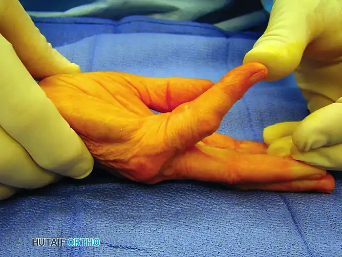
- Dynamic Assessment: With the patient's thumb abducted and the wrist flexed, lift the APL and the EPB tendons from their osseous groove using a blunt hook or Freer elevator.
🚨 Surgical Warning: The Missed Compartment
If the tendons cannot be easily retracted from the radial styloid, or if the EPB appears absent, you must actively look for additional "aberrant" tendons and separate sub-compartments. A separate fibro-osseous tunnel for the EPB is present in up to 60% of patients and is the leading cause of surgical failure if left unreleased.
- Septum Management: Releasing the EPB tendon first, followed by the APL, usually reveals the presence or absence of an intervening septum. Sometimes this septum is quite pronounced and thick; its complete resection is warranted to ensure adequate decompression.
- The Brachioradialis Footprint: If there is no EPB present within the primary compartment, inspect the floor of the compartment for the footprint of the brachioradialis tendon. The brachioradialis has a distinct Y-shaped tendinous insertion on the radial styloid. If this Y-shaped insertion is clearly visualized, you can be assured that the floor of the compartment has been reached and the release is anatomically complete.
- Arterial Protection: When resecting a thick septum or exploring the distal aspect of the compartment, take extreme care not to injure the underlying radial artery, which courses dorsally just beyond the radial styloid toward the anatomical snuffbox.
Complications and Management of Tendon Subluxation
Failure to obtain complete relief after primary surgery is a recognized complication and may result from four primary etiologies:
1. Neural adhesions or neuroma formation (injury to the SRN or LABCN).
2. Volar tendon subluxation (due to over-resection of the retinaculum or failure to preserve a volar flap).
3. Failure to find and release a separate aberrant tendon within a separate compartment (missed EPB).
4. Scar hypertrophy and deep fibrotic tethering.
Reconstructive Techniques for Recurrent Subluxation
If volar tendon subluxation occurs (often presenting as a painful, palpable snap over the radial styloid during wrist flexion and thumb motion), surgical reconstruction of the first dorsal compartment is required. Several techniques have been described to create a nonstenosing restraint:
- Brachioradialis Slip Reconstruction: A distally based slip of the brachioradialis tendon can be harvested and routed over the APL and EPB to reconstruct a nonstenosing compartment.
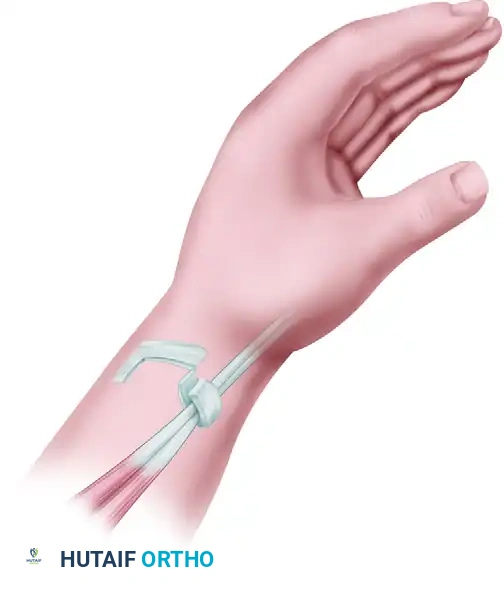
- Extensor Retinaculum Sling (Ramesh and Britton): This technique utilizes a portion of the extensor retinaculum to create a U-shaped sling that retains the tendons while allowing frictionless glide.
- Littler Reconstruction: Littler et al. described a highly effective reconstruction in which the septum dividing the first extensor compartment is entirely removed, the EPB is transposed out of the compartment, and the retinacular sheath is reapproximated loosely over the APL tendon alone to prevent subluxation.
- Radial Forearm Fascial Flap: For severe, recurrent De Quervain disease with extensive scarring, a radial forearm fascial flap can be utilized to provide healthy, gliding tissue. However, this requires a significantly more extensive forearm dissection and is reserved for complex salvage cases.
Closure and Postoperative Care
- Closure: Make sure that the volarly based retinacular flap remains positioned over the released tendons to prevent volar tendon subluxation. Close the skin incision only using non-absorbable monofilament sutures (e.g., 4-0 or 5-0 nylon) or absorbable sutures based on surgeon preference. Deep dermal or subcutaneous sutures are avoided to prevent tethering of the superficial radial nerve.
- Dressing: Apply a small, bulky pressure dressing to minimize hematoma formation and dead space.
- Rehabilitation: The small pressure dressing is removed after 48 hours; an additional light dressing or adhesive bandage can be applied if needed. Thumb and hand motion is immediately encouraged to prevent tendon adhesions. Activity is increased as tolerated, with one critical exception: forceful wrist flexion must be avoided during the first 2 weeks after surgery, as this specific motion predisposes the newly released tendons toward volar subluxation before the retinacular tissues have adequately healed.
SURGICAL TREATMENT OF TRIGGER FINGER AND THUMB
Trigger digit (stenosing tenosynovitis of the flexor tendon sheath) is a common cause of hand pain and disability, leading to an inability to smoothly extend a flexed digit (the classic "triggering" or "catching" phenomenon).
Pathophysiology and Clinical Presentation
Trigger thumb in adults is a distinctly separate clinical entity from "congenital" trigger thumb (which typically presents with a fixed flexion deformity of the interphalangeal joint in infants and requires different management).
In adults, stenosing tenosynovitis is usually seen in individuals older than 45 years of age. The pathology localizes to the first annular (A1) pulley of the flexor tendon sheath.
* Systemic Associations: When associated with a collagen vascular disease, inflammatory arthritis (such as Rheumatoid Arthritis), or diabetes mellitus, several fingers may be involved simultaneously or sequentially. The long and ring fingers are the most frequently affected digits.
* Physical Examination: Patients frequently note a painful lump or knot in the distal palm. This lump may represent the thickened, fibrotic area of the A1 pulley itself, or a reactive nodule/fusiform swelling of the flexor tendon just distal to it.
* Palpation: The nodule can be easily palpated by the examiner’s fingertip and will be felt to move longitudinally with the tendon during active flexion and extension. The tendon nodule is usually located just proximal to the A1 anulus at the level of the metacarpophalangeal (MCP) joint.
* Rheumatoid Variations: In a patient with rheumatoid arthritis, a nodule distal to the A1 pulley (often at the A3 pulley or within the superficialis decussation) may cause triggering. This requires careful preoperative assessment, as a standard A1 pulley release will not resolve the mechanical block.
* Mislocalization: A critical clinical pearl is that patients frequently mislocalize the source of their pain and catching. They will often state with conviction that the problem is in the proximal interphalangeal (PIP) joint (for trigger fingers) or in the interphalangeal (IP) joint (for trigger thumbs). The examiner must rely on palmar palpation, as pressure directly over the A1 pulley accentuates the apparent snapping of the more distal joints.
Differential Diagnosis
While trigger digit is a clinical diagnosis, other conditions can cause similar symptoms and must be considered to determine effective treatment for idiopathic trigger finger:
* Intraarticular disorders (e.g., loose bodies, advanced degenerative joint disease, and occult fractures).
* Common extensor tendon subluxation (sagittal band rupture) at the MCP joint, which can mimic the sudden "snap" of a trigger finger.
* Post-traumatic nodules: Occasionally, a partially lacerated flexor tendon at the level of the A1 pulley heals with a nodule sufficiently large to cause mechanical triggering.
Nonoperative Management
Treatment of trigger digits usually begins with nonoperative modalities, especially in uncomplicated conditions in patients with a short duration of symptoms.
* Conservative Measures: Nonoperative methods include gentle stretching, night splinting (to prevent the digit from locking in flexion during sleep), and combinations of heat and ice.
* Corticosteroid Injections: Injection of a corticosteroid preparation into the flexor sheath (not directly into the tendon substance) is highly effective. Studies indicate that approximately 60% of patients achieve long-term success after a single injection.
* Diabetic Considerations: Patients with diabetes mellitus are notably more refractory to nonoperative management and have higher recurrence rates post-injection. Furthermore, corticosteroid injections may significantly elevate serum glucose levels for 5 days or more. Patients with poorly controlled or unstable diabetes may be better treated with primary surgical release to avoid glycemic dysregulation.
Surgical Technique: Open Release
Surgical release of the A1 pulley reliably relieves the mechanical impingement for the vast majority of patients. Literature demonstrates that approximately 97% of patients have complete resolution of symptoms after operative treatment. When surgical failure occurs, persistence of triggering (due to incomplete release) is far more common than true recurrence.
🔪 Surgical Pearl: Awake Testing
Trigger release should always be performed with a local anesthetic block (WALANT technique) rather than general anesthesia or heavy sedation. This allows the surgeon to ask the patient to actively flex and extend the digit intraoperatively, ensuring that the cessation of triggering can be definitively evaluated before skin closure.
- Incision: A transverse or oblique incision is made in the distal palmar crease (for the long and ring fingers) or over the MCP flexion crease (for the thumb).
- Dissection: Blunt dissection is used to spread the subcutaneous fat, carefully protecting the neurovascular bundles which lie on either side of the flexor sheath.
- Pulley Release: The A1 pulley is identified and longitudinally divided in its midline using a scalpel or fine scissors. The release must extend from the proximal edge of the A1 pulley to its distal margin.
- Intraoperative Assessment: The patient is asked to actively make a full fist and fully extend the digits.
- Unmasking: Some adjacent finger triggering may become obvious only after a given finger is released; both can be released at the same surgical setting if consented preoperatively.
- Palmar Aponeurosis Catching: Patients may experience persistent triggering after operative release of the A1 pulley because of the catching of the tendon nodule on the transverse fibers of the palmar aponeurosis. If active triggering persists despite a complete A1 release, these fascial fibers must be carefully excised.
Percutaneous Trigger Finger Release
The safety and effectiveness of percutaneous trigger finger release using a specialized needle (e.g., 18-gauge) or a push knife have been well documented in the literature. This technique offers the advantage of no skin incision and faster immediate recovery.
However, significant caveats exist. Incomplete pulley release remains a primary concern. More importantly, iatrogenic damage to the flexor tendons (causing longitudinal scoring or fraying) and injury to the digital nerves are severe risks. The digital nerves of the index finger and the thumb are particularly vulnerable due to their anatomical proximity to the flexor sheath and their more central trajectory. Therefore, percutaneous release is generally reserved for the long and ring fingers by experienced practitioners, while open release remains the gold standard for the thumb and index finger to ensure direct visualization and neurovascular protection.
📚 Medical References
- stenosing tenosynovitis of the fi rst dorsal compartment, J Occup Environ Med 39:990, 1997.
- Ramesh R, Britton JM: A retinacular sling for subluxing tendons of the fi rst extensor compartment: a case report, J Bone Joint Surg 82B:424, 2000.
- Rayan GM: Distal stenosing tenosynovitis, J Hand Surg 15A:973, 1990.
- Rayan GM: Stenosing tenosynovitis in bowlers, Am J Sports Med 18:214, 1990.
- Stein AH, Ramsey RH, Key JA: Stenosing tendovaginitis at the radial styloid process (de Quervain’s disease), Arch Surg 63:216, 1951.
- Weiss AP, Akelman E, Tabatabai M: Treatment of de Quervain’s disease, J Hand Surg 19A:595, 1994.
- Wilson IF, Schubert W, Benjamin CI: The distally based radial forearm fascia-fat fl ap for treatment of recurrent de Quervain’s tendonitis, J Hand Surg 26A:506, 2001.
- Witczak JW, Masear VR, Meyer RD: Triggering of the thumb with de Quervain’s stenosing tendovaginitis, J Hand Surg 15A:265, 1990.
- Witt J, Pess G, Gelberman RH: Treatment of de Quervain tenosynovitis: a prospective study of the results of injection of steroids and immobilization in a splint, J Bone Joint Surg 73A:219, 1991.
- Trigger Thumb and Trigger Finger Bain GI, Turnbull J, Charles MN, et al: Percutaneous A1 pulley release: a cadaver study, J Hand Surg 20A:781, 1995.
- Benson LS, Ptaszek AJ: Injection versus surgery in the treatment of trigger fi nger, J Hand Surg 22A:138, 1997.
- Blumberg N, Arbel R, Dekel S: Percutaneous release of trigger digits, J Hand Surg 26B:256, 2001.
- Boyner M: Percutaneous release with steroid injection was more effective than steroid injection alone for trigger thumb, J Hand Surg 28B:586, 2003.
- Cihantimur B, Akin S, Özcan M: Percutaneous treatment of trigger fi nger, 34 fi ngers followed 0.5-2 years, Acta Orthop Scand 69:167, 1998.
- DeSmet L, Van Ransbeeck H, Fabry G: Bowler’s thumb treated by translocation of the digital nerve, Acta Orthop Belg 65:367, 1999.
- Dunn MJ, Pess GM: Percutaneous trigger fi nger release: a comparison of a new push knife and a 19-gauge needle in a cadaver model, J Hand Surg 24A:860, 1999.
- Finsen V, Hagen S: Surgery for trigger fi nger, Hand Surg 8:201, 2003.
- Gaffi eld JW, Mackay DR: A-3 pulley trigger fi nger, Ann Plast Surg 46:352, 2001.
- Gilberts EC, Beekman WH, Stevens HJ, et al: Prospective randomized trial of open versus percutaneous surgery for trigger digits, J Hand Surg 26A:497, 2001.
- Griggs SM, Weiss A-PC, Lane LB, et al: Treatment of trigger fi nger in patients with diabetes mellitus, J Hand Surg 20A:787, 1995.
- Ha KI, Park MJ, Ha CW: Percutaneous release of trigger digits, J Bone Joint Surg 83B:75, 2001.
- Itsubo T, Uchiyama S, Takahara K, et al: Snapping wrist after surgery for carpal tunnel syndrome and trigger digit: a case report, J Hand Surg 29A:384, 2004.
- Le Viet D, Tsionos I, Boulouednine M, et al: Trigger fi nger treatment by ulnar superfi cialis slip resection (U.S.S.R.), J Hand Surg 29B:368, 2004.
- Maneerit J, Sriworakun C, Budhraja N, et al: Trigger thumb: results of a prospective randomised study of percutaneous release, J Bone Joint Surg 86A:1103, 2004.
- Moore JS: Flexor tendon entrapment of the digits (trigger fi nger and trigger thumb), J Occup Environ Med 42:526, 2000.
- Park MJ, Oh I, Ha KI: A1 pulley release of locked trigger digit by percutaneous technique, J Hand Surg 29B:502, 2004.
- Pope DF, Wolfe SW: Safety and effi cacy of percutaneous trigger fi nger release, J Hand Surg 20A:280, 1995.
- Saldana MJ: Trigger digits: diagnosis and treatment, J Am Acad Orthop Surg 9:246, 2001.
- Sherman PJ, Lane LB: The palmar aponeurosis pulley as a cause of trigger fi nger, J Bone Joint Surg 78A:1753, 1996.
- Turowski GA, Zdankiewicz PD, Thomson JG: The results of surgical treatment of trigger fi nger, J Hand Surg 22A:145, 1997.
- Wilhelmi BJ, Mowlavi A, Neumeister MW, et al: Safe treatment of trigger fi nger with longitudinal and transverse landmarks: an anatomic study of the border fi ngers for percutaneous release, Plast Reconstr Surg 112:993, 2003.
- Wilhelmi BJ, Snyder N 4th, Verbesey JE, et al: Trigger fi nger release with hand surface landmark ratios: an anatomic and clinical study, Plast Reconstr Surg 109:2606, 2002.
- Young L, Holtmann B: Trigger fi nger and thumb secondary to amyloidosis, Plast Reconstr Surg 65:68, 1980.
- Miscellaneous Tenosynovitis Finkelstein H: Stenosing tendovaginitis at the radial styloid process, J Bone Joint Surg 30:509, 1930.
- Mackinnon SE, Hudson AR, Gentili F, et al: Peripheral nerve injection with steroid agents, Plast Reconstr Surg 69:482, 1982.
- Rettig AC: Wrist and hand overuse syndromes, Clin Sports Med 20:591, 2001.
- Steuber JB, Klineman WB: Flexor carpi radialis tunnel syndrome, J Hand Surg 6:293, 1981.
You Might Also Like