Surgical Management & Advanced Rehabilitation of Foot & Ankle Pathologies: An Academic Guide
Key Takeaway
Complex foot and ankle rehabilitation entails a comprehensive approach beginning with strategic surgical intervention for conditions like fractures or arthritis. Post-operative protocols are then guided by detailed surgical anatomy, including bones, ligaments, muscles, and nerves, coupled with biomechanical principles to restore optimal function and mobility.
Introduction & Epidemiology
Foot and ankle pathologies represent a significant burden on the healthcare system, leading to substantial morbidity, functional impairment, and socioeconomic impact. Rehabilitation, in its broadest sense, encompasses the entire continuum of care aimed at restoring function and mobility, ranging from acute injury management to advanced functional recovery. This guide focuses on the surgical intervention as a critical component of this rehabilitative journey for complex foot and ankle conditions, followed by structured post-operative protocols. The goal of surgical intervention, in conjunction with targeted rehabilitation, is to facilitate the patient's return to pre-morbid activity levels and optimize long-term functional outcomes.
Epidemiologically, foot and ankle injuries are exceedingly common. Ankle fractures are among the most prevalent lower extremity fractures, with an incidence estimated at 100-200 per 100,000 person-years, demonstrating a bimodal distribution affecting young, active individuals and older, osteoporotic populations. Pilon fractures, while less common (approximately 7-10% of tibia fractures), represent high-energy injuries with significant soft tissue compromise and high complication rates. Hindfoot and midfoot injuries, including calcaneal and talar fractures, and Lisfranc injuries, are often associated with axial loading mechanisms and carry a high risk of long-term arthrosis and functional deficits if not anatomically reduced and stably fixed. Degenerative conditions, such as primary and post-traumatic ankle and subtalar arthritis, also frequently necessitate surgical intervention (e.g., arthrodesis, arthroplasty) to alleviate pain and restore functional alignment. Chronic soft tissue pathologies, including tendon ruptures (e.g., Achilles tendon, posterior tibial tendon) and ligamentous instabilities, further contribute to the caseload requiring surgical management and structured rehabilitation. The complexity and diverse etiology of these conditions underscore the necessity for a comprehensive understanding of surgical principles and subsequent rehabilitative strategies.
Surgical Anatomy & Biomechanics
A thorough understanding of the intricate anatomy and biomechanics of the foot and ankle complex is paramount for successful surgical planning, execution, and post-operative management.
Osseous Structures
The ankle joint (tibiotalar joint) is a hinge joint formed by the distal tibia (plafond and medial malleolus), distal fibula (lateral malleolus), and the talus. The malleoli create a mortise that stabilizes the talus. The subtalar joint (talocalcaneal joint) is crucial for hindfoot motion, allowing inversion and eversion. The midfoot comprises the navicular, cuboid, and three cuneiforms, articulating with the hindfoot and forefoot. The forefoot includes the five metatarsals and phalanges. Key articulations include the talonavicular, calcaneocuboid (Chopart's joint), and tarsometatarsal (Lisfranc's joint) joints, which together provide flexibility and rigidity during the gait cycle.
Ligamentous Structures
-
Ankle Joint:
- Lateral Collateral Ligament Complex: Anterior talofibular ligament (ATFL), calcaneofibular ligament (CFL), posterior talofibular ligament (PTFL). The ATFL is the weakest and most commonly injured.
- Deltoid Ligament (Medial Collateral Ligament): Superficial and deep layers. The deep posterior tibiotalar ligament is critical for ankle stability.
- Syndesmosis: Composed of the anterior inferior tibiofibular ligament (AITFL), posterior inferior tibiofibular ligament (PITFL), interosseous ligament, and inferior transverse ligament. Maintains tibiofibular congruity.
- Subtalar Joint: Interosseous talocalcaneal ligament, cervical ligament.
- Midfoot: Lisfranc's ligament (medial cuneiform to base of second metatarsal) is the key stabilizer of the TMT joint complex. The spring ligament (plantar calcaneonavicular ligament) supports the medial longitudinal arch.
- Plantar Fascia: A thick aponeurosis extending from the calcaneus to the phalanges, critical for arch support and gait mechanics.
Musculotendinous Units
The foot and ankle musculature is organized into four compartments in the leg and intrinsic muscles within the foot:
*
Anterior Compartment:
Tibialis anterior (dorsiflexion, inversion), extensor hallucis longus (EHL), extensor digitorum longus (EDL) (toe extension, dorsiflexion), peroneus tertius (dorsiflexion, eversion).
*
Lateral Compartment:
Peroneus longus, peroneus brevis (eversion, plantarflexion).
*
Deep Posterior Compartment:
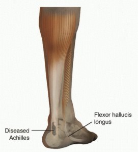
Tibialis posterior (inversion, plantarflexion, arch support), flexor digitorum longus (FDL), flexor hallucis longus (FHL) (toe flexion, plantarflexion).
*
Superficial Posterior Compartment:
Gastrocnemius, soleus (plantarflexion), plantaris. These unite to form the Achilles tendon.
*
Intrinsic Foot Muscles:
Contribute to arch support and fine motor control of the toes.
Neurovascular Structures
-
Nerves:
- Tibial Nerve: Branches into medial and lateral plantar nerves in the foot. Supplies posterior compartment muscles and plantar sensation. Vulnerable in tarsal tunnel, medial ankle approaches.
-
Common Peroneal Nerve:
Divides into superficial and deep peroneal nerves.
- Superficial Peroneal Nerve: Supplies lateral compartment muscles, sensation to dorsum of foot. Vulnerable laterally, especially during fibular approaches.
- Deep Peroneal Nerve: Supplies anterior compartment muscles, sensation to 1st web space. Vulnerable anteriorly, in anterior ankle approaches.
- Sural Nerve: Sensory nerve supplying lateral foot and ankle. Vulnerable in posterolateral approaches and fibular incisions.
- Saphenous Nerve: Sensory nerve for medial leg and foot. Vulnerable in medial ankle approaches.
-
Vessels:
- Posterior Tibial Artery: Becomes medial and lateral plantar arteries. Pulse palpable posteromedially.
- Dorsalis Pedis Artery: Continuation of anterior tibial artery. Pulse palpable dorsally, lateral to EHL.
- Peroneal Artery: Supplies lateral compartment and fibula.
Biomechanics
The foot and ankle complex functions as a rigid lever during propulsion and a flexible adapter to uneven surfaces. Key biomechanical principles include:
*
Kinetic Chain:
Motion at one joint influences others.
*
Weight-Bearing:
Distribution of forces through the tibia, talus, and calcaneus, then through the midfoot to the metatarsal heads.
*
Arch Mechanics:
The medial longitudinal arch, supported by ligaments (spring ligament, plantar fascia) and tendons (tibialis posterior, FHL), is crucial for shock absorption and propulsion.
*
Gait Cycle:
Defined phases (heel strike, stance, toe-off) involve complex interplay of muscle contractions and joint movements, emphasizing controlled motion.
Indications & Contraindications
The decision for surgical intervention in foot and ankle pathology is multifactorial, balancing the potential benefits of improved function and pain relief against the risks of surgery and potential complications.
General Indications for Operative Intervention
- Failure of conservative management despite adequate trial (e.g., persistent pain, instability, progressive deformity).
- Acute limb-threatening conditions (e.g., open fractures, compartment syndrome, irreducible dislocations).
- Conditions with high risk of poor outcome with non-operative management (e.g., unstable fractures, severe deformities, certain tendon ruptures).
- Neurological deficits directly correctable by surgery (e.g., nerve entrapment).
- Unacceptable functional limitation impacting daily activities or desired lifestyle.
Specific Operative Indications (Examples)
- Fractures: Unstable ankle fractures (displaced bimalleolar, trimalleolar, syndesmotic disruption), displaced pilon fractures, displaced calcaneal fractures (intra-articular depression, widening), displaced talar fractures (neck, body), unstable Lisfranc injuries, displaced metatarsal/phalangeal fractures interfering with function.
- Ligamentous Instability: Chronic lateral ankle instability unresponsive to physical therapy, acute syndesmotic injuries (high ankle sprains) with instability on stress radiographs/MRI.
- Degenerative Conditions: Severe, symptomatic arthritis of the ankle, subtalar, or midfoot joints (failed conservative measures, pain at rest, significant functional limitation), often requiring arthrodesis or arthroplasty (for ankle).
- Deformities: Progressive adult acquired flatfoot deformity (AAFD) with flexible hindfoot valgus and midfoot collapse, severe cavus foot, severe hallux valgus with pain and functional limitations, symptomatic hammertoe deformities.
- Tendon Pathologies: Acute Achilles tendon rupture (often in active individuals), chronic posterior tibial tendon insufficiency with progressive deformity, peroneal tendon ruptures.
- Infections: Septic arthritis of foot/ankle joints, osteomyelitis requiring debridement and culture-directed therapy.
Specific Non-Operative Indications (Examples)
- Stable, non-displaced fractures (e.g., stable unimalleolar ankle fractures, non-displaced distal fibula fractures, non-displaced stress fractures).
- Mild to moderate sprains and strains without significant instability.
- Tendonitis, fasciitis, and other inflammatory conditions responsive to rest, anti-inflammatory medications, physical therapy, and orthotics.
- Mild degenerative changes or early-stage arthritis with good response to activity modification, bracing, NSAIDs, and injections.
- Asymptomatic or mildly symptomatic deformities.
- Non-operative management may also be chosen based on patient preference, despite surgical indications, after thorough discussion of risks and benefits.
Contraindications
-
Absolute Contraindications:
- Severe, uncontrolled medical comorbidities precluding safe anesthesia and surgery (e.g., acute myocardial infarction, uncontrolled heart failure, severe pulmonary disease).
- Active infection in the surgical field (e.g., cellulitis, severe soft tissue infection, unless surgery is for source control).
- Critical limb ischemia with compromised healing potential (unless revascularization is planned).
- Anesthetic contraindications.
-
Relative Contraindications:
- Poor skin integrity or significant soft tissue swelling precluding safe incision and closure, especially in acute trauma (often requires delayed surgery).
- Uncontrolled diabetes mellitus (increased risk of infection, wound healing complications, Charcot arthropathy).
- Active smoking (significantly impairs wound healing and increases non-union rates).
- Poor nutritional status.
- Unrealistic patient expectations regarding surgical outcome or recovery timeline.
- Significant neurological deficits or cognitive impairment that prevent adherence to post-operative protocols.
Summary of Operative vs. Non-Operative Indications
| Category | Operative Indications | Non-Operative Indications |
|---|---|---|
| Fractures | Unstable, displaced (>2mm), open, syndesmotic disruption, intra-articular step-off, significant comminution | Stable, non-displaced (<2mm), closed, hairline, stress fractures |
| Ligamentous | Chronic symptomatic instability, syndesmotic instability | Acute sprains (Grade I/II), mild instability, good response to PT |
| Degenerative | Severe pain/functional limitation, failed conservative Tx, advanced arthrosis | Mild-moderate pain, early OA, good response to conservative Tx |
| Deformity | Progressive, symptomatic, rigid, failed bracing, neurological deficit | Mild, asymptomatic, flexible, accommodative |
| Tendon Injury | Acute rupture (Achilles, PTT), chronic insufficiency with progressive deformity | Tendonitis, partial tears (responsive to PT/bracing) |
| Infection | Septic arthritis, osteomyelitis (source control, debridement) | Cellulitis (antibiotics only), superficial wound infection |
| General Patient | Otherwise healthy, compliant, realistic expectations | Significant comorbidities, poor healing potential, non-compliance |
Pre-Operative Planning & Patient Positioning
Meticulous pre-operative planning and appropriate patient positioning are critical for optimizing surgical outcomes, minimizing complications, and ensuring operative efficiency.
Clinical Assessment
- History: Detailed medical history focusing on comorbidities (diabetes, peripheral vascular disease, neurological conditions), medication use (anticoagulants, steroids), smoking status, allergies, previous surgeries, and social support. Assess functional limitations, pain characteristics, and patient expectations.
- Physical Examination: Comprehensive evaluation of the affected limb including skin integrity, soft tissue envelope (swelling, blistering, open wounds), neurovascular status (pulses, sensation, motor function), range of motion, stability, and assessment for deformity. For acute trauma, serial examinations are crucial to monitor swelling and watch for compartment syndrome.
Imaging
- Radiographs: Standard views (AP, lateral, mortise for ankle; AP, lateral, oblique for foot) are essential. Stress radiographs (e.g., external rotation stress views for syndesmosis, gravity stress for Lisfranc) can assess instability. Specific views (e.g., Broden's for subtalar joint, Harris heel view for calcaneus) are used for specific pathologies.
- Computed Tomography (CT): Gold standard for assessing complex fracture patterns (pilon, calcaneus, talus), intra-articular involvement, non-unions, and pre-operative templating for arthrodesis. Often performed with 3D reconstructions.
- Magnetic Resonance Imaging (MRI): Best for evaluating soft tissue injuries (ligaments, tendons, cartilage), osteonecrosis, stress fractures, and occult pathology.
- Angiography/Doppler Ultrasound: Indicated if there are concerns for vascular compromise.
Surgical Plan Development
- Diagnosis & Pathoanatomy: Confirm diagnosis, thoroughly understand the injury pattern or deformity.
- Approach Selection: Determine optimal surgical approach based on pathology, ensuring adequate exposure, protection of neurovascular structures, and avoidance of compromised soft tissue.
- Reduction Strategy: Plan the sequence of reduction maneuvers (e.g., direct vs. indirect, ligamentotaxis) and temporary fixation methods (e.g., K-wires, external fixator).
- Implant Selection: Choose appropriate implants (screws, plates, wires, intramedullary nails) based on fracture morphology, bone quality, biomechanical stability, and patient factors.
- Pre-operative Templating: Essential for reconstructive procedures (e.g., osteotomies, arthrodesis, total ankle arthroplasty) to plan cuts, implant sizes, and desired alignment.
- Staged Procedures: For severe open fractures or pilon fractures with significant soft tissue swelling, a staged protocol (e.g., temporary external fixation followed by definitive ORIF) is often necessary.
- Pre-habilitation: For elective cases, optimizing patient health (smoking cessation, glycemic control, nutritional support, weight loss, physical therapy) can improve outcomes.
- Antibiotic Prophylaxis: Administer pre-operatively per institutional guidelines.
- DVT Prophylaxis: Assess risk factors and initiate prophylaxis (mechanical/pharmacological) as appropriate.
Patient Positioning
Proper positioning ensures surgical access, patient safety, and optimal fluoroscopic imaging.
*
General Considerations:
*
Padding:
All pressure points must be meticulously padded to prevent neuropathies (e.g., peroneal nerve at fibular head, ulnar nerve at elbow) and skin breakdown.
*
Tourniquet:
Typically applied to the proximal thigh, inflated to 100 mmHg above systolic blood pressure or a standardized pressure (e.g., 250-300 mmHg) for approximately 90-120 minutes.
*
C-Arm Access:
Ensure unrestricted access for intra-operative fluoroscopy from multiple angles.
*
Leg Holder/Support:
Often used to stabilize the operative leg and provide optimal positioning.
*
Common Positions:
*
Supine:
Most common for anterior/anteromedial ankle approaches, forefoot surgery, medial malleolus fixation. Allows access to both lower extremities if needed.
*
Lateral Decubitus:
Useful for posterolateral approaches to the ankle or hindfoot (e.g., fibular fractures, posterior malleolus, calcaneus) or for lateral hindfoot procedures. The unaffected limb is padded and positioned anteriorly.
*
Prone:
Used for posterior approaches (e.g., Achilles repair, posterior pilon fractures, posterior ankle arthroscopy). Requires careful airway management and abdominal support.
*
Beach Chair/Semi-Fowler:
Less common but may be used for certain hindfoot procedures where a more upright patient position is desired.
*
Specific Positioning for Ankle Fracture ORIF:
*
Supine:
Leg holder on the contralateral side or an ankle bump under the ipsilateral hip for internal rotation. A small bump under the ipsilateral calf may facilitate ankle dorsiflexion.
*
Tourniquet:
High thigh.
*
Fluoroscopy:
Positioned for AP, lateral, and mortise views without repositioning the limb.
Detailed Surgical Approach / Technique
Given the vast array of foot and ankle pathologies, a comprehensive description of every surgical technique is beyond the scope of this guide. Instead, we will outline general principles and illustrate a detailed example using open reduction and internal fixation (ORIF) of an unstable ankle fracture, which is a common and representative procedure.
General Principles of Foot & Ankle Surgery
- Aseptic Technique: Strict adherence to sterile protocol to minimize infection risk.
- Delicate Soft Tissue Handling: Crucial given the often superficial nature of foot and ankle anatomy and the common compromise of the soft tissue envelope, especially in trauma. Minimize retraction, avoid crushing, and ensure meticulous hemostasis.
- Anatomical Reduction: Restoration of normal joint congruity and bone alignment is paramount for long-term function and to prevent post-traumatic arthritis.
- Stable Fixation: Achieved with appropriate implants to allow for early motion and promote bone healing.
- Neurovascular Protection: Careful identification and protection of superficial nerves and vessels.
- Internervous/Intermuscular Planes: Utilizing these planes minimizes muscle damage and facilitates exposure.
- Fluoroscopic Guidance: Essential for confirming reduction and implant placement intraoperatively.
Example: Open Reduction Internal Fixation (ORIF) of a Supination-External Rotation (SER) Type IV Ankle Fracture
This common fracture pattern typically involves a spiral oblique fibular fracture at or above the level of the syndesmosis, a medial malleolus fracture or deltoid ligament rupture, and potential posterior malleolus involvement, with associated syndesmotic disruption.
1. Pre-operative Preparation
- Patient supine with ipsilateral hip bump or contralateral leg holder. Tourniquet on high thigh.
- Prepare and drape the extremity from mid-thigh to toes.
- Administer prophylactic antibiotics.
2. Incision & Exposure
-
Lateral Malleolus:
A curvilinear incision centered over the distal fibula, typically 8-10 cm long, parallel to the fibula. Start just distal to the tip of the lateral malleolus and extend proximally.
- Dissection: Incise skin and subcutaneous tissue. Carefully identify and protect branches of the superficial peroneal nerve (often anterior to the fibula). Dissect down to the fibular shaft, identifying the fracture hematoma.
- Internervous Plane: Anteriorly, between the peroneus brevis/longus and the extensor digitorum longus (deep peroneal nerve). Posteriorly, between the peroneals and the soleus/flexor hallucis longus (sural nerve, small saphenous vein).
-
Medial Malleolus (if fractured):
A separate curvilinear incision over the medial malleolus, parallel to the bone.
- Dissection: Incise skin and subcutaneous tissue. Carefully identify and protect the saphenous nerve and great saphenous vein (anterior). Dissect to the fracture site, opening the fracture hematoma.
3. Fibular Reduction & Fixation
- Cleaning: Debride hematoma and any soft tissue interposition from the fracture site.
-
Reduction:
- Restore fibular length, typically by distracting the fracture fragments and aligning the posterior border of the fibula with the posterior border of the tibia (incisura fibularis).
- Correct fibular rotation. The fibula often malrotates internally with external rotation injuries.
- Correct any medial or lateral translation.
- Temporary Fixation: Hold reduction with a bone clamp (e.g., pointed reduction clamp) or K-wires.
-
Definitive Fixation:
- Lag Screws: If the fibular fracture is amenable to interfragmentary compression (e.g., long oblique or spiral fracture), place one or two lag screws perpendicular to the fracture plane.
- Neutralization Plate: Apply a 1/3 tubular plate, locking plate, or contoured reconstruction plate to the lateral or posterior-lateral surface of the fibula. The plate neutralizes bending, rotational, and axial forces. Secure with cortical screws. Ensure distal screws capture adequate bone in the malleolus.
- Intra-operative Fluoroscopy: Confirm fibular length, rotation, and alignment on AP, lateral, and mortise views. Ensure the distal fibula is centered in the incisura.
4. Medial Malleolus Reduction & Fixation
- Cleaning: Clear hematoma from the fracture.
- Reduction: Anatomically reduce the medial malleolus fracture.
-
Fixation:
- Cancellous Screws: Typically, two 4.0 mm or 3.5 mm cancellous screws are placed perpendicular to the fracture line, often from the anterior cortex of the malleolus into the tibial shaft. Ensure adequate length and purchase.
- Tension Band Wiring: For small, comminuted medial malleolus fragments, tension band wiring can be an effective alternative.
- Deltoid Repair: If the deltoid ligament is ruptured and the medial malleolus is intact, direct repair may be indicated, often using suture anchors or transosseous sutures.
5. Syndesmotic Assessment & Fixation (if indicated)
-
Assessment:
After fibular and medial fixation, assess syndesmotic stability.
- Manual Stress Test: Apply external rotation or lateral translation stress to the talus while holding the tibia and fibula reduced. Look for widening of the clear space between the fibula and tibia (clear space > 6mm or > 2mm difference from contralateral).
- "Cotton Test" (Hook Test): A hook placed in the fibula to test for lateral translation.
- Fluoroscopic Views: Compare clear space and tibiofibular overlap to the uninjured side.
-
Fixation (if unstable):
- Positioning: Dorsiflex the ankle to neutral to prevent over-compression.
- Screw Placement: Typically 1-2 cortical syndesmotic screws (3.5 mm or 4.5 mm) are placed, usually 2-3 cm proximal to the plafond, engaging 3 or 4 cortices.
- Dynamic Fixation: Suture button devices (e.g., TightRope) are increasingly used, allowing for physiologic micromotion and avoiding routine removal.
- Fluoroscopic Confirmation: Confirm syndesmotic screw position and reduction.
6. Posterior Malleolus (if fractured)
- If the fragment is large (>25% of articular surface) or significantly displaced, fixation is indicated.
- Approach: Can be approached posterolaterally (between peroneals and Achilles, or between FHL and peroneals), or directly through the lateral incision for anterior-to-posterior screws.
- Fixation: Lag screws from anterior-to-posterior (through the fibula or through the anterolateral incision) or posterior-to-anterior buttress plating.
7. Final Check & Closure
- Perform a final fluoroscopic check in multiple views (AP, lateral, mortise) to ensure anatomical reduction of all fractures, appropriate implant placement, and restoration of ankle mortise congruity.
- Irrigate wounds thoroughly.
- Close subcutaneous tissue and skin in layers.
- Apply sterile dressings and a well-padded splint (e.g., posterior slab with or without sugar tong) in a neutral position.
Complications & Management
Despite meticulous surgical technique, foot and ankle surgery is associated with a range of potential complications. Proactive recognition and management are crucial for salvage and optimizing patient outcomes.
Common Complications
-
Surgical Site Infection (SSI):
- Incidence: 1-5% for elective cases, significantly higher for open fractures (up to 20-30%) or cases with extensive soft tissue damage. Risk factors include diabetes, smoking, poor nutrition, prolonged surgery, and immunosuppression.
-
Management:
- Superficial: Oral antibiotics, local wound care.
- Deep/Periprosthetic: Surgical debridement, irrigation, tissue culture-directed IV antibiotics. Hardware retention possible if fixation is stable and infection is superficial or acute. If chronic or hardware-related, hardware removal often necessary, potentially with staged reconstruction. Negative pressure wound therapy (NPWT) can be beneficial.
-
Wound Dehiscence/Skin Necrosis:
- Incidence: 5-10%, higher in areas of thin skin, poor vascularity, or significant soft tissue swelling (e.g., pilon fractures, calcaneus).
- Management: Local wound care, serial debridement. NPWT. If conservative measures fail, may require skin grafting or local/regional flap coverage. Delayed primary closure is an option for early dehiscence.
-
Non-Union/Delayed Union:
- Incidence: 5-15%, depending on bone, fracture type, and patient factors (smoking, NSAIDs, diabetes). Higher in pilon fractures, talar neck fractures, and comminuted fractures.
- Management: Optimize patient factors (smoking cessation). Non-operative options include prolonged immobilization, bracing, bone stimulators. Surgical options include revision ORIF with debridement of non-union site, bone grafting (autograft or allograft), biologics (e.g., PRP, BMPs), or conversion to arthrodesis.
-
Malunion:
- Incidence: 5-20%, leading to altered biomechanics, joint overload, and post-traumatic arthritis. Common in ankle fractures (fibular shortening/malrotation) and calcaneal fractures (varus, widening).
- Management: Conservative management (orthotics, bracing) for mild, asymptomatic malunions. Corrective osteotomy for symptomatic, significant malunions. Arthrodesis for severe symptomatic arthritis secondary to malunion.
-
Hardware Failure/Prominence:
- Incidence: <5% for true failure (bending, breakage) but prominence is common, especially subcutaneously.
- Management: Hardware removal if symptomatic prominence, after bone healing is complete. Revision fixation if true hardware failure contributes to non-union or instability.
-
Nerve Injury:
- Incidence: <1-5%, often transient neurapraxia due to traction or direct trauma. Superficial peroneal, sural, saphenous, and deep peroneal nerves are most vulnerable.
- Management: Observation for neurapraxia. If persistent or severe deficit, nerve conduction studies/EMG. Exploration, neurolysis, or nerve repair/grafting in severe cases. Gabapentinoids for neuropathic pain.
-
Vascular Injury:
- Incidence: Rare but serious, can lead to compartment syndrome or limb loss.
- Management: Immediate surgical exploration and vascular repair. Fasciotomy for compartment syndrome.
-
Post-Traumatic Arthritis (PTA):
- Incidence: Highly variable, 10-50% for intra-articular fractures (ankle, pilon, talus, calcaneus, Lisfranc). Can develop years after injury despite anatomical reduction.
- Management: Conservative (NSAIDs, PT, bracing, injections) for early stages. Surgical options include arthroscopic debridement, osteotomies, distraction arthroplasty, arthrodesis, or total ankle arthroplasty.
-
Complex Regional Pain Syndrome (CRPS) Type I:
- Incidence: 2-5% after lower extremity trauma/surgery.
- Management: Multidisciplinary approach: physical therapy, pain management (nerve blocks, gabapentinoids, TCAs), psychological support. Early recognition and aggressive PT are key.
-
Deep Vein Thrombosis (DVT) / Pulmonary Embolism (PE):
- Incidence: 1-10% (lower extremity trauma/surgery, especially if prolonged immobilization or hypercoagulable state).
- Management: Anticoagulation per guidelines. IVC filter in rare cases of contraindication to anticoagulation or recurrent PE. Prophylaxis (mechanical/pharmacological) is critical.
Table of Common Complications and Management Strategies
| Complication | Incidence (%) | Salvage Strategy |
|---|---|---|
| Surgical Site Infection (SSI) | 1-5 (elective), 10-30 (open Fx) | Debridement, antibiotics (IV/PO), hardware removal (if chronic), NPWT, wound closure/flaps |
| Wound Dehiscence/Necrosis | 5-10 (higher in high-energy trauma) | Local wound care, debridement, NPWT, skin graft, flap reconstruction |
| Non-Union/Delayed Union | 5-15 (bone/Fx dependent) | Revision ORIF, bone grafting, biologics, external fixation, arthrodesis |
| Malunion | 5-20 | Orthotics, bracing, corrective osteotomy, arthrodesis |
| Hardware Failure/Prominence | <5 (failure), common (prominence) | Hardware removal (if symptomatic), revision fixation (if unstable) |
| Nerve Injury | <1-5 (usually transient) | Observation, neurolysis, nerve repair/graft, gabapentinoids |
| Post-Traumatic Arthritis | 10-50 (intra-articular Fx) | Conservative Tx, arthroscopy, osteotomy, arthrodesis, total ankle arthroplasty |
| CRPS Type I | 2-5 | Multidisciplinary pain management, PT, nerve blocks, psychological support |
| DVT/PE | 1-10 | Anticoagulation, IVC filter (rare), mechanical/pharmacological prophylaxis |
Post-Operative Rehabilitation Protocols
Post-operative rehabilitation is a cornerstone of recovery following foot and ankle surgery, directly impacting functional outcomes. Protocols are tailored to the specific surgery, bone/soft tissue healing constraints, implant stability, and individual patient factors. A phased approach is generally employed.
Phase I: Protection & Early Motion (Typically Weeks 0-6)
- Goals: Protect surgical repair/fixation, manage pain and edema, maintain motion in non-operated joints, initiate early protected range of motion (ROM) as indicated.
-
Immobilization:
- Initial period in a bulky soft dressing, posterior splint, or rigid cast to minimize swelling and protect the surgical site.
- Transition to a controlled ankle motion (CAM) boot, short leg cast, or similar protective device.
-
Weight-Bearing (WB) Status:
- Non-Weight Bearing (NWB): Standard for most fracture fixations, fusions, or significant soft tissue repairs, particularly in the initial 4-6 weeks to allow for initial soft tissue and bone healing. Crutches, walker, or knee scooter are used.
- Touchdown Weight Bearing (TDWB) / Partial Weight Bearing (PWB): May be initiated earlier for stable fixation, certain arthroplasties, or stable non-operative injuries, to encourage muscle activation and proprioception without excessive load.
- Edema & Pain Management: Elevation, ice, compression, analgesics (opioid/non-opioid), NSAIDs (caution with bone healing in NWB phase).
-
Range of Motion:
- Passive ROM (PROM) / Active-Assisted ROM (AAROM): May be initiated very early for certain arthroplasties (e.g., total ankle arthroplasty) or stable osteotomies.
- Active ROM (AROM): For adjacent joints (e.g., knee, hip, toes) to prevent stiffness. Avoid direct stress on the surgical site.
- Strengthening: Gentle isometric exercises for thigh and gluteal muscles. Early core strengthening.
- Patient Education: Emphasize adherence to WB restrictions, signs of complications, and importance of elevation.
Phase II: Progressive Strengthening & Range of Motion (Typically Weeks 6-12)
- Goals: Restore full pain-free ROM, progressively increase strength and endurance, normalize gait pattern, improve proprioception.
-
Weight-Bearing Progression:
- Gradual transition from NWB/TDWB to full weight-bearing (FWB) in a CAM boot or supportive shoe. This is typically guided by radiographic evidence of healing and clinical stability.
- Use of assistive devices (crutches/cane) is weaned as strength and balance improve.
- Range of Motion: Aggressive focus on restoring ankle dorsiflexion, plantarflexion, inversion, and eversion. Manual therapy, stretching, and self-mobilization techniques.
-
Strengthening:
- Isometric: Progress to isotonic exercises (resistance bands, light weights) for ankle dorsiflexors, plantarflexors, invertors, and evertors.
- Concentric & Eccentric: Focus on controlled movements.
- Gastroc-Soleus Complex: Calf raises (bilateral, then unilateral) are critical.
- Proprioception & Balance: Single-leg stance, wobble board, balance beam exercises.
- Gait Training: Focus on normal heel-toe gait pattern, stride length, and cadence.
Phase III: Functional Integration & Return to Activity (Typically Weeks 12+)
- Goals: Maximize strength, power, endurance, agility, and sport-specific function. Gradual return to desired activities.
-
Activity Progression:
- Cardiovascular: Stationary cycling, swimming, elliptical trainer.
- Impact Loading: Progress from walking to light jogging, then running, incorporating changes in direction.
- Plyometrics: Box jumps, hopping, jumping drills (advanced).
- Advanced Strengthening: Increased resistance training, sport-specific drills, agility ladders.
- Dynamic Balance & Proprioception: Advanced balance exercises, complex motor tasks.
- Orthotics/Bracing: Custom or off-the-shelf orthotics may be prescribed to support arches, correct alignment, or reduce stress on healing tissues. Ankle bracing may be used for sport.
-
Return to Work/Sport Criteria:
- Full pain-free ROM.
- Strength within 85-90% of the uninjured limb.
- Good balance and proprioception.
- Ability to perform activity-specific tasks without pain or compensatory mechanisms.
- Psychological readiness.
- Often requires objective testing (e.g., isokinetic dynamometry, functional hop tests).
Specific Considerations
- Arthrodesis: Prolonged NWB (10-12 weeks) until radiographic evidence of fusion, followed by gradual WB in a boot. Emphasize adjacent joint mobility.
- Total Ankle Arthroplasty (TAA): Early motion protocols are common to preserve implant motion, with protected WB initially.
- Achilles Tendon Repair: Controlled ankle motion in a boot with progressive plantarflexion from equinus to neutral over several weeks, followed by gradual WB and strengthening.
- Ligament Reconstruction: Immobilization followed by progressive ROM and strengthening, with particular emphasis on proprioception and dynamic stability.
Summary of Key Literature / Guidelines
Evidence-based practice forms the foundation of modern orthopedic surgery and rehabilitation. Several key studies and guidelines have shaped the management of foot and ankle pathologies.
Ankle Fractures
- Weber Classification: Remains a cornerstone for describing fibular fractures relative to the syndesmosis, guiding stability assessment.
- Lauge-Hansen Classification: While complex, provides insight into injury mechanisms (e.g., supination-external rotation, pronation-abduction) for surgical planning.
- Syndesmotic Injury: Management remains an area of ongoing debate. While traditional fixation with tricortical screws was common, recent literature increasingly supports quadricortical screws and dynamic fixation devices (e.g., TightRope) due to potential benefits in preserving physiological motion and reducing the need for hardware removal (Naqvi et al., 2012; Schepers et al., 2017). The role of routine syndesmotic screw removal is debated, with many studies suggesting it is not always necessary unless symptomatic.
- Early Weight-Bearing: Studies have shown that early protected weight-bearing in stable, operatively treated ankle fractures (e.g., in a CAM boot) can lead to faster functional recovery and potentially improved patient satisfaction without increasing complication rates (Lehtonen et al., 2018).
Pilon Fractures
- Staged Protocol: Due to severe soft tissue compromise, a staged approach with initial external fixation for soft tissue healing followed by definitive ORIF (often 7-14 days later) is the accepted standard (Sirkin et al., 1999).
- Minimally Invasive Techniques: ORIF using limited open approaches or arthroscopically-assisted techniques are emerging to minimize soft tissue dissection and improve outcomes (Helfet et al., 2005).
- Outcomes: Recognize the high rates of post-traumatic arthritis, malunion, and infection regardless of optimal treatment, emphasizing the need for robust counseling.
Lisfranc Injuries
- Anatomic Reduction: Crucial for good outcomes. Any displacement >2mm significantly increases the risk of post-traumatic arthritis (Kuo et al., 2000). ORIF or primary arthrodesis for purely ligamentous injuries are common.
- Fixation: Traditionally, transarticular screw fixation was used, but controversies exist regarding optimal screw placement, number, and timing of removal. Dorsal plate fixation or suture-button devices are gaining popularity for better stability and allowing earlier weight-bearing.
Tendon Repairs (e.g., Achilles)
- Early Mobilization vs. Immobilization: Meta-analyses demonstrate that early functional rehabilitation (protected weight-bearing, controlled motion) following Achilles tendon repair leads to improved functional outcomes, reduced re-rupture rates, and quicker return to activity compared to prolonged immobilization (Kearney & Costa, 2010).
Arthrodesis vs. Arthroplasty
- Ankle Arthritis: For end-stage ankle arthritis, both ankle arthrodesis and total ankle arthroplasty (TAA) are viable options. TAA is increasingly favored in appropriate patients (lower demand, good bone stock, minimal deformity) due to motion preservation. However, arthrodesis remains the gold standard for high-demand patients, failed TAA, or severe deformities/bone loss (Saltzman et al., 2005).
- Subtalar Arthrodesis: A common procedure for painful subtalar arthritis, often performed percutaneously or with limited incision. Outcomes are generally good for pain relief, but adjacent joint arthritis is a long-term concern.
General Guidelines
- Orthopedic Trauma Association (OTA) and American Academy of Orthopaedic Surgeons (AAOS): These organizations regularly publish evidence-based clinical practice guidelines and instructional courses covering a wide range of foot and ankle pathologies.
- Cochrane Reviews: Provide systematic reviews and meta-analyses, often highlighting gaps in current literature and guiding future research.
The continuously evolving literature underscores the importance of staying abreast of new techniques, implant technologies, and rehabilitation strategies to provide optimal care for patients with foot and ankle conditions. Individualized treatment plans, based on sound biomechanical principles, robust evidence, and patient-specific factors, remain paramount for successful outcomes.
You Might Also Like