Elbow Cases Biceps: Your Guide to Tendonitis Diagnosis & Relief
Key Takeaway
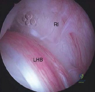
In this comprehensive guide, we discuss everything you need to know about Elbow Cases Biceps: Your Guide to Tendonitis Diagnosis & Relief. The Long head of the biceps tendonitis is characterized by anterior shoulder pain radiating to the biceps, worsened by overhead movements. Often observed in young athletes or those with repetitive use, diagnosis relies on specific physical findings. While this focuses on shoulder pain, a comprehensive understanding of biceps pathology is vital for assessing various musculoskeletal conditions, including **elbow cases biceps** pain and other associated issues.
A 44-year-old manual laborer presents with an acute elbow injury following a sudden eccentric load to a flexed, supinated forearm. Describe your clinical assessment and the significance of the "Biceps Crease Interval".
Candidate: I would assess for the classic signs of a distal biceps rupture: pain, swelling, and ecchymosis in the antecubital fossa. I'd perform a Hook Test to look for discontinuity. The Biceps Crease Interval is the distance from the antecubital crease to the distal biceps muscle belly; a normal value is under 6cm. A measurement of 8cm suggests a complete rupture with significant proximal retraction.
Failing to mention the specific functional deficits—specifically that the biceps is the primary supinator. A weak candidate might focus only on flexion strength. Failing to perform a neurovascular exam, particularly of the Lateral Antebrachial Cutaneous nerve (LABCN) and Posterior Interosseous Nerve (PIN), is a critical omission in an FRCS setting.
I would perform a focused clinical exam: 1. Visual: Look for the "reverse Popeye" deformity and ecchymosis tracking along the fascial planes. 2. Palpation: Specifically assess the Biceps Crease Interval; >6cm is indicative of rupture. 3. Provocative testing: Use the Hook Test and passive pronation test. 4. Functional: Quantify the supination strength deficit, which is pathognomonic as the distal biceps is the primary supinator. 5. NV Status: Explicitly check LABCN sensation (lateral forearm) and PIN motor function (thumb extension) as baseline before surgical intervention.
Given the patient's MRI findings, which confirm a complete rupture with retraction, evaluate this radiograph and discuss the surgical considerations regarding fixation.
Candidate: The MRI shows a full-thickness avulsion with significant proximal retraction of the tendon stump. I would opt for an anatomic reattachment to the radial tuberosity. I would use a cortical button and an interference screw to provide high load-to-failure strength and early mobilization.
Ignoring the "two-bundle" anatomy. The examiner wants to know that you understand the short head (flexion) and long head (supination) footprints. Failing to discuss the risk of PIN injury during drilling or mentioning the need for "maximal supination" during the procedure is a major oversight.
In this active patient, anatomic repair is indicated to restore supination torque. I would favor a single-incision technique using a cortical button and interference screw (tension-slide construct). - Biomechanical rationale: Provides the highest load to failure and aperture-level fixation. - Technical points: Maximal forearm supination during the approach and drilling is non-negotiable to protect the PIN. - Footprint: I would ensure the reattachment encompasses the native footprints for both the short and long heads. - Complications: Meticulous irrigation to avoid heterotopic ossification and careful retraction of the LABCN.
What are the primary risks associated with the single-incision versus the two-incision approach, and how do you mitigate the risk of radioulnar synostosis?
Candidate: The two-incision (Boyd-Anderson) approach carries a higher risk of radioulnar synostosis because of the subperiosteal dissection of the ulna. The single-incision approach has a risk of LABCN injury. To prevent synostosis, I use irrigation to remove bone debris and avoid over-drilling.
Candidates often miss the specific mechanism of synostosis: the creation of a "cross-talk" of bone marrow and osteoprogenitor cells between the radius and ulna. They also fail to mention the importance of the "posterior cortical bridge" when drilling.
The two-incision approach provides excellent anatomic exposure but is inherently linked to higher rates of heterotopic ossification (HO) due to the ulnar dissection. The single-incision approach is now preferred but risks damage to the LABCN and the PIN.
Mitigating Synostosis: 1. Copious irrigation: Flushes out bone debris and osteoprogenitor cells. 2. Construct management: Using an interference screw to seal the socket prevents the extravasation of marrow contents. 3. Technique: Avoid subperiosteal stripping of the ulna; keep the dissection limited. 4. Pharmacology: Consider a short course of prophylactic NSAIDs, balancing the risk of HO against the potential for delayed tendon-to-bone healing.
