Distal Femoral Fractures: Epidemiology, Anatomy, Biomechanics, and Surgical Principles
Key Takeaway
Distal femoral fractures (DFX) occur near the knee. Management involves anatomical reduction, stable fixation, and alignment restoration, guided by AO/OTA classification. Understanding surgical anatomy, biomechanics, and neurovascular risks is crucial for optimizing outcomes, restoring joint mechanics, and preventing post-traumatic arthritis and stiffness.
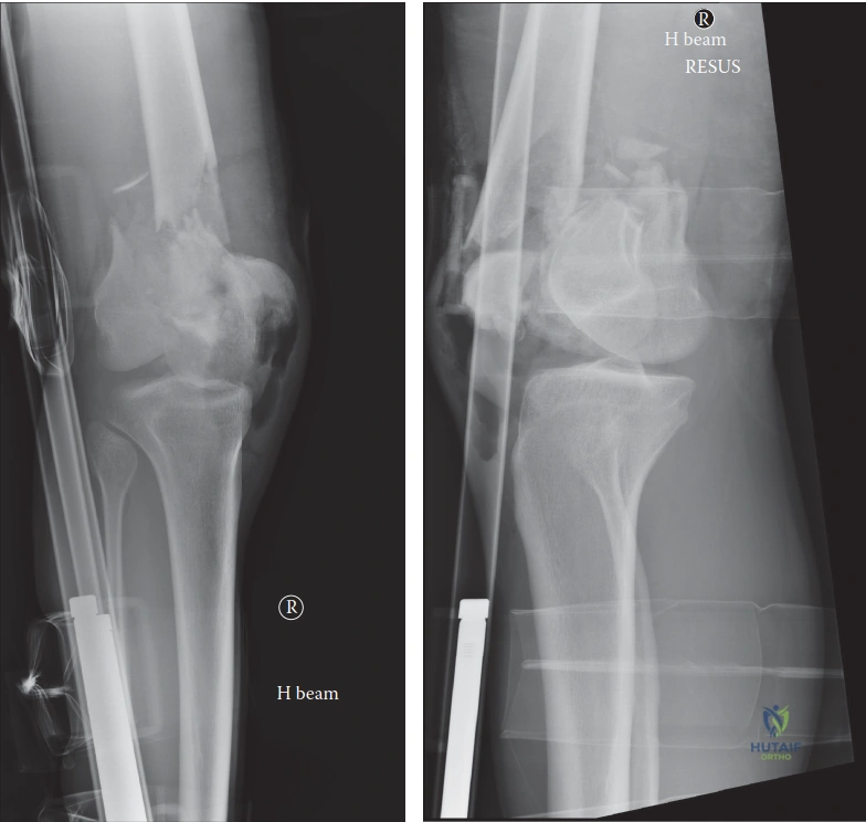
A 78-year-old patient presents following a ground-level fall. They are currently on the ward with a distal femoral fracture. Looking at the radiographs, how would you classify this injury, and what are the specific clinical implications of this fracture pattern in this patient demographic?
Candidate: I would classify this using the AO/OTA system. Based on the radiographs, it is a Type C complete articular fracture. In this elderly patient, the main implications are poor bone quality (osteoporosis), which makes stable fixation challenging, and the need for early mobilization to avoid systemic complications like VTE or chest infections.
Focusing purely on the fracture anatomy (e.g., "it's a supracondylar fracture") without addressing the "geriatric context." Failing to mention the mechanical challenges of locking screw purchase in osteoporotic bone or the physiological necessity of early weight-bearing in the elderly.
A structured response: 1. **Classification:** AO/OTA Type C (complete articular involvement). 2. **Biological/Mechanical Factors:** Highlight the "dual challenge"—osteoporotic bone stock reducing screw purchase and comminution making anatomical reduction difficult. 3. **Functional Goals:** Emphasize that in the elderly, the goal is "fixation for mobilization." 4. **Clinical Strategy:** Mention the potential need for augmentation (bone graft/cement) or, in extreme cases of comminution, the consideration of primary distal femoral replacement if the bone is too poor for stable internal fixation.
You have decided to proceed with open reduction and internal fixation (ORIF) using a lateral locking plate. Describe the surgical approach, the internervous plane, and how you would specifically manage the gastrocnemius muscle's deforming force during your reduction.
Candidate: I would use a lateral approach, specifically the subvastus or lateral parapatellar approach. The internervous plane is between the vastus lateralis (femoral nerve) and the lateral intermuscular septum (sciatic/peroneal nerve). To manage the gastrocnemius, which pulls the distal fragment posteriorly, I would use either a femoral distractor or knee flexion to relax the muscle and allow for reduction of the posterior tilt.
Forgetting to mention the specific deforming force of the gastrocnemius. Candidates often suggest manual traction alone, which is rarely sufficient to overcome the strong pull of the gastrocnemius on the distal segment.
Detail the **subvastus** approach for its biological advantages. Clearly state the **internervous plane** (Vastus lateralis vs. Lateral intermuscular septum). For the **gastrocnemius pull**: emphasize the use of a "joystick" K-wire in the condylar fragment or a femoral distractor to restore length and correct the apex-posterior angulation before applying the plate. Mention using the patella as a clinical reference for rotational alignment.
Six months post-operatively, the patient returns. The radiographs show a "non-union" at the supracondylar level with hardware breakage. How do you approach the management of this failed fixation?
Candidate: I would first rule out an occult infection with inflammatory markers and potentially a biopsy. If not infected, I would revise the fixation. I'd likely use a longer, stiffer plate or consider exchange nailing, and I would definitely add a bone graft, either autograft from the iliac crest or a bone substitute, to address the biological deficit.
Proceeding straight to "more metal" without ruling out infection. Hardware breakage is often a sign of mechanical failure secondary to biological failure (non-union) or infection; you must exclude sepsis before attempting revision.
A systematic "Diamond Concept" approach: 1. **Exclude Infection:** Mandatory bloods (CRP/ESR) and discussion of aspiration/biopsy. 2. **Mechanical Stability:** Address why the original failed—likely too short a plate or inadequate working length. Suggest a longer plate to bridge the defect. 3. **Biology:** The necessity of bone grafting (autograft is gold standard) to convert an atrophic non-union into a healing environment. 4. **Patient Factors:** Metabolic workup (Vitamin D, PTH, smoking status) to ensure the patient can heal this time.
