Midshaft Clavicle Fractures: An Evidence-Based Guide to Diagnosis, Management, and Outcomes
Key Takeaway
Operative management for midshaft clavicle fractures is indicated for open fractures, neurovascular compromise, or severe skin compromise. Relative indications include significant displacement (>100%), shortening (>1.5-2cm), severe comminution, and nonunion, especially in high-demand patients, aiming to restore anatomical alignment and optimize functional outcomes.
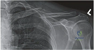
A 28-year-old male cyclist presents to the Emergency Department following a high-energy fall onto his right shoulder. He has pain and palpable deformity in his mid-clavicular region. Radiographs are provided below.
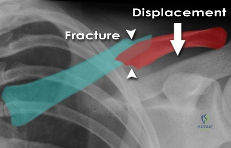
How do you classify this fracture, and what are the specific clinical implications of the displacement seen here?
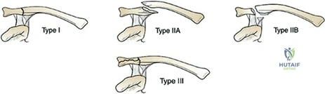
Candidate: This is a midshaft clavicle fracture. Based on the Robinson classification, it looks like a Type 2B fracture because it is displaced. The displacement is caused by the SCM pulling the medial fragment up and the weight of the arm pulling the lateral fragment down. The main concern is shortening, which can lead to nonunion or poor shoulder function.
The candidate identifies the fracture correctly but fails to provide a systematic, high-level analysis. They miss quantifying the shortening (the "1.5 to 2.0 cm" threshold is critical), ignore the biomechanical implications (scapular dyskinesia), and fail to mention the specific Robinson sub-classification (2B1 vs 2B2).
This is a Robinson Type 2B midshaft clavicle fracture. Type 2 indicates midshaft, and 'B' denotes displacement. Specifically, it appears to be a 2B1 (single butterfly) or 2B2 (comminuted) depending on the fragments. Clinically, I am assessing for >1.5 to 2.0 cm of shortening, which is a key threshold in the literature associated with increased nonunion rates and poor functional outcomes. The deforming forces—the SCM pulling the medial fragment superiorly and the deltoid/arm weight pulling the lateral fragment inferiorly—result in shortening that can compromise glenohumeral and scapulothoracic kinematics, potentially leading to rapid fatigability and weakness. I would also perform a neurovascular exam to rule out injury to the underlying subclavian vessels and brachial plexus.
The patient elects for operative management. You are planning the surgical approach. Please discuss your choice of plate placement and the relevant anatomical risks during dissection.
Candidate: I would use a pre-contoured locking plate. Superior plating is good for tension, but anteroinferior is safer for the nerves. I need to watch out for the supraclavicular nerves when I make the incision and be careful with the drill so I don't hit the subclavian vein.
The candidate is vague on the biomechanical trade-offs. Examiners look for a discussion of the "tension side" (superior) vs. "neurovascular safety" (anteroinferior). They also must mention the platysma closure and the specific risks of nerve injury causing post-op patient dissatisfaction.
I would choose between superior and anteroinferior plating based on fracture morphology. Superior plating is biomechanically advantageous as it is on the tension side, but it is associated with a higher risk of symptomatic hardware prominence and soft tissue irritation. Anteroinferior plating is safer because the screws are directed posterosuperiorly, away from the subclavian vessels and pleura. During dissection, the primary risk is injury to the supraclavicular nerves, which run perpendicular to the clavicle; I would protect these to minimize post-operative chest wall numbness. I would avoid excessive periosteal stripping to preserve the blood supply and minimize nonunion risk. Finally, I would use at least three bicortical screws per fragment for a stable construct.
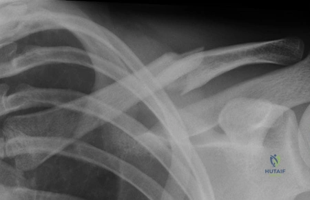
Six months post-operatively, the patient returns. His fracture has not healed, and there is persistent pain at the fracture site. What is your management, and how do you prevent this in future cases?
Candidate: If it's a nonunion, I would suggest revising the surgery. I'd perform an ORIF again, maybe use some bone graft if needed, and make sure the hardware is tight.
The answer is too simplistic. It fails to distinguish between the biology of the nonunion (atrophic vs. hypertrophic) and lacks a structured plan for revision, such as the use of autologous bone grafting (iliac crest) or ensuring the mechanical stability of the construct.
First, I would classify the nonunion. If it is atrophic, the primary issue is biology; I would perform debridement of the nonunion site, rigid internal fixation, and add autologous bone graft, typically from the iliac crest. If it is hypertrophic, the issue is mechanical instability; I would focus on more rigid fixation—perhaps upgrading to a larger locking plate or adding orthogonal plates. To prevent this, I ensure meticulous preservation of the periosteal blood supply during the index procedure, avoid over-stripping fragments, achieve a minimum of 6 cortices of purchase on each side, and ensure the patient understands the post-operative rehabilitation protocol to avoid premature loading.
