Subtrochanteric Femur Fractures: Comprehensive Guide to Epidemiology, Surgical Anatomy, and Biomechanics
Key Takeaway
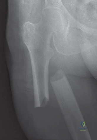
Subtrochanteric femur fractures are complex proximal femoral injuries located 5cm below the lesser trochanter. They are challenging due to bimodal epidemiology (high-energy/young, low-energy/elderly), powerful deforming muscular forces causing significant displacement, and their unique biomechanical environment. Understanding surgical anatomy is paramount for successful management and preventing complications like nonunion.
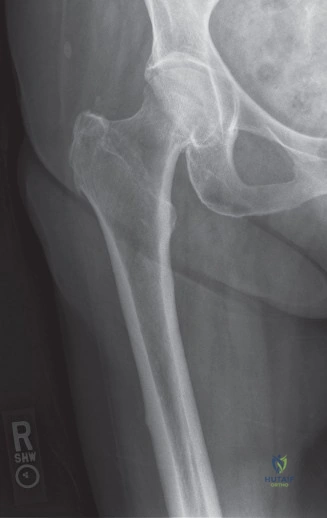
A 78-year-old female presents with an isolated, closed, low-energy subtrochanteric femur fracture after a ground-level fall. Her radiographs demonstrate a transverse fracture pattern with medial cortical thickening of the subtrochanteric region. What is your primary diagnosis, and what are the immediate clinical management priorities?
Candidate: The primary diagnosis is an Atypical Femoral Fracture (AFF), likely secondary to long-term bisphosphonate use. My priorities are to assess the contralateral femur for similar radiographic features, check the patient's medication history, and plan for surgical stabilization with a long intramedullary nail. I would also order a CT scan to delineate the comminution and initiate metabolic bone workup.
The candidate focuses solely on the fracture fixation (e.g., "I'd put a nail in it"). They fail to mention the high risk of bilaterality, the diagnostic criteria for AFFs, or the need to stop the offending medication, missing the systemic "bone health" aspect of the pathology.
Systematically address: 1) Diagnosis: Recognize this as an AFF based on the lateral cortical thickening and transverse pattern. 2) Bilateral Assessment: MANDATORY full-length femur imaging of the contralateral side, as there is a high risk of synchronous or metachronous fracture. 3) Management: Prophylactic or definitive fixation with a long cephalomedullary nail. 4) Systemic: Discontinuation of bisphosphonates, referral to metabolic bone/endocrinology, and checking vitamin D/calcium levels.
During your attempt at closed reduction of a comminuted subtrochanteric fracture, the proximal fragment remains flexed and abducted. Despite traction and adduction, you cannot achieve a satisfactory reduction. What anatomical deforming forces are at play, and what technical maneuvers can you employ to achieve reduction?
Candidate: The iliopsoas flexes the proximal fragment, while the abductors cause abduction and external rotation. To achieve reduction, I would increase hip flexion on a radiolucent table, use a proximal Schanz pin as a joystick, or employ blocking (Poller) screws to guide the nail.
Failing to mention the specific mechanism of the blocking screw. A failing candidate might suggest "using an open approach" too early, ignoring indirect reduction techniques that preserve biology.
Structure the response by anatomy: Deforming Forces: Iliopsoas (flexion), Glutei (abduction), External rotators (ER). Reduction Maneuvers: 1) Positioning (flexing the hip to relax the iliopsoas). 2) Percutaneous "joysticking" using a Schanz pin in the proximal fragment. 3) Blocking Screws: Medial or anterior Poller screws to prevent the nail from malaligning the fragment. 4) If these fail, limited open reduction with minimal soft-tissue stripping, potentially using a cerclage wire (though note the risk to periosteal blood supply).
You have successfully nailed a subtrochanteric fracture. Describe the specific biomechanical reasons why you would choose a long intramedullary nail over a short nail in this patient.
Candidate: Long nails are preferred because they span the entire femoral shaft. This avoids stress concentration at the tip of the nail, which is a common site for periprosthetic or iatrogenic shaft fractures, especially in a long-term, high-load-bearing area like the subtrochanteric zone.
Focusing only on "it's stronger." Examiners want to hear the specific concept of "stress risers" at the tip of short implants and the biomechanical benefit of spanning the diaphysis.
The long nail is superior because: 1) Stress Riser: A short nail terminates in the middle of the diaphysis, creating a significant stress riser susceptible to future fracture. 2) Load Sharing: It provides a continuous load-sharing construct across the entire femoral length. 3) Stability: It controls rotation more effectively than a short nail. 4) Distal Fixation: It allows for distal locking in the more metaphyseal bone of the distal femur, which is biomechanically advantageous.
