of Arthroscopy: Instrumentation, Techniques, and Complication Management
Key Takeaway
Arthroscopy has revolutionized orthopedic surgery by offering high diagnostic accuracy and low morbidity. This comprehensive guide details the fundamental principles of arthroscopy, including essential instrumentation, fluid management, and the critical triangulation technique. It also provides an evidence-based overview of indications, contraindications, and the management of intraoperative and postoperative complications, serving as an essential resource for orthopedic residents and practicing consultants.
Introduction to Arthroscopic Surgery
During the past five decades, arthroscopy has dramatically changed the orthopaedic surgeon’s approach to the diagnosis and treatment of a variety of joint ailments. A high degree of clinical accuracy, combined with low patient morbidity, accelerated rehabilitation, and decreased postoperative pain, has encouraged the widespread use of arthroscopy. Today, it is utilized not only to assist in diagnosis and determine prognosis but primarily to provide definitive therapeutic intervention.
Clinical Pearl: Arthroscopic procedures must serve as adjuncts to—and not as replacements for—thorough clinical evaluation. Advanced imaging and endoscopic visualization are never substitutes for meticulous history-taking and comprehensive physical examination skills.
Progressive improvements in the Hopkins rod-lens systems of arthroscopes, high-definition fiberoptic imaging, instrument miniaturization, and specialized accessory operative tools have made advanced operative arthroscopic techniques possible for virtually every joint in the body. This includes the knee, shoulder, hip, ankle, elbow, wrist, hand, and foot. Furthermore, endoscopic techniques have successfully transitioned into spinal surgery. However, while many arthroscopic procedures have proven superior to traditional open techniques, surgical outcomes and patient safety must never be sacrificed merely to expand the indications for minimally invasive surgery.
Instruments and Equipment
The modern arthroscopic suite requires a complex array of highly specialized equipment. Mastery of this equipment is mandatory for the safe and efficient execution of arthroscopic procedures.
The Arthroscope and Television Cameras
The standard arthroscope utilizes a Hopkins rod-lens system, providing a wide field of view and exceptional clarity.
* Diameters: The 4.0-mm arthroscope is the standard for large joints (knee, shoulder, hip). Smaller joints (ankle, elbow, wrist) require 2.7-mm or 1.9-mm scopes.
* Angles of Inclination: The 30-degree angled arthroscope is the workhorse of orthopedic endoscopy, allowing the surgeon to sweep the visual field by rotating the light post. The 70-degree arthroscope is essential for visualizing hard-to-reach areas, such as the posterior compartments of the knee or the inferior capsule of the shoulder.
High-definition (HD) and 4K television cameras, coupled with high-intensity xenon or LED light sources, transmit the intraarticular image to surgical monitors. Proper white-balancing and focusing prior to insertion are critical steps that must not be overlooked.
Accessory Instruments
The tactile feedback and mechanical precision of accessory instruments are the extensions of the surgeon's hands.
- The Probe: Often described as the "arthroscopist’s finger," the blunt hook probe is the most critical diagnostic tool. It is used to palpate articular cartilage for malacia, test the tension of ligaments (e.g., the ACL), and manipulate meniscal or labral tears to determine their depth and configuration.
- Scissors: Miniature scissors (straight and curved) are utilized for delicate tissue resection, such as dividing synovial plicae or trimming capsular adhesions.
- Basket Forceps (Punches): Available in various angles (straight, up-biting, rotary), basket forceps are used to resect meniscal tissue or labral fraying. The biting mechanism must be sharp to avoid tearing or pulling normal tissue.
- Grasping Forceps: Essential for retrieving loose bodies, removing resected tissue, and managing sutures during reconstructive procedures.
- Knife Blades: Disposable, shielded arthroscopic knives (e.g., retrograde knives, banana blades) are used for precise capsular releases or meniscal excisions.
Motorized Shaving Systems
Motorized shavers and burrs are indispensable for efficient tissue resection and bone debridement. These systems feature interchangeable blades (e.g., full-radius resectors, aggressive cutters) and operate in forward, reverse, or oscillating modes.
Surgical Warning: Always maintain direct visualization of the shaver tip when the motor is engaged. Blind shaving is a primary cause of iatrogenic injury to normal articular cartilage and ligamentous structures.
Electrosurgical, Laser, and Radiofrequency Instruments
Radiofrequency (RF) wands and electrocautery devices are utilized for hemostasis, capsuloligamentous ablation, and tissue shrinkage. Bipolar RF devices operate efficiently in a conductive saline environment.
* Pitfall: Excessive use of thermal energy can lead to chondrolysis or thermal necrosis of adjacent neurovascular structures. Continuous fluid flow must be maintained to dissipate heat.
Implants and Miscellaneous Equipment
The evolution of arthroscopy has birthed a vast array of specialized implants, including bioabsorbable and biocomposite suture anchors, interference screws, and meniscal repair devices. Miscellaneous equipment includes specialized cannulas, switching sticks, and knot pushers, all designed to facilitate complex intraarticular reconstructions.
Operating Room Setup and Patient Preparation
Care and Sterilization of Instruments
Arthroscopic instruments are delicate and highly susceptible to damage. The rod-lens system of the arthroscope can shatter if dropped or bent. Sterilization is typically achieved via autoclaving (for compatible instruments), ethylene oxide gas, or low-temperature hydrogen peroxide gas plasma systems. Strict adherence to institutional sterilization protocols is mandatory to prevent surgical site infections.
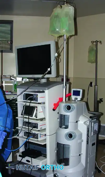
Irrigation Systems
A clear visual field depends entirely on adequate joint distention and continuous fluid irrigation. Normal saline or lactated Ringer's solution is typically used.
* Gravity Flow vs. Mechanical Pumps: While gravity flow (bags hung high above the patient) is safe and inexpensive, mechanical infusion pumps provide superior control over intraarticular pressure and flow rates.
* Hemostasis: Epinephrine (typically 1 mg per 3 liters of fluid) is frequently added to the irrigation fluid to induce local vasoconstriction and minimize bleeding.
Surgical Warning: High pump pressures can lead to massive fluid extravasation, potentially resulting in compartment syndrome. Pump pressures should generally be maintained below 40-50 mm Hg, or just high enough to overcome venous pressure and maintain a clear field.
Tourniquet and Leg Holders
For knee arthroscopy, a pneumatic tourniquet is routinely placed on the proximal thigh, though it is often left uninflated unless visualization is compromised by bleeding.
* Leg Holders: A circumferential leg holder or a lateral post is used to apply valgus or varus stress, opening the medial or lateral compartments of the knee. Care must be taken to pad the thigh adequately to prevent tourniquet paresis or crushing injuries to the soft tissues.
Anesthesia and Documentation
Arthroscopy can be performed under general, regional (spinal/epidural), or even local anesthesia, depending on the joint, the complexity of the procedure, and patient comorbidities.
Meticulous documentation is a medicolegal and clinical necessity. High-quality intraoperative photographs and video recordings should be captured, demonstrating the pathology identified and the definitive treatment rendered.
Clinical Application: Indications and Contraindications
Advantages
- Decreased postoperative pain and reduced need for narcotic analgesia.
- Lower morbidity and faster return to functional activities or sports.
- Superior visualization of intraarticular structures compared to open arthrotomy.
- Lower rates of surgical site infection and reduced scar formation.
Disadvantages
- Steep learning curve requiring specialized psychomotor skills.
- Dependence on complex, expensive, and potentially temperamental equipment.
- Risk of unique complications, such as fluid extravasation and instrument breakage.
Indications and Contraindications
Indications: Arthroscopy is indicated for the diagnosis and treatment of meniscal tears, ligamentous ruptures (e.g., ACL/PCL reconstruction), articular cartilage defects, loose bodies, synovial diseases, and labral pathology in the shoulder or hip.
Contraindications: Absolute contraindications include active superficial soft-tissue infection (cellulitis) overlying the portal sites, and severe joint ankylosis that prevents safe instrument insertion. Relative contraindications include severe degenerative joint disease where arthroscopy offers little therapeutic benefit.
Basic Arthroscopic Techniques
The Triangulation Technique
The fundamental psychomotor skill required for successful arthroscopy is triangulation. This technique involves bringing the arthroscope and the operating instrument together at a specific target within the joint, forming a triangle.
- Spatial Awareness: The surgeon must adapt to a two-dimensional monitor while operating in a three-dimensional space.
- The Fulcrum Effect: The skin portal acts as a fulcrum. To move the tip of the instrument down inside the joint, the surgeon’s hand outside the joint must move up.
- Execution: The arthroscope is introduced through a viewing portal and focused on the pathology. A spinal needle is often used to localize the optimal trajectory for the working portal. Once the working portal is established, the instrument is introduced blindly but directed toward the light of the arthroscope until it enters the visual field.
Clinical Pearl: Never advance an instrument forcefully if it is not in the field of view. If you lose sight of the instrument, withdraw it slightly, re-center the arthroscope, and slowly advance the instrument again.
Complications and Management
While arthroscopy is minimally invasive, it is not without risk. The overall complication rate is low (typically less than 1-2%), but complications can be devastating if not promptly recognized and managed.
Damage to Intraarticular Structures
- Damage to Menisci and Fat Pad: Iatrogenic scuffing of the articular cartilage or accidental resection of the anterior horn of the meniscus can occur during portal placement or aggressive shaving. The infrapatellar fat pad is highly vascular; aggressive debridement here can lead to postoperative scarring and anterior knee pain.
- Damage to Cruciate Ligaments: The anterior cruciate ligament (ACL) is vulnerable during intercondylar notch debridement. Motorized shavers must be used with extreme caution near the ACL and PCL to avoid partial or complete iatrogenic transection.
Damage to Extraarticular Structures
- Blood Vessels: Vascular injury is the most catastrophic complication in arthroscopy. In the knee, the popliteal artery is at risk during posterior horn meniscectomy, PCL reconstruction, or posterior capsular releases. In the shoulder, the axillary artery is at risk during inferior capsular work. Any suspicion of arterial injury mandates immediate vascular surgery consultation and angiography.
- Compartment Syndromes: Extravasation of irrigation fluid into the fascial compartments can cause acute compartment syndrome. This is most common during capsular releases, when capsular tears are present, or when high pump pressures are used. If the calf or thigh becomes tense and non-compressible, the procedure must be aborted, and fasciotomies may be required.
- Nerves: Portal placement puts superficial nerves at risk. The infrapatellar branch of the saphenous nerve is frequently injured during anteromedial or accessory medial portal placement in the knee, leading to numbness or painful neuromas. In the shoulder, the axillary and suprascapular nerves are at risk.
- Ligaments and Tendons: The medial collateral ligament (MCL) can be ruptured if excessive valgus force is applied to a stiff knee while attempting to open the medial compartment.
Hemarthrosis and Thrombophlebitis
Postoperative hemarthrosis is common following extensive synovectomy or lateral retinacular release. It presents as a tense, painful, swollen joint. Management includes aspiration, compressive wrapping, and ice.
Deep vein thrombosis (DVT) and thrombophlebitis are rare but recognized complications. Prophylaxis should be considered in patients with a history of hypercoagulability, prolonged tourniquet times, or those undergoing complex reconstructive procedures.
Infection
Septic arthritis following arthroscopy is rare (incidence < 0.5%) but represents a surgical emergency. It typically presents 3 to 7 days postoperatively with severe, out-of-proportion pain, erythema, and a dramatic decrease in range of motion. Diagnosis is confirmed via joint aspiration (cell count > 50,000 WBCs/mm³ with > 90% polymorphonuclear leukocytes). Treatment requires emergent arthroscopic irrigation and debridement (I&D) and targeted intravenous antibiotic therapy.
Tourniquet Paresis
Prolonged tourniquet inflation or excessive pressure can lead to neuropraxia or tourniquet paresis. Tourniquet time should ideally be kept under 90 to 120 minutes. If more time is required, the tourniquet should be deflated for 15 to 20 minutes to allow tissue reperfusion before reinflation.
Synovial Herniation and Fistulas
Failure to adequately close capsular defects or skin portals can result in synovial fistulas (persistent fluid drainage) or synovial herniation. Persistent fistulas increase the risk of intraarticular infection and may require secondary surgical closure if conservative management (immobilization and compression) fails.
Instrument Breakage
The breakage of delicate arthroscopic instruments (e.g., the tip of a basket forceps or a probe) inside the joint is a highly stressful complication.
Surgical Pitfall: If an instrument breaks, the surgeon must immediately stop all movement and turn off the fluid outflow/suction to prevent the fragment from migrating.
The arthroscope should be kept focused on the broken fragment. A magnetic retriever or a sturdy grasping forceps should be introduced through the working portal to extract the piece. If the fragment is lost, intraoperative fluoroscopy must be utilized to locate it. A joint must never be closed with a retained broken instrument fragment unless exhaustive, documented attempts at retrieval have failed and the patient is subsequently informed.
You Might Also Like