ABOS Part I Orthopaedic Review: TKA Revision & Hallux Valgus Surgical Management | Part 21607

Key Takeaway
This ABOS Part I Orthopaedic Review module provides 20 advanced MCQs for ABOS Part I and AAOS OITE exams. It covers critical topics in Total Knee Arthroplasty (TKA) Revision, such as aseptic loosening and bone loss, and comprehensive Hallux Valgus surgical management, including osteotomies and complication strategies, derived from high-yield clinical cases.
ABOS Part I Orthopaedic Review: TKA Revision & Hallux Valgus Surgical Management | Part 21607
A 65-year-old patient presents with increasing pain and instability in their left knee, 10 years after a primary total knee arthroplasty. The provided radiographs are shown.

Correct Answer: C
The radiographs clearly demonstrate widespread lucency around both the femoral and tibial components, indicating loss of fixation. There is significant subsidence of the tibial component and extensive osteolysis, particularly in the proximal tibia and distal femur. These findings are classic for aseptic loosening. While periprosthetic joint infection (PJI) is always in the differential for a painful TKA, the radiographic findings alone are more definitively characteristic of aseptic loosening and associated bone loss. There is no clear evidence of a periprosthetic fracture, and the extent of loosening and osteolysis goes beyond early polyethylene wear or isolated patellar issues.
A 72-year-old patient presents with a painful left total knee arthroplasty. Radiographs are provided. The surgeon notes extensive bone loss involving a major portion of both the distal femoral condyles and the proximal tibial plateau, requiring significant augmentation and likely long-stemmed components.
Correct Answer: C
The Anderson Orthopaedic Research Institute (AORI) classification for bone loss is crucial in revision TKA planning. Type 1 involves intact metaphyseal bone with minor defects. Type 2 involves damaged metaphyseal bone with loss of cancellous bone, categorized as 2A (one condyle/plateau) or 2B (both condyles/plateaux). Type 3 describes deficient metaphyseal bone where bone loss comprises a major portion of either condyle or plateau, often associated with ligament detachment and requiring long-stemmed implants, bone grafts, or custom prostheses. The case description explicitly states "extensive bone loss involving a major portion of both the distal femoral condyles and the proximal tibial plateau," which directly corresponds to AORI Type 3 for both the femur and tibia. The radiographs support this with widespread osteolysis and significant bone defects.
Following the diagnosis of aseptic loosening, the candidate expresses concern about the state of the collateral ligaments, particularly the MCL, and the extensive bone loss. The patient is planned for revision surgery.
Correct Answer: C
The case explicitly states, "The collateral ligaments are likely to be dysfunctional and especially the MCL therefore a constrained knee replacement may be required." The constraint ladder for knee implants progresses from PCL retaining (CR) to PCL substituting (PS), then to unlinked constrained condylar (VVC), and finally to linked, constrained condylar (RHK). A VVC implant, such as an LCCK or TC3, is specifically designed to provide anteroposterior and varus-valgus stability, substituting for deficient collateral ligaments. While an RHK (Linked, Constrained Condylar) is used for global instability or severe bone loss/fracture, the primary concern highlighted is dysfunctional collateral ligaments, making VVC the most appropriate initial step up in constraint for this specific issue. CR and PS designs do not adequately address significant collateral ligament insufficiency.
The patient, described as "reasonably young," has extensive bone loss around the failed total knee replacement. The surgeon is planning the revision procedure.
Correct Answer: C
The case explicitly states, "In this patient who is reasonably young restoration of bone stock is preferable, because of likelihood of further revision surgery." For younger patients with extensive bone loss, preserving and restoring bone stock is paramount to facilitate potential future revisions. This is best achieved through the use of bone grafting (structural or morselized) and/or modular metaphyseal sleeves, which allow for biological ingrowth and reconstruction of the metaphyseal bone. While cement, modular augments, or even hinged prostheses can address defects, they do not primarily restore bone stock. Avoiding complex bone grafting for the sake of surgical time would compromise the long-term outcome and future revision potential in a young patient.
The lateral radiograph of the failed total knee replacement clearly demonstrates anterior femoral cortical notching.
Correct Answer: C
Anterior femoral cortical notching is a well-recognized complication of total knee arthroplasty, typically resulting from over-resection of the distal femur or improper sizing of the femoral component during the primary surgery. This notching creates a stress riser in the anterior femoral cortex, significantly weakening the bone and increasing the risk of a supracondylar periprosthetic femoral fracture, especially with minor trauma. It is not directly indicative of infection, polyethylene wear, or PCL insufficiency, nor is it a benign finding.
The candidate initially suggests that the radiographs are "suggestive of infection until proven otherwise," despite the examiner later confirming aseptic loosening.
Correct Answer: A
In the initial workup of a painful total knee arthroplasty, differentiating between aseptic loosening and periprosthetic joint infection (PJI) is critical. Elevated inflammatory markers such as erythrocyte sedimentation rate (ESR) and C-reactive protein (CRP) are highly sensitive indicators of PJI. While not definitive on their own, they are the most indicative of infection among the given options. A continuous radiolucent line greater than 2mm and progressive component subsidence are classic radiographic signs of aseptic loosening. Pain with activity that improves with rest is a common symptom of mechanical loosening. Anterior femoral cortical notching is a technical complication related to fracture risk, not directly to infection.
The examiner asks about the advantages of PCL substituting (PS) over PCL retaining (CR) designs.

Correct Answer: C
The case explicitly lists several advantages of PCL substituting (PS) designs over PCL retaining (CR) designs. These include: conforming surfaces allowing roll-back, no component slide, providing a degree of VVC (varus-valgus constraint), and the cam-post mechanism improving anterior-posterior stability. PS designs also facilitate deformity correction, use more congruent joint surfaces (reducing wear), offer better range of motion, and are often considered technically easier and more reproducible. Options A and B are generally associated with CR designs (PCL preservation potentially leading to less bone resection and more natural kinematics), while options D and E are not recognized advantages of PS designs; in fact, PS designs can sometimes have specific patellofemoral issues related to the box cut, and are generally considered technically simpler than CR in terms of balancing.
The patient has extensive bone loss, concerns about collateral ligament integrity, and calcified soft tissues. The surgeon is planning a single-stage revision.
Correct Answer: C
The candidate in the case explicitly states, "The soft tissues appear contracted and calcified which may lead to wound complications." This highlights a critical concern for revision surgery, where soft tissue quality is often compromised. Meticulous soft tissue management, including careful dissection, debridement, and thoughtful wound closure techniques, is essential to minimize the risk of wound complications, infection, and skin necrosis. A standard primary TKA system would be inadequate for extensive bone loss and ligamentous instability. A minimally invasive approach is generally not suitable for complex revisions with significant bone loss and soft tissue issues. Avoiding stemmed components would compromise stability and fixation in the presence of extensive bone loss. Relying solely on cement fixation might not be sufficient for severe bone defects, especially in a young patient where bone ingrowth is desired for long-term stability.
The radiographs demonstrate calcification of soft tissues, including the popliteal vessels.
Correct Answer: C
The presence of soft tissue calcification, particularly involving the popliteal vessels, is a significant finding. The candidate in the case notes that "The soft tissues appear contracted and calcified which may lead to wound complications." Beyond wound complications, calcification of major vessels like the popliteal artery increases the risk of intraoperative vascular injury during dissection, especially in a revision setting with distorted anatomy. It also suggests underlying peripheral vascular disease, which can impair wound healing and increase the risk of other postoperative complications. While heterotopic ossification is a possibility, the immediate and most critical concern for surgical planning relates to vascular integrity and wound healing.
The patient is described as "reasonably young" with extensive bone loss, and the examiner emphasizes the importance of long-term planning.
Correct Answer: C
The case explicitly states, "In this patient who is reasonably young restoration of bone stock is preferable, because of likelihood of further revision surgery." For younger patients, the expectation of a longer lifespan means a higher probability of requiring additional revision surgeries in the future. Therefore, the primary long-term goal in the current revision is to preserve and, if possible, restore the existing bone stock. This approach ensures that there is adequate bone available for subsequent procedures, making future revisions technically feasible and potentially more successful. While range of motion, cost, and hospital stay are important, they are secondary to the fundamental goal of bone stock management in a young patient with extensive defects. An RHK is not indicated in all cases of extensive bone loss; the choice depends on the specific type of instability and bone defect.
A 65-year-old lady presents with bilateral hallux valgus. You obtain weight-bearing radiographs. Based on the provided AP radiograph of the right foot, which of the following findings is most consistent with the described deformity (intermetatarsal angle 15°, hallux valgus angle 35°, and minimal passive correction)?
Correct Answer: C
The case describes a significant hallux valgus deformity with an intermetatarsal angle of 15° (normal < 9°) and a hallux valgus angle of 35° (normal < 15°). These severe angular deformities are typically associated with significant lateral subluxation of the sesamoid complex, which is a hallmark of first metatarsal pronation and hallux valgus progression. The AP radiograph (Figure 4.7) would visually confirm this lateral displacement of the sesamoids relative to the first metatarsal head.
Option A (Normal sesamoid position) is incorrect because severe hallux valgus invariably involves sesamoid subluxation.
Option B (Medial sesamoid subluxation) is incorrect; sesamoid subluxation in hallux valgus is typically lateral.
Option D (Widening of the first metatarsophalangeal joint space) is generally not seen in hallux valgus; rather, joint space narrowing might indicate arthritic changes, or the joint space might appear normal or slightly compressed laterally.
Option E (Absence of arthritic changes) is not guaranteed. While the case doesn't explicitly state arthritic changes on the right foot, a long-standing deformity of this magnitude often has some degree of degenerative change, and the question asks for the finding most consistent with the *described deformity*, which is primarily angular and rotational, leading to sesamoid subluxation.
For the 65-year-old lady with an intermetatarsal angle of 15°, a hallux valgus angle of 35°, and minimal passive correction of the hallux on her right foot, assuming a normal first tarsometatarsal joint, what is the most appropriate surgical plan?
Correct Answer: C
The case explicitly states that for an intermetatarsal angle of 15° and a hallux valgus angle of 35° with minimal passive correction, the candidate would plan a scarf osteotomy combined with a lateral release and an Akin osteotomy of the proximal phalanx if necessary. This combination addresses the severe intermetatarsal angle, the significant hallux valgus angle, and the lack of passive correction. The Scarf osteotomy is chosen for its versatility and ability to achieve substantial correction without significant shortening, which is crucial for severe deformities.
Option A (Chevron osteotomy) is typically reserved for mild to moderate deformities (IM angle < 13-15°, HV angle < 30-35°) and would likely be inadequate for the described severe deformity.
Option B (Mitchell osteotomy) is also a distal osteotomy and, as mentioned in the case, produces shortening of the first metatarsal, which could lead to transfer metatarsalgia, making it less desirable for severe corrections.
Option D (First metatarsophalangeal joint arthrodesis) is a salvage procedure or indicated for severe arthritis, recurrent deformity, or in specific cases like the patient's left foot with a missing second toe, but not as a primary procedure for a correctable deformity with a normal TMT joint.
Option E (Keller resection arthroplasty) is generally reserved for elderly, low-demand patients with significant MTP joint arthritis and is associated with complications like hallux cock-up deformity and loss of push-off strength, making it less suitable for this active patient with a correctable deformity.
Following the decision to perform a Scarf osteotomy for the patient's right foot, the examiner asks why this procedure was chosen over simpler distal osteotomies. Which of the following is the most compelling reason for selecting a Scarf osteotomy in this specific case?
Correct Answer: C
The case explicitly states the advantages of a Scarf osteotomy: 'It is a very versatile procedure with stable fixation allowing postoperative mobilization without a cast. It maintains length of the metatarsal but allows translation, angulation and depression of the metatarsal head as necessary. It can also be used to shorten or even lengthen the metatarsal.' The candidate further explains that for the described degree of deformity (IM 15°, HV 35°) and lack of passive correction, a distal osteotomy would provide inadequate correction, and a Mitchell osteotomy specifically produces shortening, which could lead to transfer metatarsalgia. Therefore, the Scarf's versatility in achieving greater correction while maintaining metatarsal length is the key advantage.
Option A (Simpler procedure with faster recovery) is incorrect. Scarf osteotomy is a more complex procedure than distal osteotomies, though it does allow early weightbearing due to stable fixation.
Option B (Primarily addresses metatarsal shortening) is incorrect. Scarf osteotomy *maintains* metatarsal length and can even lengthen it, which is an advantage over procedures like Mitchell that cause shortening. Shortening is generally undesirable as it can lead to transfer metatarsalgia.
Option D (Less stable fixation) is incorrect. The case states it provides 'stable fixation allowing postoperative mobilization without a cast.'
Option E (Only osteotomy that can be combined with an Akin) is incorrect. An Akin osteotomy is a proximal phalangeal osteotomy and can be combined with various metatarsal osteotomies to address residual hallux valgus interphalangeus.
When discussing the proposed Scarf osteotomy with the patient, which of the following complications is specifically mentioned in the case as a potential risk that is difficult to treat?
Correct Answer: D
The candidate explicitly states, 'I would mention the possibility of hallux varus as a complication as this is difficult to treat.' This directly answers the question.
Option A (Superficial infection) is mentioned as a risk, but not specifically highlighted as 'difficult to treat' in the same context as hallux varus.
Option B (Recurrence of the deformity) is mentioned as possible, especially in adolescent cases, but not described as 'difficult to treat' in the same way as hallux varus.
Option C (Significant stiffness of the MTP joint) is mentioned as a possibility for a minority of patients, but again, not characterized as 'difficult to treat' in the same manner as hallux varus.
Option E (Intraoperative metatarsal fracture) is mentioned as a possibility, but it's an acute intraoperative event, not a chronic post-operative complication described as 'difficult to treat' in the long term.
A few months post-Scarf osteotomy, the patient develops a flexible hallux varus deformity. Which of the following is the most appropriate initial management strategy for this complication, as discussed in the case?
Correct Answer: D
The case states, 'A subtle varus may improve as the patient returns to normal foot wear.' This indicates that for a flexible or subtle varus, initial non-operative management with observation and appropriate footwear is the first step. The case then describes soft tissue procedures (EHL slip transfer, abductor hallucis and medial capsular release) for *flexible* deformity, and arthrodesis for *significant stiffness or arthrosis*. Since the question specifies a 'flexible' deformity, the initial approach would be the least invasive.
Option A (Immediate revision surgery with MTP joint arthrodesis) is incorrect. Arthrodesis is reserved for significant stiffness or arthrosis, not for a flexible deformity, especially as an initial step.
Option B (Transfer of a slip of the Extensor Hallucis Longus (EHL) tendon) and Option C (Abductor hallucis and medial capsular release) are soft tissue procedures described for *flexible* deformity, but they would typically follow initial conservative measures if the varus does not resolve with footwear changes.
Option E (Resection of the medial eminence and lateral capsular plication) is a procedure for hallux valgus, not hallux varus. Hallux varus involves the hallux deviating medially, so a lateral capsular plication would worsen it, and medial eminence resection is for the bunion deformity.
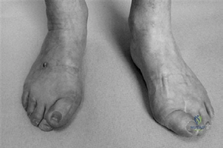
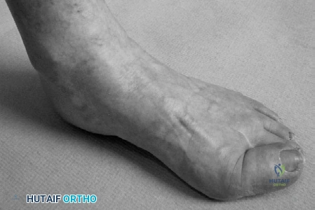
After successful treatment of the right foot, the patient's left foot, which previously underwent second toe removal and has recurrent hallux valgus symptoms, requires surgical intervention. Considering the oblique view of the left foot and the patient's history, what is the most appropriate surgical recommendation for the left hallux?
Correct Answer: C
The case explicitly addresses this scenario: 'So you have successfully treated this lady’s right foot and she is pleased with the result. Would you go ahead and do the same on the left? CANDIDATE: No. The absence of the second toe predisposes to recurrence and I would propose arthrodesis of the hallux MTP joint.' The absence of the second toe significantly alters the biomechanics of the forefoot, increasing the risk of recurrence with traditional hallux valgus correction procedures. Arthrodesis provides a stable, definitive correction in such complex cases.
Option A (Repeat Scarf osteotomy) is incorrect because the case specifically advises against repeating the same procedure due to the altered anatomy and high recurrence risk.
Option B (Chevron osteotomy) is a less powerful osteotomy and would also be prone to recurrence in this setting.
Option D (Proximal phalangeal osteotomy (Akin) alone) only addresses the hallux valgus interphalangeus component and would be insufficient for the overall hallux valgus deformity, especially with the added complexity of a missing second toe.
Option E (Resection arthroplasty) is generally reserved for very low-demand patients or those with severe arthritis and is associated with significant functional limitations and potential for hallux cock-up deformity, making arthrodesis a more reliable option for stability and function in this context.
During the physical examination for hallux valgus, the candidate outlines several key assessment points. Which of the following is NOT explicitly mentioned as a component of the physical examination in the case discussion?
Correct Answer: D
The candidate's description of the physical examination includes: 'I would examine the gait and the posture of the weighted foot... I would palpate for areas of tenderness, paying particular attention to the hallux MTP joint and lesser metatarsal heads. I would assess the degree of active and passive correction possible and the range of movement of the involved joints and look for gastrocnemius tightness. I would also perform a grind test to assess pain from loading the MTP joint. Neurovascular status must also be assessed.'
While 'gastrocnemius tightness' is mentioned, which involves the ankle, a general 'assessment of ankle range of motion' beyond this specific test is not explicitly listed as a primary component of the hallux valgus examination in the provided text. The 'range of movement of the involved joints' refers specifically to the hallux MTP and first TMT joints.
Options A, B, C, and E are all explicitly mentioned in the candidate's detailed description of the physical examination.
The examiner asks about the normal range of key radiographic angles in hallux valgus. Based on the AP radiograph and the case discussion, which statement accurately describes the normal values for the intermetatarsal angle (IMA), hallux valgus angle (HVA), and distal metatarsal articular angle (DMAA)?
Correct Answer: B
The case explicitly states the normal ranges for these angles: 'The intermetatarsal angle is normally less than 9. The hallux valgus angle should be less than 15. The distal metatarsal articular angle is normally a maximum of 15 from perpendicular to the axis of the first metatarsal.'
Options A, C, D, and E present incorrect combinations or values for the normal ranges as stated in the case.
The candidate emphasizes obtaining a detailed history, including questioning about relevant conditions. Which of the following conditions is NOT specifically mentioned in the case as a relevant condition to inquire about during the history for a patient presenting with hallux valgus?
Correct Answer: D
The candidate states: 'I would complete the history, including questioning about relevant conditions such as diabetes, inflammatory arthritis, vascular disease and neuropathy...'
Options A, B, C, and E are all explicitly mentioned in the case as relevant conditions to inquire about during the history.
Option D (Gout) is a common condition that can affect the MTP joint and mimic or exacerbate hallux valgus symptoms, and would typically be part of a comprehensive history. However, it is not explicitly listed in the specific conditions mentioned by the candidate in this teaching case, making it the correct answer to the question 'NOT specifically mentioned'.
The candidate describes securing the Scarf osteotomy with two headless compression screws. Which of the following statements accurately describes a key mechanical advantage or characteristic of the Scarf osteotomy, as discussed in the case?
Correct Answer: D
The case describes the Scarf osteotomy as 'a very versatile procedure with stable fixation allowing postoperative mobilization without a cast. It maintains length of the metatarsal but allows translation, angulation and depression of the metatarsal head as necessary. It can also be used to shorten or even lengthen the metatarsal.'
Option A (Primarily achieves correction through shortening) is incorrect. The case emphasizes that it 'maintains length' and avoids the shortening seen with procedures like Mitchell osteotomy.
Option B (Simple distal osteotomy, limiting application to mild deformities) is incorrect. The case highlights its versatility and suitability for more severe deformities, contrasting it with distal osteotomies that would be 'inadequate' for the described deformity.
Option C (Provides unstable fixation, necessitating prolonged cast immobilization) is incorrect. The case states it provides 'stable fixation allowing postoperative mobilization without a cast.'
Option E (Its main benefit is the ability to lengthen the first metatarsal without affecting other planes of correction) is partially correct in that it *can* lengthen, but the primary benefit highlighted is its versatility in allowing translation, angulation, and depression while *maintaining* length, which is a more comprehensive description of its advantages for correction.
Intraoperatively, the surgeon notes AORI Type III bone loss on both the femur and tibia, with complete incompetence of the medial collateral ligament. Which of the following implant constraints is most appropriate?
Which of the following procedures is most appropriate to address the interphalangeal deformity?
Which of the following is the most biomechanically sound fixation method for the tibial component?
Which of the following is the most appropriate corrective action?
None
