Surgical Management of the Rheumatoid Foot and Ankle: A Comprehensive Operative Guide
Key Takeaway
The surgical management of the rheumatoid foot and ankle requires a comprehensive understanding of altered biomechanics, progressive deformity, and compromised soft tissue envelopes. This guide details evidence-based operative strategies, including forefoot reconstruction via first metatarsophalangeal arthrodesis and lesser metatarsal head resection, as well as hindfoot arthrodesis techniques. Mastery of these procedures ensures optimal pain relief, restoration of plantigrade alignment, and improved functional outcomes in patients with advanced rheumatoid arthritis.
Introduction and Pathomechanics
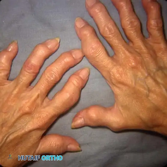
Rheumatoid arthritis (RA) is a systemic autoimmune disease that profoundly affects the musculoskeletal system, with foot and ankle involvement occurring in up to 90% of patients over the course of their disease. The hallmark of RA is chronic synovial inflammation, which leads to capsular distension, ligamentous laxity, cartilage destruction, and eventual periarticular bone erosion.
In the foot and ankle, these destructive processes result in predictable patterns of deformity. The forefoot is most commonly affected, characterized by severe hallux valgus, dorsal subluxation or dislocation of the lesser metatarsophalangeal (MTP) joints, and distal migration of the plantar fat pad. This exposes the prominent metatarsal heads to unmitigated ground reaction forces, leading to intractable plantar keratoses and ulceration. In the hindfoot, tenosynovitis of the posterior tibial tendon combined with subtalar joint destruction frequently precipitates a progressive, rigid planovalgus deformity.
Clinical Pearl: The surgical reconstruction of the rheumatoid foot must be approached holistically. A fundamental axiom in orthopedic reconstruction is to correct proximal deformities before addressing distal ones. Therefore, severe hindfoot valgus must be stabilized prior to, or concurrent with, forefoot reconstruction to prevent the rapid recurrence of forefoot deformity driven by abnormal hindfoot biomechanics.
Preoperative Evaluation and Optimization
Clinical Assessment
A meticulous clinical examination is paramount. The surgeon must assess the flexibility of deformities, the presence of rheumatoid nodules, and the integrity of the soft tissue envelope. Vascular assessment is critical; patients with RA frequently suffer from concomitant vasculitis or atherosclerosis. Palpable pedal pulses or a Doppler assessment with an Ankle-Brachial Index (ABI) > 0.6 is required before considering elective reconstruction.
Radiographic Evaluation
Standard weight-bearing radiographs (anteroposterior, lateral, and oblique views of the foot; AP, lateral, and mortise views of the ankle) are mandatory.
* Forefoot: Look for periarticular osteopenia, marginal erosions, joint space narrowing, and MTP joint subluxation/dislocation.
* Hindfoot/Ankle: Assess for subtalar collapse, talonavicular unroofing, and tibiotalar tilt.
* Advanced Imaging: Magnetic Resonance Imaging (MRI) is highly sensitive for detecting early tenosynovitis (particularly of the posterior tibial tendon) and bone marrow edema before radiographic erosions become apparent.
Medical Optimization
Coordination with the patient's rheumatologist is essential. The perioperative management of Disease-Modifying Antirheumatic Drugs (DMARDs) and biologic agents requires a delicate balance between preventing disease flare-ups and mitigating infection risks. Generally, conventional synthetic DMARDs (e.g., Methotrexate) can be continued, while biologic agents (e.g., TNF-alpha inhibitors) should be withheld for one to two dosing cycles prior to surgery.
Forefoot Reconstruction
The primary goals of rheumatoid forefoot reconstruction are pain relief, restoration of a plantigrade weight-bearing surface, and the ability to wear commercially available accommodative footwear.
The First Metatarsophalangeal (MTP) Joint
Historically, procedures such as the Keller resection arthroplasty or silicone implant arthroplasty were popularized for the rheumatoid first MTP joint. However, long-term follow-up has demonstrated high rates of implant failure, silicone synovitis, and recurrent deformity. Today, arthrodesis of the first MTP joint is universally recognized as the gold standard. It provides a stable medial column, restores the windlass mechanism, and prevents the recurrence of hallux valgus.
The Lesser MTP Joints
For the lesser toes, joint-preserving osteotomies are rarely indicated in advanced RA due to the complete destruction of the articular cartilage and severe soft tissue contractures. The Hoffmann procedure (resection of the lesser metatarsal heads) remains the workhorse technique.
Surgical Warning: When performing lesser metatarsal head resections, it is critical to re-establish a smooth, cascading metatarsal parabola. Failure to do so will result in transfer metatarsalgia beneath the longest remaining metatarsal stump.
Step-by-Step Surgical Technique: Forefoot Reconstruction
1. Positioning and Preparation
* The patient is placed supine with a bump under the ipsilateral hip to internally rotate the leg to a neutral position.
* A thigh or calf tourniquet is applied.
* Prophylactic intravenous antibiotics are administered.
2. Surgical Approaches
* First MTP Joint: A dorsal-medial longitudinal incision is made over the first MTP joint, taking care to protect the dorsal medial cutaneous nerve.
* Lesser MTP Joints: Two dorsal longitudinal incisions are typically utilized—one in the second webspace (accessing the 2nd and 3rd MTP joints) and one in the fourth webspace (accessing the 4th and 5th MTP joints). Alternatively, a transverse plantar approach (Tillmann incision) can be used, which allows direct access to the prominent metatarsal heads and facilitates excision of plantar rheumatoid nodules, though it carries a higher risk of delayed wound healing.
3. First MTP Arthrodesis
* The joint capsule is incised, and aggressive synovectomy is performed.
* The articular cartilage of the metatarsal head and the base of the proximal phalanx is denuded using cup-and-cone reamers to expose bleeding subchondral bone while preserving the native contour.
* Positioning: The hallux is provisionally fixed with K-wires in 10 to 15 degrees of valgus and 10 to 15 degrees of dorsiflexion relative to the floor (or 20-25 degrees relative to the first metatarsal shaft). Neutral rotation is imperative.
* Fixation: Rigid internal fixation is achieved using a low-profile dorsal titanium plate with a lag screw crossing the arthrodesis site.
4. Lesser Metatarsal Head Resection
* Through the dorsal webspace incisions, the extensor tendons are retracted or lengthened (Z-plasty).
* A dorsal capsulotomy is performed, and the collateral ligaments are released to allow plantarflexion of the metatarsal head.
* Using an oscillating saw, the metatarsal heads are resected at the surgical neck. The cut should be angled from dorsal-distal to plantar-proximal to ensure the plantar aspect of the stump does not become a weight-bearing prominence.
* The resection must follow a smooth cascade: the 2nd metatarsal is the longest, with progressive shortening of the 3rd, 4th, and 5th metatarsals.
* The bases of the proximal phalanges may be minimally resected if severe contractures persist.
* The toes are aligned and stabilized with longitudinal 1.25 mm or 1.6 mm Kirschner wires driven retrograde through the toe and antegrade into the metatarsal shafts.
5. Closure
* Meticulous hemostasis is achieved after tourniquet deflation.
* The skin is closed with non-absorbable monofilament sutures using a tension-free technique (e.g., Allgöwer-Donati stitches).
Hindfoot and Ankle Reconstruction
In the rheumatoid hindfoot, the combination of ligamentous laxity, posterior tibial tendon dysfunction, and erosive arthritis leads to a severe, rigid planovalgus deformity. When conservative measures (e.g., custom ankle-foot orthoses) fail, arthrodesis is indicated.
Subtalar and Triple Arthrodesis
Isolated subtalar arthrodesis may be sufficient for early, localized disease. However, advanced rheumatoid flatfoot typically involves the talonavicular and calcaneocuboid joints, necessitating a triple arthrodesis.
- Biomechanics: Fusing the talonavicular joint eliminates nearly all hindfoot motion. The primary goal is to restore the talus over the calcaneus and realign the midfoot to create a plantigrade foot.
- Approach: A standard lateral approach (Ollier incision) provides access to the subtalar and calcaneocuboid joints. A separate medial incision is required for the talonavicular joint. In patients with severe valgus and compromised lateral skin, a single extended medial approach can be utilized to access all three joints, minimizing lateral wound complications.
Tibiotalocalcaneal (TTC) and Pantalar Arthrodesis
When the ankle joint (tibiotalar) is concomitantly destroyed, a TTC arthrodesis is required. If the midfoot is also involved, a pantalar arthrodesis (fusion of the tibiotalar, subtalar, talonavicular, and calcaneocuboid joints) may be considered as a salvage procedure.
Pitfall: Pantalar arthrodesis creates an entirely rigid lever arm below the knee. This places immense stress on the midfoot and knee joints. It should be reserved for severe, end-stage pan-articular destruction where amputation is the only alternative.
Step-by-Step Surgical Technique: TTC Arthrodesis via Retrograde Nailing
1. Positioning and Approach
* The patient is positioned in the lateral decubitus position.
* A transfibular approach is highly effective. A longitudinal incision is made over the distal fibula.
* The distal 5-7 cm of the fibula is resected. This fibular strut is preserved for use as autograft.
2. Joint Preparation
* The tibiotalar and subtalar joints are widely exposed.
* Aggressive synovectomy is performed.
* All articular cartilage and sclerotic subchondral bone are removed using osteotomes and high-speed burrs until punctate bleeding bone is visualized.
* Deformity correction is achieved by translating the talus posteriorly and correcting the valgus tilt.
3. Fixation
* A plantar incision is made in line with the sustained axis of the tibia and talus.
* A guide wire is passed retrograde through the calcaneus, talus, and into the tibial canal.
* The canal is sequentially reamed.
* A retrograde intramedullary nail is inserted. Internal compression is applied across the arthrodesis sites before locking the nail proximally and distally.
* The morselized fibular autograft is packed tightly into the arthrodesis sites to promote osteogenesis.
Postoperative Protocols
The rheumatoid patient requires a highly structured and conservative postoperative rehabilitation protocol due to compromised bone quality and delayed soft tissue healing.
Forefoot Protocol
- Weeks 0-2: The patient is placed in a bulky, non-weight-bearing Jones dressing. Strict elevation is mandated to minimize edema and protect the fragile dorsal skin bridges.
- Weeks 2-6: Sutures are removed at 2 to 3 weeks. The K-wires in the lesser toes are typically removed in the clinic at 4 to 6 weeks. The patient transitions to a rigid-soled postoperative shoe, allowing heel-weight-bearing only.
- Weeks 6-12: Progressive weight-bearing is initiated as tolerated. Radiographs are obtained to confirm the consolidation of the 1st MTP arthrodesis.
- Long-term: Patients are transitioned into wide-toe-box, accommodative footwear with custom orthoses to distribute plantar pressures evenly.
Hindfoot/Ankle Protocol
- Weeks 0-2: The leg is immobilized in a well-padded, non-weight-bearing short leg splint.
- Weeks 2-6: Sutures are removed. The patient is transitioned to a non-weight-bearing short leg cast.
- Weeks 6-12: The cast is exchanged for a Controlled Ankle Motion (CAM) boot. The patient remains strictly non-weight-bearing until radiographic evidence of bridging trabecular bone is observed (typically 8-10 weeks).
- Weeks 12+: Gradual progression to full weight-bearing in the CAM boot, followed by a transition to supportive footwear with an ankle-foot orthosis (AFO) if necessary.
Complications and Salvage Strategies
Operating on the rheumatoid foot and ankle is fraught with potential complications. The surgeon must be prepared to manage these aggressively.
- Wound Dehiscence and Infection: The combination of thin skin, vasculitis, and immunosuppressive medications makes wound healing precarious. If dehiscence occurs, aggressive local wound care, culture-directed antibiotics, and negative pressure wound therapy (NPWT) are indicated.
- Nonunion: The rate of nonunion in rheumatoid arthrodesis is higher than in the osteoarthritic population. Asymptomatic nonunions (fibrous unions) may be observed. Symptomatic nonunions require revision surgery with structural bone grafting (autograft or allograft) and revision of hardware, often utilizing orthobiologics such as Bone Morphogenetic Protein (BMP).
- Hardware Prominence: Due to the paucity of subcutaneous fat, plates and screws frequently become symptomatic once swelling subsides. Routine hardware removal is not recommended, but targeted removal of prominent screws may be necessary after solid fusion is achieved.
Conclusion
The surgical management of the rheumatoid foot and ankle is a complex but highly rewarding endeavor. By adhering to strict biomechanical principles—prioritizing hindfoot stability, achieving rigid first MTP arthrodesis, and meticulously realigning the lesser metatarsal parabola—the orthopedic surgeon can reliably alleviate pain, restore function, and significantly improve the quality of life for patients suffering from this debilitating disease.
You Might Also Like