Acute Ankle Ligamentous Injuries: Anatomy, Biomechanics, and Surgical Management
Key Takeaway
Acute ankle ligamentous injuries are among the most common orthopaedic pathologies, with lateral inversion sprains comprising the vast majority. This comprehensive guide details the intricate anatomy of the lateral and deltoid ligament complexes, biomechanical principles of ankle stability, and evidence-based management strategies. From clinical evaluation and advanced imaging to non-operative rehabilitation and precise surgical repair techniques, it provides essential knowledge for orthopaedic surgeons managing complex acute ankle instability.
ACUTE LIGAMENTOUS INJURIES OF THE ANKLE
Acute ankle ligamentous injuries represent a significant proportion of musculoskeletal trauma encountered by orthopaedic surgeons, sports medicine physicians, and emergency departments. Sprains constitute approximately 85% of all ankle injuries, and 85% of those involve a lateral inversion mechanism. The epidemiological burden is particularly high in the athletic population, where ankle injuries account for 14% to 21% of all sports-related injuries.
Sport-specific kinematics play a crucial role in injury patterns; approximately 40% of basketball injuries and 25% of soccer injuries involve the ankle. Volleyball and football also carry exceptionally high risks of ankle ligament disruption due to frequent jumping, landing on uneven surfaces (often another player's foot), and rapid cutting maneuvers.
Demographic variables also influence injury patterns. Compared with men, women have a slightly higher overall incidence of ankle injuries in similar sports activities, likely due to differences in neuromuscular control, ligamentous laxity, and landing biomechanics. Men, however, demonstrate a higher incidence of medial ankle (deltoid) and syndesmotic sprains. Furthermore, greater mean height and weight, increased body mass index (BMI), and participation in specific athletic activities (e.g., basketball, cheerleading, rugby) are established independent risk factors for acute ankle sprains.
CLASSIFICATION SYSTEMS
Accurate classification of acute ligamentous injuries is essential for prognosticating recovery and guiding treatment algorithms.
The O’Donoghue Classification
Historically, ankle ligamentous injuries have been classified by O’Donoghue based on the degree of macroscopic tissue damage:
* Type I Sprain: Minor ligamentous “stretch” injuries with microscopic tearing but no macroscopic disruption. The joint remains clinically stable.
* Type II Sprain: Incomplete or partial ligamentous tears. This presents with moderate pain, swelling, ecchymosis, and mild to moderate joint instability.
* Type III Sprain: Complete disruption of the ligament or ligaments, resulting in profound swelling, hemorrhage, and gross mechanical instability.
The Clanton and McGarvey Classification
Clanton and McGarvey suggested that a more practical, clinically relevant classification should be based on the dynamic stability of the ankle—as determined by stress testing—and the functional level of the patient. This functional classification dictates whether a patient requires aggressive surgical intervention or functional rehabilitation, particularly distinguishing between the elite athlete requiring immediate mechanical restoration and the sedentary individual who will tolerate mild laxity.
Clinical Pearl: While grading systems provide a framework, the decision to operate on an acute ankle sprain should heavily weigh the patient's functional demands, the presence of concomitant intra-articular pathology (e.g., osteochondral lesions of the talus), and the specific ligaments involved.
PATHOANATOMY AND BIOMECHANICS
Diagnosis and treatment depend fundamentally on a precise understanding of the ligamentous and muscular structures around the ankle. The mechanism of injury dictates the pathoanatomy: eversion and abduction of the foot typically result in disruption of the medial deltoid ligament, whereas the far more common inversion stress results in ligamentous disruption on the lateral side of the ankle.
The Medial Complex: The Deltoid Ligament
Stabilizing the medial side of the ankle anteriorly and posteriorly is the strong, flat, triangular deltoid ligament, which consists of five distinct components divided into deep and superficial layers.
The Deep Deltoid Ligament:
The deep portion of the deltoid ligament is the most critical biomechanical stabilizer of the medial ankle, providing the greatest restraint against lateral translation of the talus. It consists of two components:
1. Anterior Deep Tibiotalar Ligament: Attaches to the anterior aspect of the medial malleolus and inserts on the medial talus.
2. Posterior Deep Tibiotalar Ligament: The thickest and strongest component of the entire deltoid complex. It originates from the posterior aspect of the medial malleolus and inserts broadly onto the medial surface of the talus.
Both deep components are intra-articular but extra-synovial. Disruption of the deep deltoid is a prerequisite for lateral talar shift in bimalleolar equivalent ankle fractures.
The Superficial Deltoid Ligament:
The superficial portion of the deltoid ligament consists of the remaining three components, which cross two joints (the ankle and the subtalar joint):
1. Tibionavicular Component: Located anteriorly, inserting onto the navicular tuberosity.
2. Tibiocalcaneal Component: Located centrally, inserting onto the sustentaculum tali of the calcaneus.
3. Superficial Posterior Tibiotalar Component: Located posteriorly.
Together, the superficial and deep components equally resist valgus tilting of the talus and act as secondary restraints against anterior translation of the talus.
The Lateral Complex
Laterally, the ankle is stabilized by three primary ligaments that vary significantly in structure, orientation, and biomechanical function.
- Anterior Talofibular Ligament (ATFL): The ATFL is the most frequently injured ligament in the human body. It is a thickening of the anterior joint capsule, measuring approximately 2.0 to 2.5 mm thick, 15 to 20 mm long, and 6 to 8 mm wide. It attaches posteriorly to the anterior border of the lateral malleolus and runs anteromedially to insert on the talar neck. The ATFL is the primary restraint to inversion when the ankle is in plantarflexion.
- Calcaneofibular Ligament (CFL): An extra-articular, cord-like structure that originates from the tip of the lateral malleolus and courses posteroinferiorly to insert on the lateral calcaneus. It is the primary restraint to inversion when the ankle is in dorsiflexion.
- Posterior Talofibular Ligament (PTFL): The strongest of the lateral ligaments, running horizontally from the posterior digital fossa of the fibula to the posterior talar tubercle. It is rarely injured except in cases of severe trauma resulting in frank ankle dislocation.
Surgical Warning: The ATFL is intimately blended with the joint capsule. During surgical exploration or repair, it may be difficult to distinguish the ATFL from the capsule itself. Imbrication of the capsule is often necessary to restore ATFL tension.
CLINICAL EVALUATION
History and Mechanism of Injury
A meticulous history is paramount. The classic presentation of a lateral ankle sprain involves a plantarflexion and inversion mechanism. Patients often report an audible "pop" or tearing sensation, followed by immediate swelling and difficulty weight-bearing. Medial injuries typically involve an eversion/external rotation mechanism and are frequently associated with syndesmotic disruption (high ankle sprain).
Physical Examination
- Palpation: Systematic palpation of the ATFL, CFL, PTFL, deltoid complex, anterior inferior tibiofibular ligament (AITFL), and the entire length of the fibula is required to rule out a Maisonneuve fracture.
- Anterior Drawer Test: Evaluates the integrity of the ATFL. With the ankle in 10 to 15 degrees of plantarflexion, the heel is drawn anteriorly while stabilizing the tibia. Excessive anterior translation or a lack of a firm endpoint indicates ATFL incompetence.
- Talar Tilt Test: Evaluates the CFL. With the ankle in neutral dorsiflexion, an inversion force is applied to the hindfoot. Increased laxity compared to the contralateral side suggests CFL disruption.
- External Rotation Stress Test & Squeeze Test: Utilized to assess for concomitant syndesmotic injury.
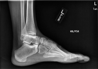
DIAGNOSTIC IMAGING
Radiography
Standard radiographic evaluation should follow the Ottawa Ankle Rules to minimize unnecessary radiation. When indicated, a standard trauma series includes:
* Anteroposterior (AP) View: Assesses the medial and lateral malleoli and the talar dome.
* Lateral View: Evaluates the posterior malleolus, talar neck, and base of the fifth metatarsal.
* Mortise View: An AP view taken with the leg internally rotated 15 to 20 degrees. Crucial for evaluating the medial clear space (normal is < 4 mm). A widened medial clear space indicates deep deltoid disruption and lateral talar shift.
Advanced Imaging
- Magnetic Resonance Imaging (MRI): While not routinely indicated for acute, uncomplicated sprains, MRI is the gold standard for evaluating suspected osteochondral lesions of the talus (OCLs), peroneal tendon pathology, syndesmotic tears, or chronic instability failing conservative management.
- Ultrasound: A dynamic, cost-effective modality that is highly sensitive and specific for ATFL and CFL tears in the hands of an experienced musculoskeletal ultrasonographer.
NON-OPERATIVE MANAGEMENT
The vast majority of acute lateral ankle sprains (Grades I, II, and most Grade III) are successfully managed non-operatively. The traditional "RICE" (Rest, Ice, Compression, Elevation) protocol has evolved into the "POLICE" principle (Protection, Optimal Loading, Ice, Compression, Elevation), emphasizing the importance of early functional rehabilitation.
- Phase I (Acute): Focuses on reducing pain and edema. Short-term immobilization in a CAM (Controlled Ankle Motion) boot or rigid stirrup brace may be used for 3 to 7 days for severe injuries.
- Phase II (Proliferation): Initiates early, protected range of motion (ROM), particularly emphasizing Achilles tendon stretching and active dorsiflexion.
- Phase III (Remodeling): Focuses on proprioceptive neuromuscular facilitation (PNF), peroneal strengthening, and sport-specific functional training. Proprioceptive deficits are the primary driver of chronic subjective instability.
OPERATIVE MANAGEMENT
Indications for Acute Surgical Repair
Acute surgical repair of lateral ankle ligaments remains controversial. However, primary repair is indicated in specific scenarios:
1. Elite or professional athletes with massive Grade III tears where rapid, predictable return to play is mandated.
2. Large bony avulsion fractures of the fibula or talus that are displaced and amenable to internal fixation.
3. Severe medial (deltoid) ligament ruptures associated with syndesmotic disruption or lateral malleolar fractures (bimalleolar equivalents) where the medial clear space cannot be anatomically reduced or maintained.
Surgical Technique: Acute Lateral Ligament Repair (Modified Broström-Gould)
The modified Broström-Gould procedure is the gold standard for anatomic repair of the lateral ligamentous complex.
Positioning and Preparation:
The patient is positioned supine with a sandbag placed under the ipsilateral gluteus to internally rotate the leg, bringing the lateral malleolus into profile. A thigh tourniquet is applied.
Surgical Approach:
1. A curvilinear incision is made over the anterior border of the lateral malleolus, extending from the distal fibula toward the sinus tarsi.
2. Pitfall Avoidance: Meticulous dissection is required to identify and protect the intermediate dorsal cutaneous branch of the superficial peroneal nerve anteriorly, and the sural nerve posteroinferiorly.
Deep Dissection and Repair:
1. The extensor retinaculum is identified and mobilized.
2. The joint capsule is incised vertically, leaving a 2-3 mm cuff of tissue on the fibula for repair. The joint is inspected for osteochondral loose bodies.
3. The torn ends of the ATFL and CFL are identified. In acute settings, these may be primarily repaired using non-absorbable sutures (e.g., 0 or 2-0 FiberWire).
4. If the tissue is avulsed from the fibula (most common), suture anchors (typically 2.5 mm to 3.0 mm) are placed into the anterior border of the distal fibula.
5. The ligaments and capsule are imbricated and tied with the ankle held in neutral dorsiflexion and slight eversion to restore appropriate tension.
6. The Gould Modification: The inferior extensor retinaculum is mobilized and advanced over the repaired ATFL/CFL complex, suturing it to the periosteum of the fibula. This reinforces the repair, limits inversion, and helps correct subtalar instability.
Surgical Technique: Acute Deltoid Ligament Repair
While historically managed non-operatively even in the setting of fractures, acute deltoid repair is increasingly advocated to restore medial column stability, particularly in high-demand athletes or when the ligament blocks anatomic reduction of the talus.
- A medial longitudinal incision is made centered over the medial malleolus.
- Care is taken to protect the saphenous nerve and vein.
- The superficial and deep components of the deltoid are inspected. Deep deltoid avulsions from the medial malleolus are most common.
- Suture anchors are placed into the medial malleolus. The deep posterior tibiotalar ligament is repaired first to restore the primary restraint to lateral talar shift.
- The superficial deltoid is then repaired or imbricated over the deep repair.
POSTOPERATIVE PROTOCOL
Successful surgical outcomes rely heavily on strict adherence to postoperative rehabilitation.
* Weeks 0-2: The patient is placed in a non-weight-bearing posterior splint or cast with the ankle in neutral dorsiflexion and slight eversion.
* Weeks 2-6: Transition to a CAM boot. Weight-bearing is progressively advanced as tolerated. Active dorsiflexion and plantarflexion exercises are initiated out of the boot, but inversion/eversion is strictly prohibited to protect the repair.
* Weeks 6-12: Transition to a functional lace-up ankle brace. Aggressive physical therapy commences, focusing on peroneal strengthening, proprioception (e.g., balance board training), and restoration of full ROM.
* Months 3-6: Gradual return to sport-specific activities and cutting maneuvers.
COMPLICATIONS
- Nerve Injury: The most common surgical complication is neuropraxia or neuroma formation of the superficial peroneal nerve or sural nerve due to aggressive retraction or entrapment in sutures.
- Stiffness: Overtightening of the lateral complex, particularly if tied in excessive eversion or plantarflexion, can lead to a permanent loss of inversion and subtalar stiffness.
- Recurrent Instability: Failure of the repair can occur due to non-compliance with postoperative protocols, unrecognized generalized ligamentous laxity (e.g., Ehlers-Danlos syndrome), or missed concomitant pathology such as a subtle cavovarus foot alignment that places excessive stress on the lateral repair.
-
Complex Regional Pain Syndrome (CRPS): A rare but debilitating complication requiring early recognition and aggressive multidisciplinary management.
You Might Also Like