Masterclass in Fundamental Orthopaedic Surgical Techniques and Approaches
Key Takeaway
Mastering fundamental orthopaedic surgical techniques and anatomical approaches is paramount for achieving optimal patient outcomes. This comprehensive guide details essential perioperative protocols, including tourniquet application, patient positioning, and bone grafting principles. Furthermore, it provides a step-by-step analysis of major surgical approaches to the hip, knee, and shoulder, emphasizing biomechanics, internervous planes, and evidence-based postoperative care for orthopaedic surgeons and residents.
Introduction to Advanced Operative Orthopaedics
The field of orthopaedic surgery is defined by a continuous evolution of innovative techniques, advanced instrumentation, and a deepening understanding of musculoskeletal biomechanics. For the orthopaedic resident, fellow, and practicing consultant, mastery of surgical approaches and fundamental operative techniques is the bedrock of successful patient outcomes. This comprehensive guide synthesizes the core principles of operative orthopaedics, transitioning from essential perioperative management—such as tourniquet application and patient positioning—to the intricate, step-by-step execution of foundational surgical approaches.
Strict adherence to anatomical internervous planes, meticulous soft tissue handling, and evidence-based postoperative protocols are paramount. The following sections provide a postgraduate-level analysis of these critical components, designed to serve as an authoritative reference for FRCS and AAOS candidates.
Fundamental Perioperative Principles
Tourniquet Application and Management
The pneumatic tourniquet is an indispensable tool in extremity surgery, providing a bloodless operative field that facilitates meticulous dissection and precise implant placement. However, its use is not benign and requires strict adherence to physiological principles to prevent ischemic and compressive complications.
- Biomechanics and Pressure Settings: Tourniquet pressure should be individualized based on the patient's Limb Occlusion Pressure (LOP). Modern practice dictates adding a safety margin to the LOP (e.g., 40 mm Hg for LOP <130 mm Hg; 60 mm Hg for LOP 131–190 mm Hg). Alternatively, standard pressures of 250 mm Hg for the upper extremity and 300 mm Hg for the lower extremity are frequently utilized, though tailored pressures reduce the risk of neurapraxia.
- Time Limits: Ischemic time should ideally not exceed 120 minutes. If prolonged surgery is anticipated, the tourniquet should be deflated for 15 to 20 minutes to allow for reperfusion and clearance of metabolic byproducts before reinflation.
- Application Technique: The limb must be exsanguinated using an Esmarch bandage prior to inflation, except in cases of infection or malignancy, where simple elevation for 3 to 5 minutes is preferred to prevent systemic dissemination.
Surgical Warning: Tourniquet paralysis is a devastating complication typically resulting from excessive pressure rather than ischemia. Always ensure the tourniquet is adequately padded with cast padding, avoiding wrinkles that can cause focal skin necrosis or nerve compression.
Positioning of the Patient
Optimal patient positioning is critical for surgical exposure, anesthesia access, and the prevention of perioperative complications such as pressure ulcers and peripheral nerve injuries.
- Supine Position: Used for anterior approaches to the hip, knee, and shoulder. Ensure the heels are floated and the ulnar nerve at the cubital tunnel is padded.
- Lateral Decubitus Position: Frequently utilized for posterior approaches to the hip and shoulder arthroscopy. The patient must be secured with a pelvic positioner (peg board or bean bag). An axillary roll is mandatory to prevent brachial plexus traction injuries in the dependent arm.
- Prone Position: Essential for posterior spinal approaches and posterior approaches to the knee/ankle. The abdomen must hang free to decrease intra-abdominal pressure, which in turn reduces epidural venous engorgement and intraoperative bleeding.
Infection Prevention and Site Preparation
Surgical site infections (SSIs) remain a catastrophic complication in orthopaedic surgery, particularly in the presence of hardware.
- Skin Preparation: Alcohol-based chlorhexidine solutions are the gold standard, demonstrating superior efficacy in reducing skin flora compared to aqueous povidone-iodine. The solution must be allowed to dry completely to maximize its bactericidal effect and prevent operating room fires.
- Draping: Impervious, sterile drapes must isolate the operative field. Ioban (iodine-impregnated incise drapes) is frequently used to sequester skin flora, particularly in arthroplasty.
- Irrigation: Copious irrigation with normal saline is fundamental. The addition of dilute betadine (0.35%) to irrigation solutions prior to closure has been shown in recent literature to significantly reduce SSI rates in arthroplasty and spine surgery.
Essential Operative Techniques
Principles of Bone Grafting
Bone grafting is a cornerstone of reconstructive orthopaedics, utilized in fracture nonunions, arthrodesis, and the filling of cavitary defects. A successful bone graft must possess one or more of the following biological properties:
1. Osteogenesis: The presence of surviving, viable osteoblasts and osteoprogenitor cells (found only in fresh autograft or bone marrow aspirate).
2. Osteoinduction: The ability to stimulate the differentiation of mesenchymal stem cells into osteoblasts, primarily mediated by Bone Morphogenetic Proteins (BMPs).
3. Osteoconduction: Providing a structural scaffold for the ingrowth of neovasculature and osteoprogenitor cells (e.g., allograft, synthetic ceramics).
The Iliac Crest Bone Graft (ICBG):
The anterior or posterior iliac crest remains the gold standard for autogenous bone grafting, providing all three biological properties.
* Technique: When harvesting from the anterior iliac crest, the incision should be made 2 cm posterior to the Anterior Superior Iliac Spine (ASIS) to avoid injury to the Lateral Femoral Cutaneous Nerve (LFCN).
Clinical Pearl: To minimize donor site morbidity during ICBG harvest, preserve the inner table of the ilium. This maintains structural integrity and prevents catastrophic pelvic herniation.
Fixation of Tendon to Bone
The reattachment of tendon to bone is critical in procedures ranging from rotator cuff repairs to ligament reconstructions. The biomechanical goal is to achieve rigid initial fixation to allow for biological healing at the tendon-bone interface.
- Biomechanics: Healing occurs through the formation of Sharpey's fibers. Initial fixation strength depends on bone quality, anchor design, and the suture configuration.
- Techniques: Locking suture configurations, such as the Krackow or whipstitch, provide superior pull-out strength by gripping the longitudinal collagen fibers of the tendon. Transosseous tunnels or modern suture anchors (titanium, PEEK, or biocomposite) are utilized to secure the tendon to a decorticated bony bed, maximizing the surface area for biological integration.
Masterclass: Major Orthopaedic Surgical Approaches
A profound understanding of surgical anatomy and internervous planes is what separates the master surgeon from the technician. An internervous plane is an anatomical dissection path between two muscles supplied by different peripheral nerves, allowing for extensile exposure without denervating the musculature.
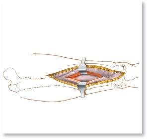
1. The Hip: Posterior Approach (Moore/Southern)
The posterior approach is the most widely utilized approach for total hip arthroplasty (THA) and hemiarthroplasty.
- Indications: THA, hemiarthroplasty, treatment of posterior acetabular fractures, and removal of loose bodies.
- Positioning: Lateral decubitus with the operative hip superior.
- Internervous Plane: There is no true internervous plane. The approach involves splitting the Gluteus Maximus, which is entirely innervated by the Inferior Gluteal Nerve. Because the split is in line with the muscle fibers, denervation does not occur.
- Step-by-Step Surgical Technique:
- Incision: A 10-15 cm curved incision centered over the greater trochanter, extending proximally toward the PSIS and distally along the femoral shaft.
- Superficial Dissection: Incise the fascia lata distally and split the gluteus maximus proximally in line with its fibers.
- Deep Dissection: Identify and retract the sciatic nerve (often palpable posterior to the short external rotators).
- Tendon Release: Tag and release the short external rotators (piriformis, superior gemellus, obturator internus, inferior gemellus) at their insertion on the greater trochanter. Reflect them posteriorly to protect the sciatic nerve.
- Capsulotomy: Perform a T-shaped or crucial capsulotomy to expose the femoral head and acetabulum.
- Postoperative Protocol: Early mobilization with weight-bearing as tolerated. Strict adherence to posterior hip precautions (avoiding flexion past 90 degrees, adduction across the midline, and internal rotation) for 6 weeks to prevent dislocation.
Pitfall: Failure to identify and protect the sciatic nerve during the release of the short external rotators or during retractor placement can lead to devastating foot drop. Always place the posterior retractor carefully beneath the transverse acetabular ligament.
2. The Knee: Medial Parapatellar Approach
The workhorse approach for total knee arthroplasty (TKA) and complex intra-articular distal femur or proximal tibia fractures.
- Indications: TKA, synovectomy, ligamentous reconstruction, and complex intra-articular fractures.
- Positioning: Supine with a tourniquet applied to the proximal thigh. A sandbag is often taped to the table to allow the knee to rest in 90 degrees of flexion.
- Internervous Plane: There is no true internervous plane. The approach utilizes an intertendinous incision through the extensor mechanism.
- Step-by-Step Surgical Technique:
- Incision: A longitudinal midline incision from 5 cm proximal to the superior pole of the patella to the tibial tubercle.
- Superficial Dissection: Elevate medial and lateral skin flaps to expose the extensor mechanism.
- Deep Dissection: Incise the quadriceps tendon longitudinally, leaving a 3-4 mm cuff of tendon on the vastus medialis for later repair. Continue the incision distally around the medial border of the patella and along the medial border of the patellar tendon down to the tibial tubercle.
- Exposure: Evert or subluxate the patella laterally. Excise the retropatellar fat pad and release the anterior horns of the menisci to fully expose the tibiofemoral joint.
- Postoperative Protocol: Immediate weight-bearing as tolerated with a walker or crutches. Aggressive early range of motion (ROM) exercises are critical to prevent arthrofibrosis.
3. The Shoulder: Deltopectoral Approach
The standard anterior approach for shoulder arthroplasty and proximal humerus fracture fixation.
- Indications: Total shoulder arthroplasty (TSA), hemiarthroplasty, open reduction internal fixation (ORIF) of proximal humerus fractures, and anterior instability repairs.
- Positioning: Beach-chair position with the head secured and the operative arm draped free.
- Internervous Plane: Between the Deltoid (Axillary Nerve) and the Pectoralis Major (Medial and Lateral Pectoral Nerves).
- Step-by-Step Surgical Technique:
- Incision: A linear incision from the tip of the coracoid process extending distally and laterally along the deltopectoral groove.
- Superficial Dissection: Identify the cephalic vein, which marks the internervous plane. The vein is typically retracted laterally with the deltoid to preserve its major venous tributaries, though medial retraction is acceptable if lateral tributaries are sparse.
- Deep Dissection: Retract the conjoined tendon (coracobrachialis and short head of the biceps) medially.
- Subscapularis Management: To expose the glenohumeral joint, the subscapularis tendon must be managed. This can be done via a tenotomy, a peel off the lesser tuberosity, or a lesser tuberosity osteotomy, depending on the procedure and surgeon preference.
- Capsulotomy: Incise the underlying capsule to expose the humeral head and glenoid.
- Postoperative Protocol: Sling immobilization. Passive ROM begins immediately, but active internal rotation is strictly restricted for 6 weeks to protect the subscapularis repair.
Surgical Warning: The musculocutaneous nerve enters the conjoined tendon approximately 5-8 cm distal to the coracoid process. Vigorous medial retraction of the conjoined tendon can cause a traction neurapraxia.
4. The Ankle: Lateral Approach to the Fibula
The fundamental approach for the fixation of lateral malleolus fractures.
- Indications: ORIF of distal fibula fractures, lateral ligament reconstruction, and fibular osteotomies.
- Positioning: Supine with a bump under the ipsilateral hip to internally rotate the leg, bringing the lateral malleolus anteriorly.
- Internervous Plane: Proximally, between the Peroneus Tertius (Deep Peroneal Nerve) and the Peroneus Brevis (Superficial Peroneal Nerve). Distally, the approach is directly onto the subcutaneous bone.
- Step-by-Step Surgical Technique:
- Incision: A longitudinal incision centered over the posterior half of the fibula, extending distally to the tip of the lateral malleolus. Placing the incision slightly posterior prevents the hardware from lying directly beneath the surgical scar.
- Superficial Dissection: Carefully dissect through the subcutaneous tissue. Identify and protect the Sural Nerve and short saphenous vein, which cross the operative field distally and posteriorly.
- Deep Dissection: Incise the periosteum longitudinally and elevate it minimally to expose the fracture site, preserving the vascular supply to the bone fragments.
- Postoperative Protocol: Immobilization in a splint or boot. Weight-bearing status is dictated by the fracture pattern and stability of fixation, typically ranging from 2 to 6 weeks of non-weight-bearing.
Conclusion
The mastery of operative orthopaedics requires a lifelong commitment to anatomical study, biomechanical understanding, and meticulous surgical technique. From the precise application of a tourniquet to the flawless execution of an internervous plane dissection, every step in the operating room dictates the patient's functional recovery. By adhering to the evidence-based principles and step-by-step approaches detailed in this masterclass, orthopaedic surgeons can minimize perioperative complications, optimize biological healing, and consistently deliver the highest standard of musculoskeletal care.
You Might Also Like