Unlocking Every Aspect of the Hip's Surgical Approaches
Key Takeaway
Discover the latest medical recommendations for Unlocking Every Aspect of the Hip's Surgical Approaches. Surgical approaches to the hip include anterior, anterolateral, lateral, posterior, and medial. These techniques provide access to the specific aspect of the hip required for procedures such as total joint replacement, hemiarthroplasty, and tumor surgery. While anterolateral and posterior are common for total hip replacement, the minimally invasive anterior approach is gaining popularity.
Approaches to the Hip click the link below
##
Anterior Approach to the Hip
Minimally Invasive Anterior Approach to the Hip
Anterolateral Approach to the Hip
Lateral Approach to the Hip
Applied Surgical Anatomy of the Anterior, Lateral, and Anterolateral
Approaches to the Hip
Posterior Approach to the Hip
Applied Surgical Anatomy of the Posterior Approaches to the Hip and the Acetabulum
Medial Approach to the Hip
Applied Surgical Anatomy of the
Medial Approach
## Eight
The Hip
Operations on the hip joint are among the most common surgical procedures performed in orthopedics. Total joint replacement for degenerative joint disease has revolutionized the lives of millions of patients. Open approaches to the hip joint are also required for
hemiarthroplasties, tumor surgery, and for the treatment of infection around the hip joint.
The
anterior approach
The full anterior approach allows good access to the pelvis as well as to the hip joint. The
anterolateral approach,
still the most common approach for total hip replacement, has many variations because of the different requirements of the several prosthetic designs that can be inserted. The standard anterolateral approach is described; readers are advised to consult the original papers of the designers of the arthroplasty before performing a particular joint replacement. The
posterior approach
is used extensively for hemiarthroplasty as well as for total hip joint replacement. It is both safe and easy to perform with only one assistant. The
medial approach
is rarely used, and then mainly for local procedures on the lesser trochanter and surrounding bone.
Minimally invasive surgical approaches to the hip have increased in popularity. Most of these techniques utilize the classical approaches described in this book. The length of the skin incision and the underlying dissection is reduced. Minimally invasive surgery can create less soft tissue damage, but the visualization of the structures is necessarily less. The techniques, therefore, are potentially more hazardous especially in obese patients and an understanding of the underlying anatomy is even more important than in larger surgical approaches. In addition, imaging may be indicated to ensure correct implant position.
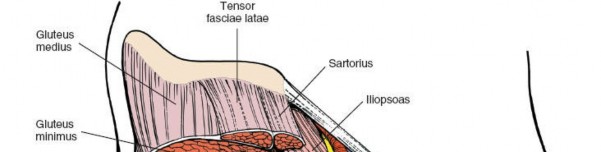
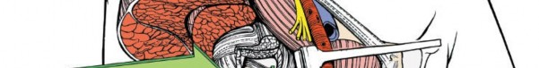
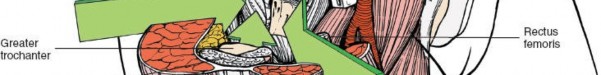
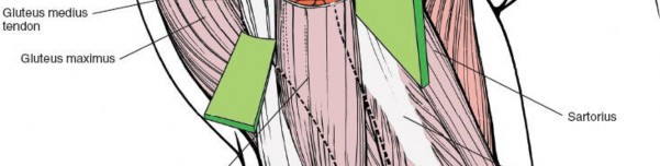
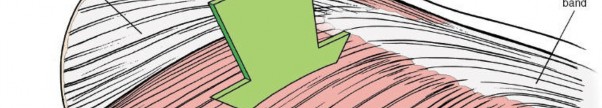
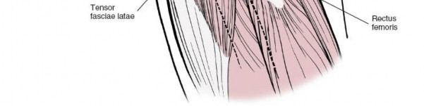 Figure 8-1 The intermuscular intervals used in the anterior, anterolateral, and posterior approaches to the hip. These four basic approaches to the hip take advantage of the muscular intervals that surround the joint. The anterior approach uses the interval between the sartorius and the tensor fasciae latae; the anterolateral approach uses the interval between the tensor fasciae latae and the gluteus medius; the posterior approach gains access either through the interval between the gluteus medius and the gluteus maximus or by splitting the gluteus maximus; and the medial approach exploits the interval between the adductor longus and the gracilis (Fig. 8-1).
Three anatomical sections augment the description of the approaches. Because the anterior and anterolateral approaches share so much anatomy, they are grouped together. The anatomy for the posterior and medial
approaches follows the appropriate approach.
Anterior Approach to the Hip
12 approach, gives safe access to the hip joint and ilium. It exploits the internervous plane between the sartorius (femoral nerve) and the tensor fasciae latae (superior gluteal nerve) to penetrate the outer layer of the joint musculature. Its uses include the following:
2. Synovial biopsies
3. Intra-articular fusions
4. Total hip replacement
5. Hemiarthroplasty
6. Excision of tumors, especially of the pelvis
The upper part of the approach may also be used for pelvic osteotomies. Note however that the approach does not expose the acetabulum as completely as other incisions unless muscles are extensively stripped off the pelvis.
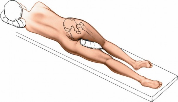
Position of the Patient
Fig. 8-2).
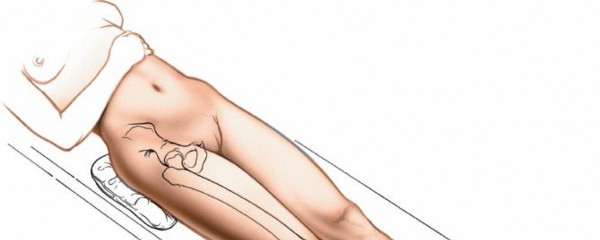
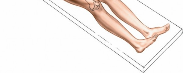 Figure 8-2 Position of the patient on the operating table for the anterior approach to the hip.
Landmarks and Incision
#### Landmarks
Figure 8-2 Position of the patient on the operating table for the anterior approach to the hip.
Landmarks and Incision
#### Landmarks
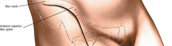
The
anterior superior iliac spine
is subcutaneous and is easily palpable in thin patients. In obese patients, it is covered by adipose tissue and is more difficult to find. You can locate it most easily if you bring your thumbs up from beneath the bony protuberance.
The
iliac crest
is subcutaneous and serves as a point of origin and insertion for various muscles. However, as none of these muscles cross the bony crest, it remains available for palpation (Fig. 8-3).
Incision
8-3).
!8-4A,B).
Superficial Surgical Dissection
8-6
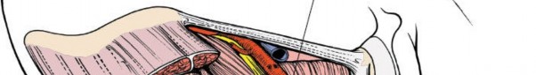
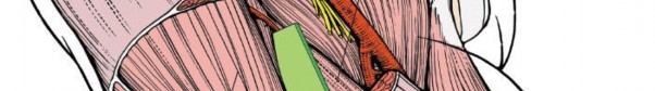
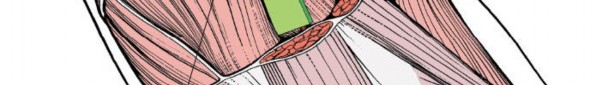

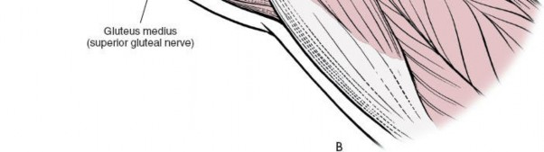 Figure 8-4A:
The internervous plane lies between the
sartorius
(femoral nerve) and the
tensor fasciae latae
(superior gluteal nerve).
B:
The deeper internervous plane lies between the
rectus femoris
(femoral nerve) and the
gluteus medius
(superior gluteal nerve).
Figure 8-4A:
The internervous plane lies between the
sartorius
(femoral nerve) and the
tensor fasciae latae
(superior gluteal nerve).
B:
The deeper internervous plane lies between the
rectus femoris
(femoral nerve) and the
gluteus medius
(superior gluteal nerve).
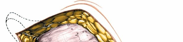
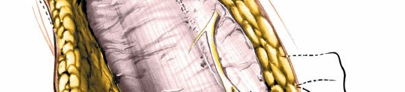
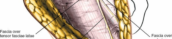
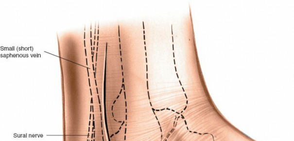
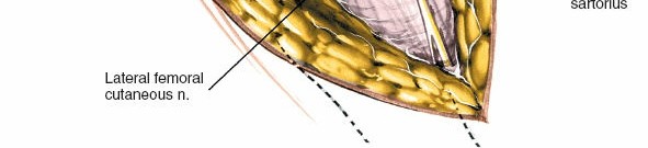 Figure 8-5 The lateral femoral cutaneous nerve (lateral cutaneous nerve of the thigh) pierces the deep fascia close to the intermuscular interval between the tensor fasciae latae and the sartorius.
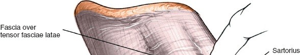

 Figure 8-6 Identify the gap between the tensor fasciae latae and the sartorius by palpation.
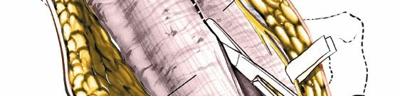
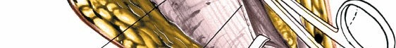
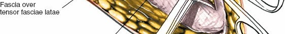
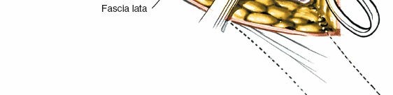 Figure 8-7 Incise the deep fascia on the medial side of the tensor fasciae latae. Retract the sartorius upward and medially and the tensor fascia downward and laterally. Incise the deep fascia on the medial side of the tensor fasciae latae. Staying within the fascial sheath of this muscle will protect you from damaging the lateral femoral cutaneous nerve because the nerve runs over the fascia of the sartorius. Retract the sartorius upward and medially and the tensor fasciae latae downward and laterally (Fig. 8-7).
Detach the iliac origin of the tensor fasciae latae to develop the internervous plane. The large ascending branch of the lateral femoral circumflex artery crosses the gap between the two muscles below the anterior superior iliac spine. It must be ligated or coagulated.
Deep Surgical Dissection
Retracting the tensor fasciae latae and the sartorius brings you on to two
Fig. 8-8).
The rectus femoris originates from two heads: the direct head, from the anterior inferior iliac spine, and the reflected head, from the superior lip of the acetabulum. The reflected head also takes origin from the anterior capsule of the hip joint. It is intimate with the capsule making dissection between the two structures difficult.
8-9).
8-10 8-1124 Inferolaterally, the shaft of the femur lies under cover of the vastus lateralis.
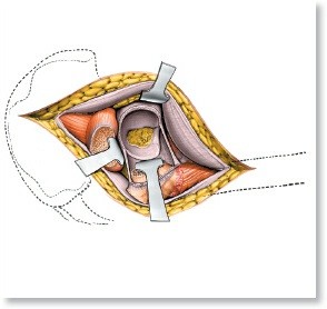
8-12). Dislocate the hip by external rotation after the capsulotomy.
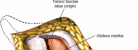
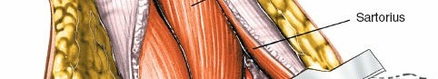
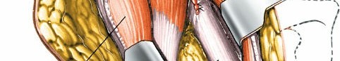
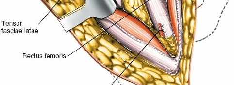
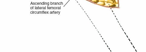 Figure 8-8 The deep layer of musculature, consisting of the rectus femoris and the gluteus medius, is now visible. The ascending branch of the lateral femoral circumflex artery must be ligated. ### Dang
Nerves
The
lateral femoral cutaneous nerve (lateral cutaneous nerve of the thigh)
reaches the thigh by passing over, behind, or through—usually over
8-14; see Fig. 8-5). Be aware that the nerve may divide into three or more branches just below the inguinal ligament and that its anatomical course is very variable. Always be on the lookout for the nerve when dissecting superficial to the deep fascia.
The
femoral nerve
lies almost directly anterior to the hip joint itself, within the femoral triangle. Because the nerve is well medial to the rectus femoris, it is not really in danger unless you stray far out of plane to the wrong side of the sartorius and the rectus femoris. If you lose the correct plane during deep dissection, locate the femoral pulse by palpation. Within the femoral triangle, the artery lies medial to the nerve (Figs. 8-15 and 8-16).
Vessels
The
ascending branch of the lateral femoral circumflex artery
8-158-168-17).
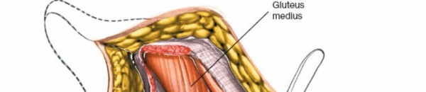
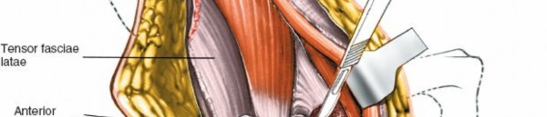
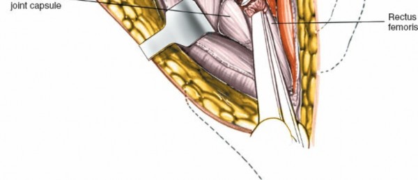 Figure 8-9 Detach the rectus femoris from both its origins, the anterior inferior iliac spine and the superior lip of the acetabulum.
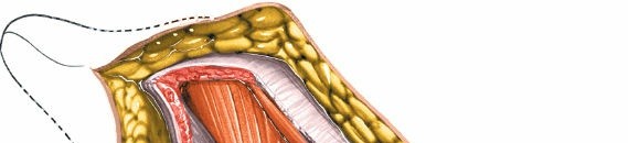
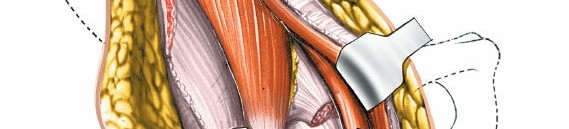
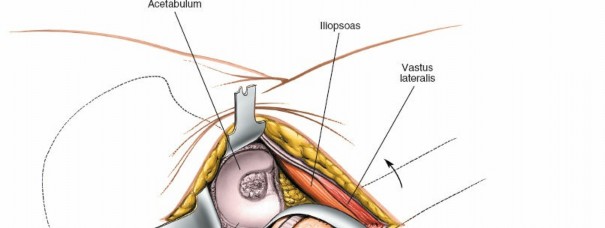
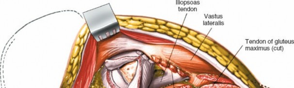
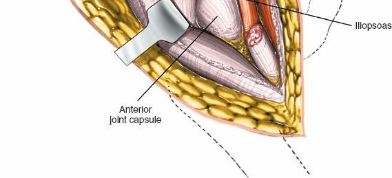 Figure 8-10 The hip joint capsule is now partly exposed. Retract the iliopsoas tendon medially.
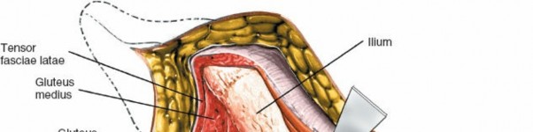
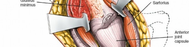
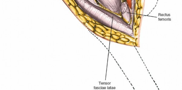 Figure 8-11 The hip joint capsule is fully exposed. Detach the muscles of the ilium if further exposure is needed.
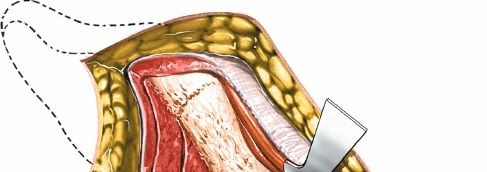
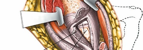
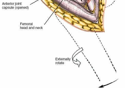 Figure 8-12 Incise the hip joint capsule.
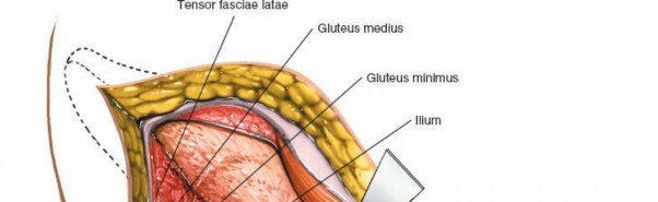
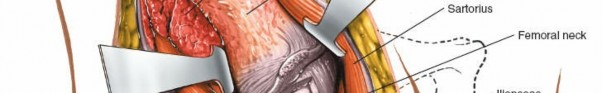
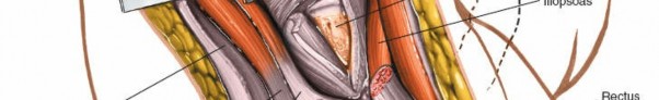
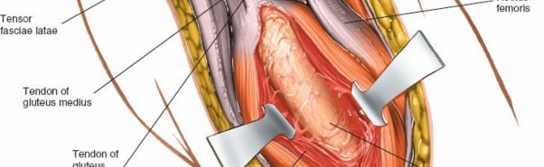
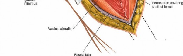 Figure 8-13 Proximal extension of the wound exposes the ilium. Distal extension of the incision exposes the anterior aspect of the femur in the interval between the vastus lateralis and the rectus femoris. It may be necessary to split muscle fibers to actually expose the lateral aspect of the femur.
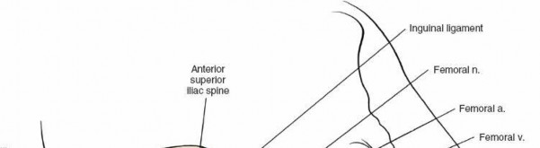
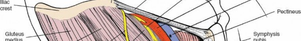
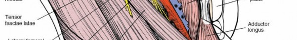
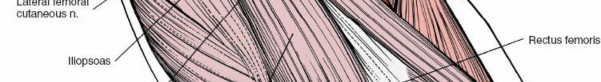

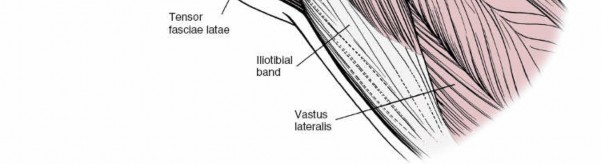 Figure 8-14 Superficial view of the muscles of the anterior region of the hip, including the femoral triangle and its contents.
Sartorius.
Origin. Anterior superior iliac spine and upper half of iliac notch. Insertion. Upper end of subcutaneous surface of tibia. Action. Flexor of thigh and knee and external rotator of hip. Nerve supply. Femoral nerve (L2-L4).
Tensor Fasciae Latae.
Origin. From outer aspect of iliac crest between the anterior superior iliac spine and the tubercle of the iliac crest. Insertion. By iliotibial tract into Gerdy’s tubercle of the tibia. Action. Maintains stability of extended knee and extended hip. Nerve supply. Superior gluteal_
Figure 8-14 Superficial view of the muscles of the anterior region of the hip, including the femoral triangle and its contents.
Sartorius.
Origin. Anterior superior iliac spine and upper half of iliac notch. Insertion. Upper end of subcutaneous surface of tibia. Action. Flexor of thigh and knee and external rotator of hip. Nerve supply. Femoral nerve (L2-L4).
Tensor Fasciae Latae.
Origin. From outer aspect of iliac crest between the anterior superior iliac spine and the tubercle of the iliac crest. Insertion. By iliotibial tract into Gerdy’s tubercle of the tibia. Action. Maintains stability of extended knee and extended hip. Nerve supply. Superior gluteal_
nerve.
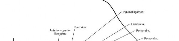

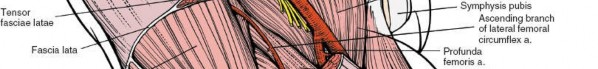
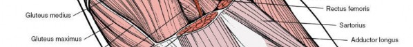
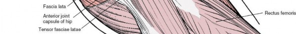
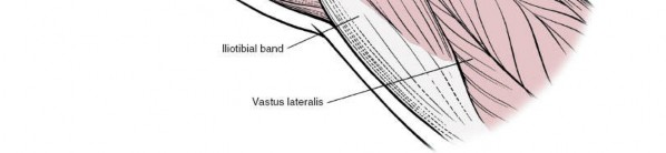 Figure 8-15 The tensor fasciae latae, the sartorius, and the fascia lata have been resected on the anterior aspect of the hip to reveal the gluteus medius, the rectus femoris, and the ascending branch of the lateral femoral circumflex artery. The hip joint capsule is visible between these two muscles. Medially, note the relationship between the iliopsoas and the rectus femoris. How to Enlarge the Approach #### Local Measures
Superficial Surgical Dissection. Detach the origins of the tensor fasciae latae and the sartorius._Deep Surgical Dissection. Detach the origins of the gluteus medius and minimus from the outer wing of the ilium by blunt dissection. (This
procedure is always necessary during pelvic osteotomies.) Bleeding from the raw exposed surface of the ilium can be controlled if you pack the wound with gauze sponges. Individual bleeding points can be controlled by the application of bone wax. There is no other way to stop bleeding.
Extensile Measures
The skin incision may be extended posteriorly along the iliac crest to expose that bone. In theory, the extension allows the taking of bone graft, but it is rarely used.
To extend the approach distally, lengthen the skin incision downward along the anterolateral aspect of the thigh. Incise the fascia lata in line with the skin incision; underneath it lies the interval between the vastus lateralis and the rectus femoris. Try to stay in the interval; you will have to split muscle fibers to expose the anterior aspect of the femur. This extension gives excellent exposure of the entire shaft of the femur (see Fig. 8-13).
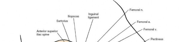
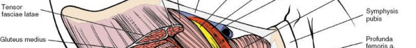
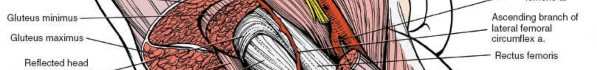
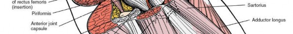
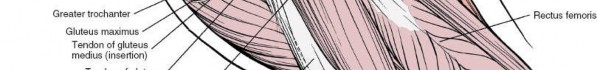
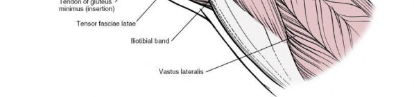 Figure 8-16 The gluteus minimus, medius, and maximus have been resected to reveal the hip joint capsule and the reflected head of the rectus femoris. The approach can be extended to allow visualization of both the inner and outer walls of the pelvis at the level of the hip joint to allow pelvis osteotomy. To obtain visualization of the outer part of the ilium, gently strip the muscular coverings from the bone at the level of the origin of the reflected head of rectus. Using blunt instruments stay in contact with bone. This dissection will lead you into the sciatic notch. Take great care that any instrument inserted into the notch remains firmly on the bone, since the sciatic nerve is also emerging through the notch. Detach the straight head of the rectus femoris from the anterior inferior iliac spine, and carefully lift off the iliacus muscle from the inside of the pelvis, again sticking very carefully to the bone. A blunt instrument will gradually lead you into the greater sciatic notch. At this stage, both instruments should be in contact with each other and with the bone of the sciatic notch. Retraction on both instruments will allow visualization of the entire thickness of the pelvis at the level of the top of the acetabulum, permitting an accurate osteotomy to be carried out.
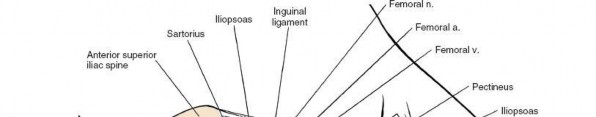


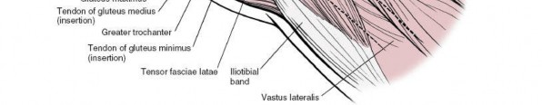 Figure 8-17 The iliopsoas tendon has been retracted medially; the rectus femoris has been resected and the joint capsule opened to reveal the joint. ## Minimally Invasive Anterior Approach to the Hip
The minimally invasive anterior approach to the hip is used mainly for elective joint replacement surgery but can also be used for reduction of displaced femoral neck fractures and drainage of hip joint infections. The approach preserves muscle and in expert hands may be associated with faster initial recovery from surgery. Because the approach gives less exposure of the joint the use of an image intensifier is advised by many to check the level of femoral neck osteotomy as well as the position of the acetabular component. The operation can be performed on a regular radiolucent table. Some surgeons however advocate the use of traction during femoral neck osteotomy and acetabular component insertion.
Position of Patient
Place the patient supine on the operating table (Fig. 8-2). This supine position permits the use of an image intensifier which is of great value in determining the position of prosthetic components in joint replacement surgery.
Landmarks and Incision
The
anterior superior iliac spine
is subcutaneous and is easily palpable in thin patients. In obese patients, it is covered by adipose tissue and is more difficult to find. To palpate it bring your thumbs up from beneath the bony protuberance.
Palpate the tip of the
greater trochanter
on the lateral aspect of the thigh.
Incision
Mark the tip of the greater trochanter and the anterior superior iliac spine with a skin marker. Make an 8-cm longitudinal incision beginning 1 cm below and 1 cm lateral to the anterior superior iliac spine. The center of
8-19). Staying within the fascial sheath will reduce the chances of damaging the lateral femoral cutaneous nerve.
Deepen this plane by blunt dissection to expose the interval between the rectus femoris and the tensor fasciae latae
The rectus femoris muscle is enclosed in a fascial sheath. Incise the anterior portion of the sheath to reveal the muscle and retract it medially. Then carefully incise the posterior aspect of the sheath to reveal the lateral circumflex vessels which are often large and need ligation.
Deep Surgical Dissection
8-20). The reflected head of the rectus muscle arises from a concavity just above the acetabulum. Develop a plane between the rectus and the hip joint capsule and carefully under direct vision place a retractor into the femoral head through the capsule. Perform an H-or L-shaped capsulotomy excising a portion of the
8-218-228-238-24).
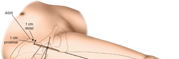
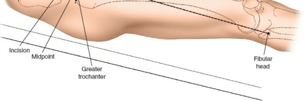 Figure 8-18 Make an 8-cm longitudinal incision beginning 1 cm below and 1 cm lateral to the anterior superior iliac spine. Aim the incision toward the head of the fibula. The center of the incision should be at the level of the tip of the greater trochanter.
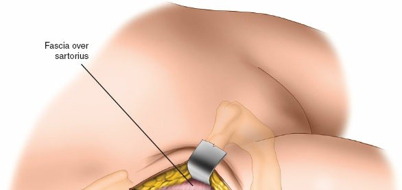
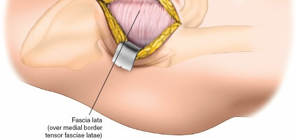 Figure 8-19 Identify the interval between sartorius and tensor fasciae latae and incise the deep fascia covering the tensor fasciae latae muscle at its medial edge.
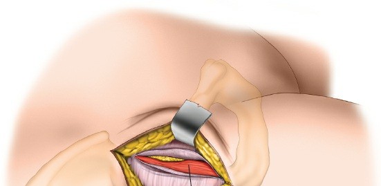
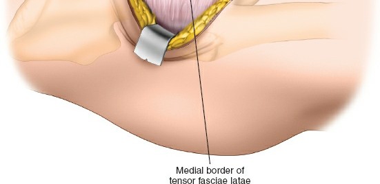 Figure 8-20 Retract the tensor fasciae latae laterally to expose the rectus muscle covered by a fascial layer.
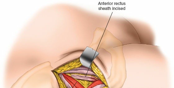
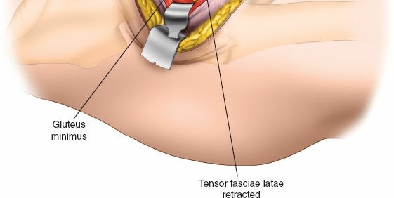 Figure 8-21 Incise the fascial sheath covering the anterior aspect of the rectus to expose the muscle.
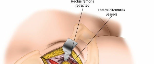
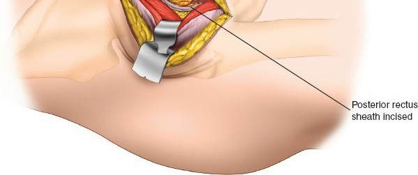 Figure 8-22 Retract the rectus muscle medially to expose the fascia covering the posterior aspect of the muscle. Incise this fascial sheath longitudinally to reveal the lateral circumflex vessels.
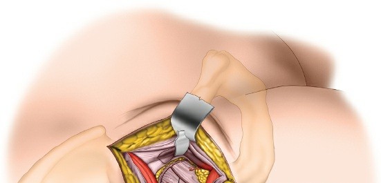
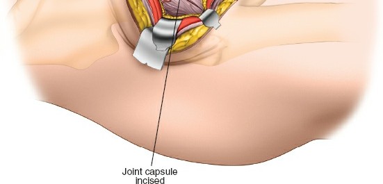 Figure 8-23 Ligate the lateral circumflex vessels. Expose the anterior hip joint capsule covered by fatty tissue. Some fibers of psoas—the iliocapsularis muscle will need to be separated from the capsule by sharp dissection. Incise the anterior capsule of the hip joint longitudinally.
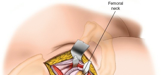
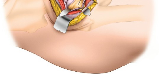 Figure 8-24 Position retractors around the superior and inferior aspects of the femoral neck. Place an anterior retractor over the anterior lip of the acetabulum under direct vision ensuring that the tip of the retractor is placed directly onto bone without any soft tissue intervention. ### Dang
Nerves
The
lateral femoral cutaneous nerve (lateral cutaneous nerve of the thigh)
reaches the thigh by passing over, behind, or through—usually over
—the sartorius muscle, about 2½ cm below the anterior superior iliac spine. Three or more branches may exist whose course is very variable. The nerve must be preserved when you incise the fascia over the medial edge of the tensor fasciae latae; cutting it may lead to the formation of a painful neuroma and may produce an area of diminished sensation on the lateral aspect of the thigh (Fig. 8-14; see Fig. 8-5). Staying within the fascial sheath of the tensor fasciae latae will protect the nerve from
damage.
The
femoral nerve
lies almost directly anterior to the hip joint itself, within the femoral triangle. Because the nerve is well medial to the rectus femoris, it is not really in danger unless you stray far out of plane to the wrong side of the sartorius and the rectus femoris. If you lose the correct plane during deep dissection, locate the femoral pulse by palpation. Within the femoral triangle, the artery lies medial to the nerve (Figs. 8-15 and 8-16).
Vessels
The
ascending branch of the lateral femoral circumflex artery
crosses the operative field, running proximally in the internervous plane between the tensor fasciae latae and the rectus femoris. It lies posterior to the posterior part of the fascial sheath that encloses the rectus femoris muscle. Ligate or coagulate it when you separate the two muscles (see Figs. 8-8, 8-15, 8-16, and 8-17).
How to Enlarge the Approach
This approach is a minimally invasive one. If you start to struggle and loose the surgical plane the approach can always be converted to the classic anterior approach. It is better to create more muscle damage than injure a vital structure or insert a prosthesis in an incorrect position.
Be aware however that this approach can be extended distally if required by splitting vastus lateralis (see Fig. 8-13).
Anterolateral Approach to the Hip
4 5 8-25).
The abductor mechanism can be released either by a trochanteric osteotomy7 or by detaching part of the anterior part of the gluteus medius
and the whole gluteus minimus from the trochanter.6 The two methods seem to offer different approaches, but they are actually variations on a theme. The differences should not obscure the fundamental fact that all anterolateral approaches exploit the same intermuscular plane, between the tensor fasciae latae and the gluteus medius.
The uses of the anterolateral approach include the following:
1. 6
2. Hemiarthroplasty
4. Synovial biopsy of the hip
5. Biopsy of the femoral neck
Position of the Patient
8-26). Tilt the table away from you as the patient lies flat. Both maneuvers allow the buttock skin and fat to fall posteriorly, away from the operative plane, and lift the skin incision clear of the table, making it easier to drape the patient. You must take the table tilt into account when you insert the acetabular portion of a total joint replacement because the guides used to position the acetabular prosthesis usually take the ground as their reference plane.
Drape the patient so that the limb can be moved freely during surgery.
Landmarks and Incision
#### Landmarks
The
anterior superior iliac spine
is subcutaneous. It is easy to palpate in all but the most obese patients, who have a thick layer of adipose tissue covering it. To palpate it, bring your thumbs up from beneath the bony protuberance.
The
greater trochanter_Fig. 8-27).
The _shaft of the femur
can be felt as a resistance through the massive vastus lateralis on the lateral side of the thigh (see Fig. 8-49).
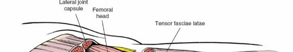
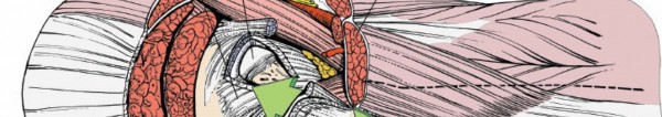
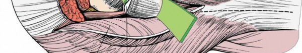
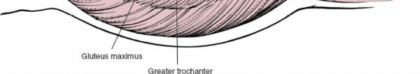 Figure 8-25 The route of the anterolateral approach to the hip joint.
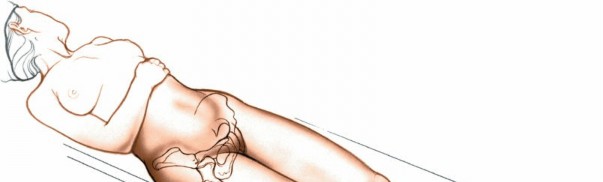
 Figure 8-26 Position of the patient on the operating table for the anterolateral approach to the hip. Bring the greater trochanter to the edge of the table, and allow the buttocks, skin, and fat to fall posteriorly, away from the operative plane.
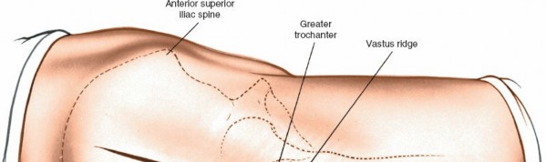
 Figure 8-27 Incision for the anterolateral approach to the hip.
The
vastus lateralis
ridge, a rough line that marks the fusion site of the greater trochanter to the lateral surface of the shaft of the femur, is easiest to palpate from distal to proximal. It is not palpable in obese patients.
Figure 8-27 Incision for the anterolateral approach to the hip.
The
vastus lateralis
ridge, a rough line that marks the fusion site of the greater trochanter to the lateral surface of the shaft of the femur, is easiest to palpate from distal to proximal. It is not palpable in obese patients.
Incision
8-29).
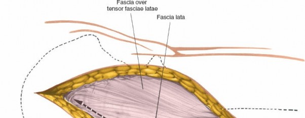
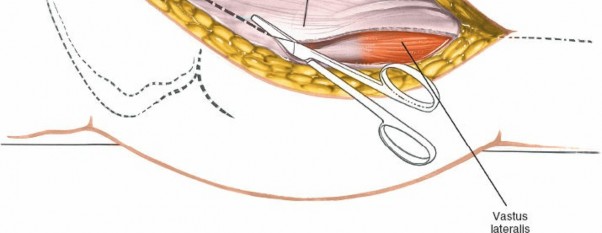 Figure 8-28 Incise the fascia lata posterior to the tensor fasciae latae.
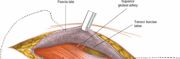
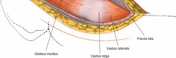 Figure 8-29 Retract the fascia lata and the tensor fasciae latae muscle, which it envelopes, anteriorly, revealing the gluteus medius and a series of vessels that cross the interval between the tensor fasciae latae and the gluteus medius.

!8-31). The safest technique is to use a swab.
Deep Surgical Dissection
Deep surgical dissection consists in detaching part or all of the abductor mechanism and then dissecting up the femoral neck superficial to the capsule of the joint until a suitable retractor can be placed over the anterior lip of the acetabulum.
Two techniques improve exposure of the acetabulum by neutralizing the abductor mechanism, allowing the femur to fall posteriorly. They also permit adduction of the leg for safe femoral reaming and accurate
positioning of prosthetic stems within the femoral shaft. The technique chosen depends on the prosthesis to be used.
1.
Trochanteric osteotomy. Performing a trochanteric osteotomy allows complete mobilization of the gluteus medius and minimus muscles, which in turn allows excellent exposure of the shaft of the femur during femoral reaming. Palpate the vastus lateralis ridge on the lateral border of the femur, from distal to proximal. Osteotomize the trochanter, using either an oscillating saw or a Gigli saw, and reflect it upward with the attached gluteus medius and minimus muscles. The base of the osteotomy should be at the base of the vastus lateralis ridge. The upper end of the osteotomy may be either intracapsular or extracapsular; the thickness of the osteotomized portion of bone varies considerably, depending on the prosthesis you intend to use. Alternatively, detach the trochanter using two cuts at right angles to one another. This will leave the trochanter looking like the roof of a Swiss chalet. This technique maximizes the bone-to-bone contact surface area and, because of its shape, also is inherently more stable after fixation than a straight osteotomy.
Figs. 8-32 8-33).
2.
Partial detachment of the abductor mechanism.8-34). In thin, nonmuscular people, you may even be able to preserve the whole of the gluteus medius attachment.
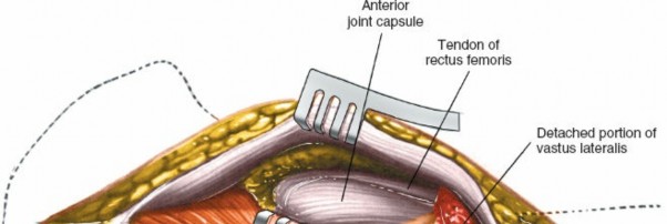
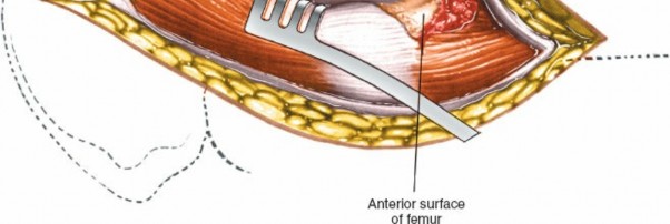 Figure 8-31 Bluntly dissect the fat pad off the anterior portion of the joint capsule to expose it and the rectus femoris tendon.
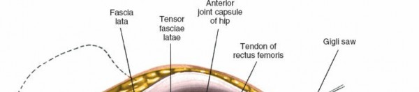
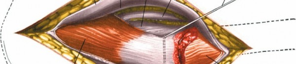
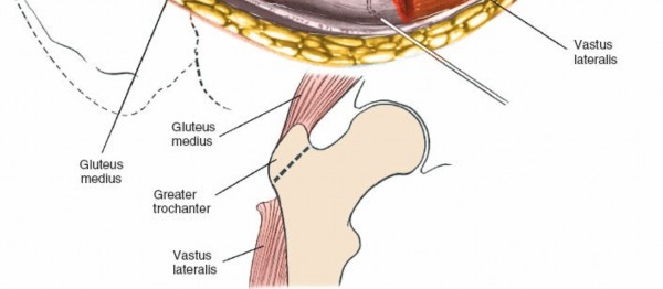 Figure 8-32 Osteotomize the greater trochanter.
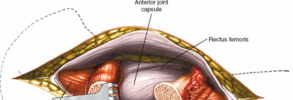
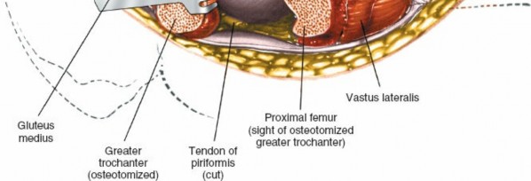 Figure 8-33 Reflect the osteotomized portion of the trochanter superiorly (with the attached gluteus medius) to reveal the joint capsule.
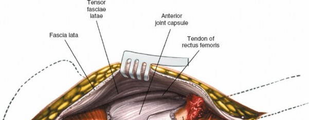
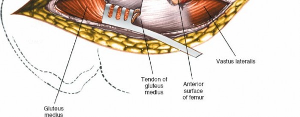 Figure 8-34 The joint capsule may also be exposed by partial resection of the gluteus medius tendon from the anterior portion of the trochanter.
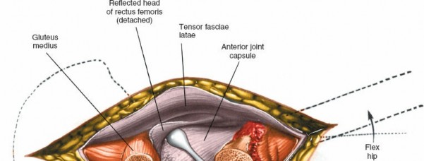
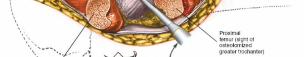
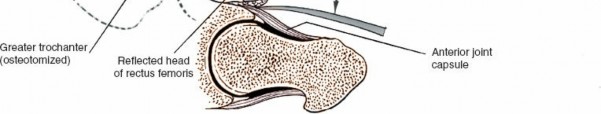 Figure 8-35 Reflect the head of the rectus femoris from the anterior portion of the joint capsule. Bluntly dissect up the anterior surface of the hip joint capsule in line with the femoral neck and head. Detach the reflected head of the rectus femoris from the joint capsule to expose the anterior rim of the acetabulum (Elevate part of the psoas tendon from the capsule. Because both the rectus femoris and the psoas may insert into the capsule, the plane between muscle and capsule is often difficult to establish.
Place a Hohmann retractor on the anterior rim of the acetabulum. Make certain that the dissection and the insertion of retractors remain beneath the rectus femoris and iliopsoas, because the neurovascular bundle lies anterior to the psoas. If you cannot develop a plane between the psoas and the capsule, incise the capsule and insert a retractor around the femoral head so that you can see the joint better.
Incise the anterior capsule of the hip joint with a longitudinal incision. Develop this into a T-shaped incision by cutting the attachment of the
8-36, and _inset_8-37).
Dang
Nerves
The femoral nerve is the most laterally placed structure in the neurovascular bundle in the femoral triangle, thus the structure closest to the operative field and most at risk. The most common problem is compression neurapraxia, caused by overexuberant medial retraction of the anterior covering structures of the hip joint. Less frequently, the nerve is directly injured by retractors placed in the substance of the iliopsoas (see Figs. 8-47 and 8-48).
Vessels
The
femoral artery and vein
may be damaged by incorrectly placed acetabular retractors that penetrate the iliopsoas, piercing the vessels as they lie on the surface of the muscle. You can avoid this complication by making sure that the tip of the retractor is placed firmly on bone, with no intervening tissue. The anterior retractor should be placed in the 1-o’clock position for the right hip and in the 11-o’clock position for the left hip. Finding the correct plane between the rectus femoris and the anterior part of the hip joint capsule is easier if the limb is in about 30 degrees of flexion. Alternatively avoid placing any retractors over the acetabular lip until after a capsulotomy has been performed and the femoral head excised when the exact position of the acetabular margin is obvious and retractors can be inserted under direct vision.
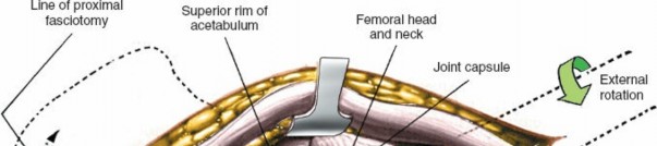

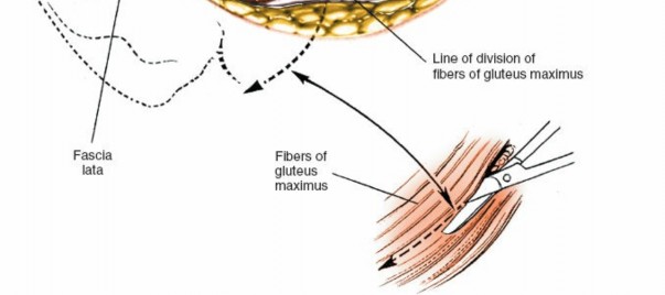 Figure 8-36
Incise the anterior joint capsule to reveal the femoral head and neck and the acetabular rim. If further proximal exposure is needed, incise the fascia lata proximally toward the iliac crest and along the iliac crest anteriorly. To facilitate dislocation of the hip, incise the tight fascia lata and the fibers of the gluteus maximus (
inset
).
Figure 8-36
Incise the anterior joint capsule to reveal the femoral head and neck and the acetabular rim. If further proximal exposure is needed, incise the fascia lata proximally toward the iliac crest and along the iliac crest anteriorly. To facilitate dislocation of the hip, incise the tight fascia lata and the fibers of the gluteus maximus (
inset
).
The
profunda femoris artery
lies on the psoas muscle, deep to the femoral artery. The artery may branch off from the femoral artery just distal to the inguinal ligament and this anatomical variant lies very close to the anterior lip of the acetabulum. The artery may be damaged by poorly placed retractors.
Be aware that damage to either of these arteries may not be obvious at the time of surgery because any bleeding that may occur may be into the retroperitoneal space and not into the surgical wound.
Fractures of the Femoral Shaft
Femoral shafts have been known to fracture while hips are being
dislocated. For that reason, it is critical that you do an adequate capsular release before attempting dislocation. To dislocate the joint, lever the femoral head out of the acetabulum with a skid (such as a Watson-Jones) while your assistant gently externally rotates the limb. Your assistant has a considerable lever arm during this procedure—if rotating the leg too forcibly, a spiral fracture of the femur can result.
In severe protrusion of the hip, you may have to osteotomize the rim of the acetabulum, which often has an osteophyte, to achieve dislocation.
If you cannot dislocate the hip without resorting to extreme force, it is safer to perform a double osteotomy of the femoral neck, excising a 1-cm portion of it, then remove the femoral head (which is lying free) with a corkscrew.
Fractures of the femoral shaft also can occur when the limb is placed in full adduction and external rotation for reaming of this femoral shaft. In order for the operator to gain a good enough view of the cut surface of the femur, the femoral shaft must be adducted. If the incision in the fascia lata has been placed too far anteriorly, then the fascia lata will resist adduction and enthusiastic assistants may cause femoral shaft fracture. This is the reason why the fascia lata should be incised initially at the posterior border of the greater trochanter. If the fascia lata gets in your way when attempting to adduct the leg, it is safest to incise it along the lines of fibers of gluteus maximus (see Fig. 8-36).
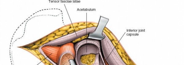
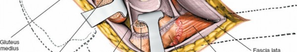
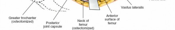 Figure 8-37 To expose the acetabulum, dislocate and resect the femoral head. Placing three or four Hohmann-type retractors around the lip of the acetabulum provides excellent exposure.
How to Enlarge the Approach
#### Local Measures
Figure 8-37 To expose the acetabulum, dislocate and resect the femoral head. Placing three or four Hohmann-type retractors around the lip of the acetabulum provides excellent exposure.
How to Enlarge the Approach
#### Local Measures
The posterior flap of the fascia lata, created during your superficial dissection, may prevent the adduction of the leg and complete dislocation of the hip needed for femoral reaming because it impinges on the trochanter. If it does, make an incision into the posterior flap of the tensor fasciae latae, heading obliquely upward and backward in line with the fibers of the gluteus maximus, which also inserts into the iliotibial tract (see Fig. 8-36). This maneuver may also be necessary to achieve reduction of the prosthetic hip when the leg has been lengthened by the operative procedure.
If anterior retraction of the tensor fasciae latae proves difficult, use a pair of scissors to incise the fascia on the muscle’s anterior aspect close to its origin from the ilium (see Fig. 8-36). Alternatively, continue the fascial incision farther down the lateral aspect of the femoral shaft. These maneuvers are seldom necessary.
The key to a full exposure of the acetabulum lies in correctly placing
the retractors. Different approaches use different retractors, but three or four Hohmann-type retractors placed around the lip of the acetabulum, directly on bone, give as good an exposure as any (see Fig. 8-37).
Extensile Measures
8-38) or when an iatrogenic femoral fracture occurs during femoral reaming.
The approach cannot usefully be extended proximally.
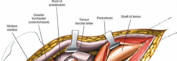
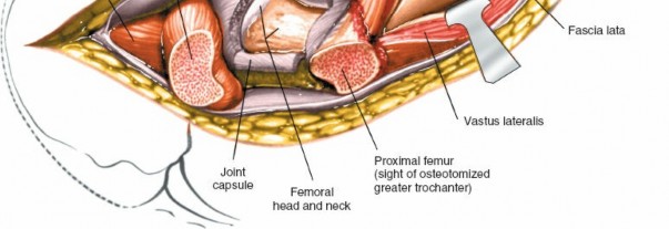 Figure 8-38 Extend the incision down the lateral aspect of the thigh, incising the deep fascia and splitting the vastus lateralis in line with its musculature to reach the lateral aspect of the femur. Lateral Approach to the Hip
The direct lateral approach (or transgluteal approach) allows excellent
25 It avoids the need for trochanteric osteotomy. Because the bulk of the gluteus medius muscle is preserved intact, it permits early mobilization of the patient following surgery. However, the approach does not give as wide an exposure as the anterolateral approach with trochanteric osteotomy. It is, therefore, difficult to perform revision surgery using this approach.
Position of the Patient
Place the patient supine on the operating table with the greater trochanter at the edge of the table. This allows the buttock muscles and gluteal fat to fall posteriorly away from the operative plane (see Fig. 8-26).
Landmarks and Incision
#### Landmarks
Palpate the anterior superior iliac spine upward from below. Palpate the lateral aspect of the greater trochanter and, below that, the line of the femur that feels like a resistance against the examining hand.
Incision
8-39).
Internervous Plane
There is no true internervous plane. The fibers of the gluteus medius muscle are split in their own line distal to the point where the superior gluteal nerve supplies the muscle. The vastus lateralis muscle is also split in its own line lateral to the point where it is supplied by the femoral nerve.
Superficial Surgical Dissection
8-40).
!8-41). You will need to detach the muscles from the greater trochanter either by sharp dissection or by lifting off a small flake of bone. Continue developing this anterior flap, following the contour of the bone onto the femoral neck, until the anterior hip joint capsule is fully exposed. You will need to detach the insertion of the gluteus minimus tendon to the anterior part of the greater trochanter (Fig. 8-42). Develop the plane between the hip joint capsule and the overlying muscles, using a swab pushed into the potential space using a blunt instrument.
8-43).
Fig. 8-44Fig. 8-45).
Dang
Nerves
The
superior gluteal nerve
8-42).
The
femoral nerve,
the most lateral structure in the anterior neurovascular bundle of the thigh, is vulnerable to inappropriately placed retractors. Anterior retractors should be placed strictly on the bone of the anterior aspect of the acetabulum and should not infringe on the substance of the psoas muscle.
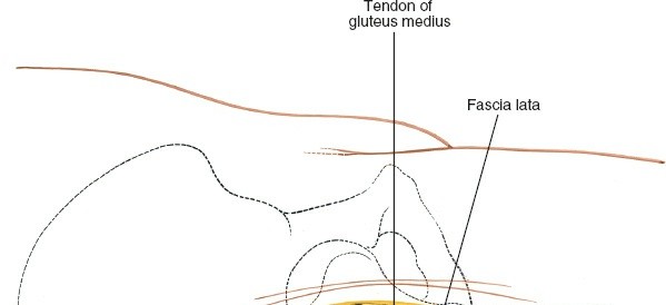
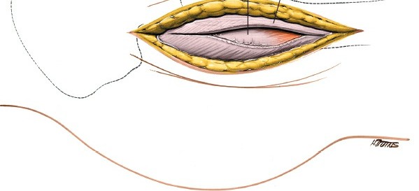 Figure 8-40 Divide the deep fascia in the line of the skin incision, retracting the fascial edges to pull the tensor fasciae latae anteriorly.
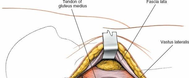
 Figure 8-41 Split the fibers of gluteus medius above the tip of the greater trochanter and extend this incision distally on the lateral aspect of the trochanter until 2 cm of the vastus lateralis is also split.
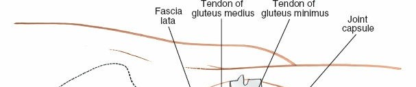
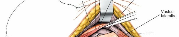
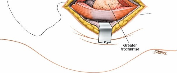 Figure 8-42 Develop this anterior flap and divide the tendon of the gluteus minimus muscle to reveal the anterior aspect of the hip joint capsule.
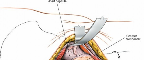
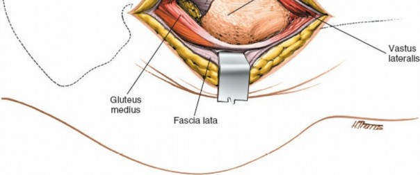 Figure 8-43 Enter the capsule using a longitudinal T-shaped incision.
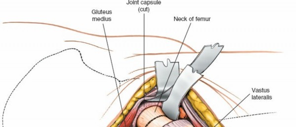
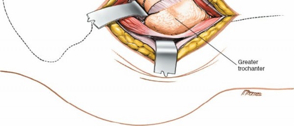 Figure 8-44 Osteotomize the femoral neck using an oscillating saw.
#### Vessels
Figure 8-44 Osteotomize the femoral neck using an oscillating saw.
#### Vessels
The
femoral artery and vein
are also vulnerable to inappropriately placed anterior retractors.
The transverse branch of the
lateral circumflex artery
of the thigh is cut as the vastus lateralis is mobilized. It must be cauterized during the approach.
How to Enlarge the Approach
#### Extensile Measures
The approach can easily be extended distally. To expose the shaft of the femur, split the vastus lateralis muscle in the direction of its fibers (see Lateral Approach in Chapter 9). The incision cannot be extended proximally.
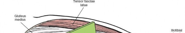
!8-46). If the iliac crest is viewed from the lateral side, the outer layer of the covering seems to consist of the fascia lata of the thigh and the muscles that it encloses. The sartorius lies farther anteriorly. The gluteus medius, which arises from the outer wing of the ilium, is covered by the fascia lata, not
enclosed by it (Fig. 8-47).
The key to the
anterolateral approach
to the hip lies in the relationship between the tensor fasciae latae and the gluteus medius. The tensor fasciae latae, a superficial structure, arises from the anterior portion of the outer lip of the iliac crest. The gluteus medius arises from the outer wall of the ilium, between the anterior and posterior gluteal lines. The origins of the two muscles are, therefore, almost continuous, but the tensor fasciae latae is slightly more superficial (lateral) and anterior than the gluteus medius (Fig. 8-48).
8-47).
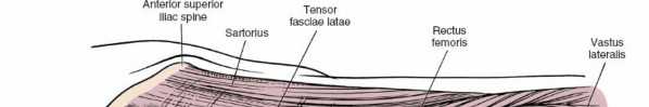
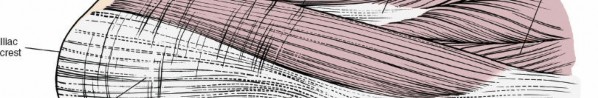
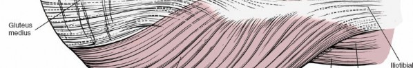
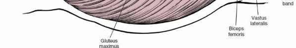 Figure 8-46 Superficial musculature of the lateral aspect of the hip.
All anterolateral approaches use this one intermuscular plane to reach the femoral neck; then they follow the joint capsule medially to expose the anterior rim of the acetabulum. The techniques used in this approach differ mainly in how they detach the abductor mechanism to allow adduction of the femur for femoral reaming and retraction of the femoral neck posteriorly for adequate exposure of the acetabulum.
Figure 8-46 Superficial musculature of the lateral aspect of the hip.
All anterolateral approaches use this one intermuscular plane to reach the femoral neck; then they follow the joint capsule medially to expose the anterior rim of the acetabulum. The techniques used in this approach differ mainly in how they detach the abductor mechanism to allow adduction of the femur for femoral reaming and retraction of the femoral neck posteriorly for adequate exposure of the acetabulum.
The
anterior approach
is more straightforward: Two distinct muscle layers must be incised. The
outer layer
consists of the tensor fasciae latae (superior gluteal nerve) and the sartorius (femoral nerve) (see Fig. 8-14). The interval between them forms a true internervous plane. Two structures, the lateral femoral cutaneous nerve and the ascending branch of the lateral femoral circumflex artery, lie between them; they must be identified and avoided during the dissection (see Figs. 8-14 and 8-15).
The
deep layer
of muscle consists of the rectus femoris (femoral nerve) and the gluteus medius (superior gluteal nerve). The interval between them is also an internervous plane; exploiting it is difficult, mainly because the short head of the rectus femoris originates partly from the anterior capsule of the hip joint, where the iliopsoas partly inserts (see Figs. 8-15 and 8-16).
The lateral approach
splits vastus lateralis and gluteus medius to provide direct access to the hip joint capsule. The approach is limited superiorly by the superior gluteal nerve, which traverses the substance of
gluteus medius.
Landmarks and Incision
#### Landmarks
The
anterior superior iliac spine
is the site of attachment of two important structures. The sartorius takes its origin from it, and the inguinal ligament uses it as a lateral attachment. The anterior superior iliac spine is rarely used as a bone graft because the lateral cutaneous nerve of the thigh lies so close to it.
The
anterior third of the iliac crest
serves as the origin for the following three muscles:
1. The
external oblique
forms the outer layer of the muscles of the anterior abdominal wall. It inserts into the outer strip of the anterior half of the iliac crest.
2. The
internal oblique
forms the middle layer of the muscles of the anterior abdominal wall. It originates from the center strip of the anterior half of the iliac crest.
3. The
tensor fasciae latae
arises from the outer lip of the anterior half of the iliac crest.
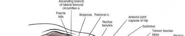


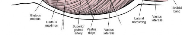 Figure 8-47 Resecting the sartorius, tensor fasciae latae, and fascia lata and reflecting the anterior portion of the gluteus maximus posteriorly reveal the gluteus medius and more anterior structures of the hip region. The fascia lata splits to envelop the tensor fasciae latae, but it only covers the gluteus medius muscle.
Gluteus Medius.
Origin. Outer aspect of ilium between anterior and posterior gluteal lines and its overlying fascia. Insertion. Lateral surface of greater trochanter. Action. Abductor and medial rotator of hip. Nerve supply. Superior gluteal nerve.
Figure 8-47 Resecting the sartorius, tensor fasciae latae, and fascia lata and reflecting the anterior portion of the gluteus maximus posteriorly reveal the gluteus medius and more anterior structures of the hip region. The fascia lata splits to envelop the tensor fasciae latae, but it only covers the gluteus medius muscle.
Gluteus Medius.
Origin. Outer aspect of ilium between anterior and posterior gluteal lines and its overlying fascia. Insertion. Lateral surface of greater trochanter. Action. Abductor and medial rotator of hip. Nerve supply. Superior gluteal nerve.
The insertion of the external oblique and origin of internal oblique are not detached during the anterior approach; the tensor fasciae latae is.
The
greater trochanter
is the traction apophysis of the proximal femur and the site of the insertion of the gluteus medius and minimus muscles.
The _vastus lateralis ridge_Figs. 8-49 and
8-50; see Fig. 8-47).
Incisions
The anterior, lateral, and anterolateral incisions largely ignore the lines of cleavage in the skin, but the scars are seldom broad and are nearly always hidden by clothing. Superficial Surgical Dissection and Its Dangers The anterior and anterolateral approaches use planes that involve the _tensor fasciae latae._8-51). The lateral approach splits gluteus medius and vastus lateralis.
Anterior Approach
The tensor fasciae latae and the sartorius run side by side from an almost continuous line of origin along the anterior end of the iliac crest. The two muscles diverge a short distance below the anterior superior iliac spine so that the rectus femoris can emerge from between them (see Fig. 8-14).
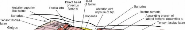
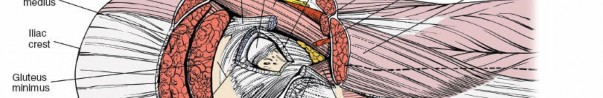
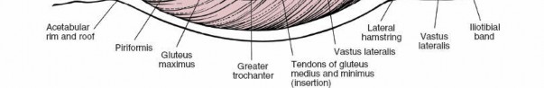 Figure 8-48 The gluteus medius, gluteus minimus, and rectus femoris have been resected to reveal the muscular layers down to the hip joint capsule. Resection of the joint capsule exposes the acetabulum and the femoral head and neck.
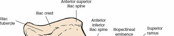
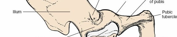
 Figure 8-49 Osteology of anterolateral aspect of the hip and pelvis.
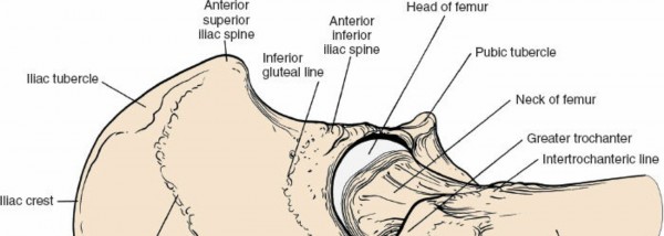
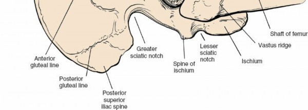 Figure 8-50 Osteology of the lateral aspect of the hip and pelvis.
The
tensor fasciae latae
itself is triangular. In cross section, it is unusually slim at its origin and thick just before it inserts into the iliotibial tract. Its action is difficult to interpret, because another large muscle, the gluteus maximus, also inserts into the iliotibial tract. In cases of poliomyelitis, dividing the iliotibial tract relieves flexion and abduction contractures of the hip joint.
Figure 8-50 Osteology of the lateral aspect of the hip and pelvis.
The
tensor fasciae latae
itself is triangular. In cross section, it is unusually slim at its origin and thick just before it inserts into the iliotibial tract. Its action is difficult to interpret, because another large muscle, the gluteus maximus, also inserts into the iliotibial tract. In cases of poliomyelitis, dividing the iliotibial tract relieves flexion and abduction contractures of the hip joint.
8
The muscle fibers of the tensor fasciae latae are considerably finer than those of the gluteus medius, but the difference in the quality of fibers rarely makes it easier to identify the plane between the two muscles.
The
sartorius
is the longest muscle in the body crossing both the hip and the knee. The individual fibers within the muscle are also the longest in the body; they leave the sartorius weak but capable of extraordinary contraction.
Two structures cross the plane between the tensor fasciae latae and the
sartorius. Both complicate the superficial surgical dissection of the anterior approach.
1. The
ascending branch of the lateral femoral circumflex artery
is a comparatively large artery that often requires ligation. It is one of a series of vessels that run circumferentially around the thigh (see Anterior Approach to the Hip above). This is one of the rare instances in which a vessel crosses an internervous plane (see Figs. 8-15 and 8-16).
2. The
lateral femoral cutaneous nerve (lateral cutaneous nerve of the thigh)
Fig. 8-14). It may divide into three or more branches just below the inguinal ligament.
Compression syndromes (meralgia paresthetica) of the lateral femoral cutaneous nerve have been reported, particularly from the section that runs behind the inguinal ligament and from the point where the nerve pierces the fascia lata. These syndromes consist of painful paresthesias on the lateral side of the thigh, conditions that may be relieved by decompressing the nerve. Occasionally, decompression may have to extend into the pelvis, since the nerve may be compressed on the surface of the iliacus.
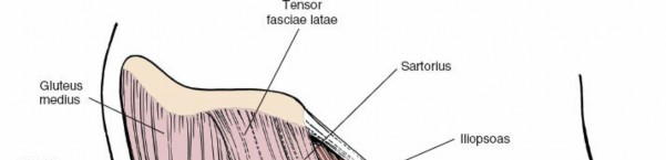
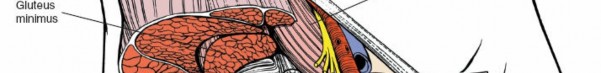
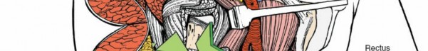
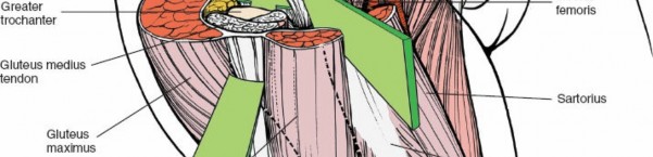
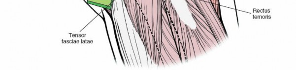 Figure 8-51 The anterior and anterolateral approaches to the hip joint, showing their muscular boundaries. #### Anterolateral Approach
One muscle, the
gluteus medius,
runs through both the superficial and the deep surgical dissection, mainly because its origin is relatively superficial while its insertion is deep (see Figs. 8-46 to 8-48). From an anterior approach, the gluteus medius appears to be one of the muscles of the inner layer over the hip joint. From an anterolateral approach, it occupies a more superficial position.
There are two important points about the structure of this muscle, knowledge of which prevents confusion during the anterolateral approach to the hip. First, fibers of the gluteus medius commonly arise from the deep surface of the fasciae latae. This means that when you elevate the fascial flap to gain access to the anterior border of gluteus medius, you
often have to detach muscular fibers from the inner surface of the fascia. Although the fascia lata encloses the tensor fasciae latae muscle and covers the gluteus medius, in many cases the fascia lata actually serves as part of the origin of the gluteus medius muscle. Second, there is often a thin fascial layer covering the gluteus medius muscle on its outer aspect just above the greater trochanter. In order to pick up the anterior border of the muscle for dissection, you frequently need to incise this fascial layer. If you do not do this, it is difficult to pick up the anterior border of the muscle since this fascial layer is continuous with the fascia covering the outer aspect of the greater trochanter.
The gluteus medius is the strongest abductor of the hip. Paralysis leads to a Trendelenburg gait in which the patient cannot prevent his hip from adducting when he puts weight on the affected leg during walking.
Note that a bursa lies between the anterior part of the muscle and the anterior part of the greater trochanter. It may become inflamed producing pain.
One nerve, the
superior gluteal nerve,
crosses the intermuscular plane between the gluteus medius and the tensor fasciae latae and must be cut if the dissection extends up to the pelvis. Whether denervation of the tensor fasciae latae muscle is clinically significant is a moot point.
Lateral Approach
The approach splits the fibers of both gluteus medius and vastus lateralis. The anterior flap of the dissection is lifted off the underlying greater trochanter until the tendon of gluteus minimus and the anterior hip joint capsule are revealed. The exposure is made possible because it utilizes the bursa beneath gluteus medius that separates the muscle from the bone superiorally. Deep Surgical Dissection and its Dangers Perhaps the most difficult part of deep dissection is finding the plane between the joint capsule and the surrounding structures, because every muscle that crosses the hip joint directly sends some of its fibers to insert into the capsule.
Anterior Approach
The deep layer of muscles consists of the rectus femoris and the gluteus medius. The rectus femoris has two heads of origin, both of which must be detached. The straight head arises from the anterior inferior iliac spine; the
reflected head arises from just above the acetabulum and from the joint capsule itself. The gluteus medius arises from the outer aspect of the ilium, between the anterior and posterior gluteal lines. While the intermuscular plane between the two muscles is easy to define and develop, the rectus is difficult to mobilize from the anterior joint capsule because part of it actually originates from the capsule itself (see Figs. 8-15 to 8-17).
The
iliopsoas muscle,
which intrudes into the inferomedial portion of the operative field, must be retracted medially to expose the anterior part of the joint. The iliopsoas crosses the hip joint directly; some of its fibers to insert into the joint capsule (iliocapsularis) (see Fig. 8-17).
The iliopsoas tendon is actually the tendon for two muscles, the psoas major and the iliacus. The iliopectineal bursa separates part of the tendon from the hip joint. The bursa, which may communicate with the joint itself, is usually obliterated in degenerative disease of the hip, leaving the tendon anchored to the anterior and medial portions of the joint capsule.
Anterolateral Approach
The
femoral vessels
enter the thigh beneath the inguinal ligament. They lie on the psoas major muscle, halfway between the anterior superior iliac spine and the pubic tubercle, the midinguinal point. The femoral artery is thus directly anterior to the hip joint, with the psoas muscle interposed. The femoral nerve lies lateral, and the femoral vein medial, to the artery. (This arrangement can be remembered through the mnemonic “VAN”—
v
ein,
a
rtery,
n
erve.)
Retractors that are placed correctly on the anterior lip of the acetabulum do not damage any of these structures, although a neurapraxia of the femoral nerve (the most lateral of the triad) may occur if retraction is prolonged and forceful.
Retractors placed more anteriorly and not directly on bones are probably biting into the substance of the psoas; they can damage any of the neurovascular structures that lie in the femoral triangle. Avoiding these complications depends on keeping to the bone of the acetabular margin and staying beneath the reflected head of the rectus femoris and the iliopsoas.
Lateral Approach
Exposure of the anterior hip joint capsule requires division of gluteus minimus tendon. This does not appear to cause a postoperative Trendelenburg gait, however.
The approach utilizes the plane between the hip joint capsule and the
overlying muscles. This plane is occupied by a small amount of fatty tissue that allows the plane to be opened up by using a swab and blunt dissection. This useful plane is always present in primary surgery, but in revision surgery, it is obliterated by scar tissue. Therefore, there is inherently more risk of damaging the femoral vessels that lie anterior to the hip joint capsule in revision surgery than in primary surgery.
Posterior Approach to the Hip
The posterior approach is the most common and practical of those used to expose the hip joint. Popularized by Moore,9 it is often called the Southern approach.
All posterior approaches allow easy, safe, and quick access to the joint and can be performed with only one assistant. Because they do not interfere with the abductor mechanism of the hip, they avoid the loss of abductor power in the immediate postoperative period. Posterior approaches allow excellent visualization of the femoral shaft, thus are popular for revision joint replacement surgery in cases in which the femoral component needs to be replaced.
Because access to the joint involves division of the posterior capsule, if dislocation of any prosthesis occurs, it will result from flexion and internal rotation of the hip. Thus, there may be a higher dislocation rate than that from anterior approaches if the posterior approach is used in fractured neck of femur surgery in elderly bedridden patients who often lie in bed with their hips in a flexed and adducted position.
Their uses include the following:
1. 10
2. Total hip replacement, including revision surgery
3. Open reduction and internal fixation of posterior acetabular fractures
4. Dependent drainage of hip sepsis
5. Removal of loose bodies from the hip joint
6. Pedicle bone grafting13
7. Open reduction of posterior hip dislocations
Position of the Patient
Place the patient in the true lateral position, with the affected limb
8-52).
Landmarks and Incision
#### Landmarks
Palpate in detail the
greater trochanter
on the outer aspect of the thigh. The posterior edge of the trochanter is more superficial than the anterior and lateral portions, and, as such, it is easier to palpate (see Fig. 8-27).
Incision
8-53).
Superficial Surgical Dissection
8-54B). (The fascial covering of the gluteus maximus varies considerably in its thickness. In the elderly, it is quite thin.)
The gluteus maximus receives its blood supply from the superior and
inferior gluteal arteries, which enter the deep surface of the muscle and ramify outward like the spokes of a bicycle wheel; hence, splitting the muscle inevitably crosses a vascular plane. In addition to the arterial bleeding, venous bleeding must be anticipated. If you split the muscle gently, you may be able to pick up, coagulate, and cut the crossing vessels before they are stretched and avulsed by the blunt dissection of the split. Obviously, vessels that are torn when stretched retract into the muscle and are more difficult to control.
--- Figure 8-52 Position of the patient on the operating table for the posterior approach to the hip joint.


!8-55).
8-56).
Internally rotate the hip to put the short external rotator muscles on a stretch (making them more prominent) and to pull the operative field farther from the sciatic nerve (Fig. 8-57A,B).
Insert stay sutures into the piriformis and obturator internus tendons
8-57C). (The upper part of the quadratus femoris rarely may also have to be divided to fully expose the posterior aspect of the joint capsule, but the muscle contains troublesome vessels that arise from the medial circumflex artery. Normally, it should be left alone.)
8-58). Posterior joint capsulotomy will have exposed the femoral head and neck.
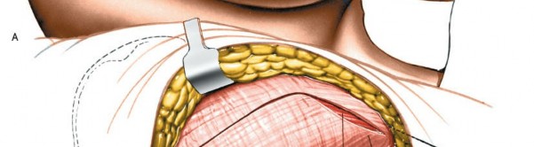
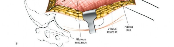 Figure 8-54A: Skin incision for the posterior approach to the hip joint. B: Incise the fascia lata.
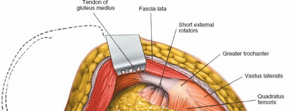
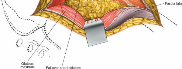 Figure 8-55 Retract the gluteus maximus to reveal the fatty layer over the short external rotators of the hip.
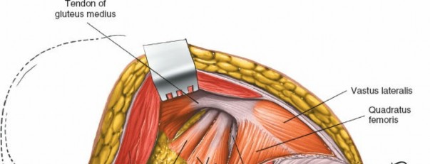
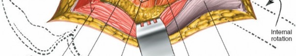
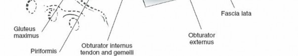 Figure 8-56 Push the fat posteromedially to expose the insertions of the short rotators. Note that the sciatic nerve is not visible; it lies within the substance of the fatty tissue. Place your retractors within the substance of the gluteus maximus superficial to the fatty tissue.
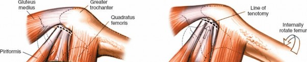
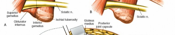
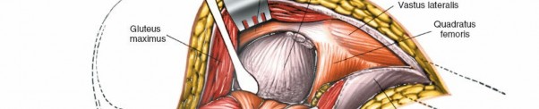
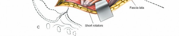 Figure 8-57A, B: Internally rotate the femur to bring the insertion of the short rotators of the hip as far lateral to the sciatic nerve as possible. C: Detach the short rotator muscles close to their femoral insertion and reflect them backward, laying them over the sciatic nerve to protect it. ### Dang
Nerves
The
sciatic nerve
is rarely exposed or transected during this approach. However, it is sometimes involved in major complications. It can be damaged if it is compressed by the posterior blade of a self-retaining retractor used to split the gluteus maximus. Always keep the retractors on the cut surfaces of the rotators; the muscles will protect the nerve.
The sciatic nerve sometimes divides into its tibial and common peroneal branches within the pelvis; on occasion, you may expose these two “sciatic nerves” during this approach. If you have identified the sciatic nerve but think that it looks too small, search for the nerve’s other branch; it is in danger if it is overlooked.
Vessels
The
inferior gluteal artery
leaves the pelvis beneath the piriformis. It spreads cephalad to supply the deep surface of the gluteus maximus. Its branches are inevitably cut when the gluteus maximus is split; you can identify and coagulate them before they are avulsed if you are dissecting carefully.
The main trunk of the artery is vulnerable as it emerges from beneath the lower border of the piriformis when pelvic fractures involve the greater sciatic notch. If it retracts into the pelvis and bleeding is brisk, turn the patient over into the supine position, open the abdomen, and tie off the artery’s feeding vessel, the internal iliac artery.
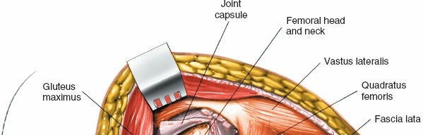
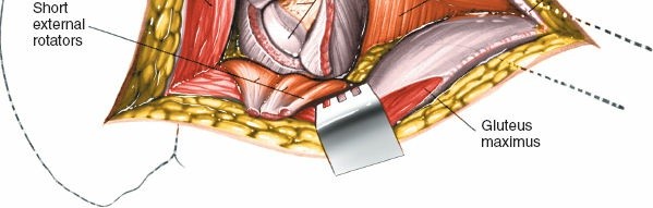 Figure 8-58 Incise the posterior joint capsule to expose the femoral head and neck. How to Enlarge the Approach #### Local Measures
1. Enlarge the skin incision. Obese patients may have a considerable layer of subcutaneous tissue over the buttock that restricts deep exposure; lengthening the skin incision and dissecting subcutaneously can compensate for this problem.
2. Extend the fascial incision superiorly and inferiorly.
3. Detach the upper half of the quadratus femoris. Because the muscle
Fig. 8-59).
4. 8-59).
!8-61).
8-6014 noted, the layer can be viewed as the “pelvic deltoid”: it covers the hip much as the deltoid muscle covers the shoulder.
15 lies at the anterior border of the gluteus maximus, between the gluteus maximus (inferior gluteal nerve) and the gluteus medius. This approach uses a true internervous plane.
9 16) involve splitting the fibers of the gluteus maximus. They are more popular than the Marcy–Fletcher approach even though they do not operate in an internervous plane, mainly because they offer excellent exposure of the hip joint.
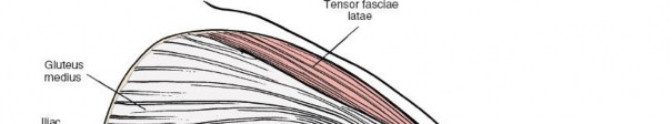
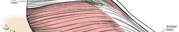
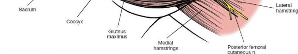 Figure 8-60 The superficial musculature of the posterior approach of the hip joint. The gluteus maximus predominates.
Gluteus Maximus.
Origin. From posterior gluteal line of ilium and that portion of the bone immediately above and behind it; from posterior surface of lower part of sacrum and from side of coccyx; and from fascia covering gluteus medius. Insertion. Into iliotibial band of fascia lata and into gluteal tuberosity. Action. Extends and laterally rotates thigh. Nerve supply. Inferior gluteal nerve.
Figure 8-60 The superficial musculature of the posterior approach of the hip joint. The gluteus maximus predominates.
Gluteus Maximus.
Origin. From posterior gluteal line of ilium and that portion of the bone immediately above and behind it; from posterior surface of lower part of sacrum and from side of coccyx; and from fascia covering gluteus medius. Insertion. Into iliotibial band of fascia lata and into gluteal tuberosity. Action. Extends and laterally rotates thigh. Nerve supply. Inferior gluteal nerve.
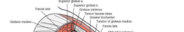
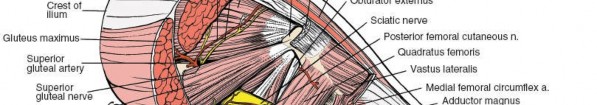


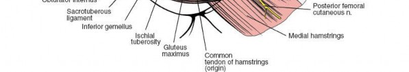 Figure 8-61 The gluteus maximus and the gluteus medius have been resected to reveal the gluteus minimus, the piriformis, and the short rotator muscles. Note the relationship of the neurovascular structures to the piriformis.
Gluteus Minimus.
Origin. From outer surface of ilium between anterior and inferior gluteal lines. Insertion. Into impression on anterior border of greater trochanter via tendon that gives expansion to joint capsule. Action. Rotates thigh medially and abducts it. Nerve supply. Superior gluteal nerve.
Piriformis.
Origin. From front of sacrum via fleshy digitations from second, third, and fourth portions of sacrum. Insertion. Into upper border of greater trochanter via round tendon. Action. Rotates thigh laterally and abducts it. Nerve supply. Branches from first and second sacral nerves.
Obturator Internus.
Origin. From inner surface of anterolateral wall of pelvis and from surfaces of greater part of obturator foramen. Insertion. Onto medial surface of greater trochanter above trochanteric fossa. Action. Rotates thigh laterally. Nerve supply. Any nerve from sacral plexus.
Quadratus Femoris.
Origin. From upper part of external border of tuberosity of ischium. Insertion. Into upper part of linea quadrata, the line that extends vertically downward from intertrochanteric crest. Action.
Figure 8-61 The gluteus maximus and the gluteus medius have been resected to reveal the gluteus minimus, the piriformis, and the short rotator muscles. Note the relationship of the neurovascular structures to the piriformis.
Gluteus Minimus.
Origin. From outer surface of ilium between anterior and inferior gluteal lines. Insertion. Into impression on anterior border of greater trochanter via tendon that gives expansion to joint capsule. Action. Rotates thigh medially and abducts it. Nerve supply. Superior gluteal nerve.
Piriformis.
Origin. From front of sacrum via fleshy digitations from second, third, and fourth portions of sacrum. Insertion. Into upper border of greater trochanter via round tendon. Action. Rotates thigh laterally and abducts it. Nerve supply. Branches from first and second sacral nerves.
Obturator Internus.
Origin. From inner surface of anterolateral wall of pelvis and from surfaces of greater part of obturator foramen. Insertion. Onto medial surface of greater trochanter above trochanteric fossa. Action. Rotates thigh laterally. Nerve supply. Any nerve from sacral plexus.
Quadratus Femoris.
Origin. From upper part of external border of tuberosity of ischium. Insertion. Into upper part of linea quadrata, the line that extends vertically downward from intertrochanteric crest. Action.
Rotates thigh laterally. _Nerve supply.
Branch from sacral plexus.
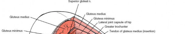
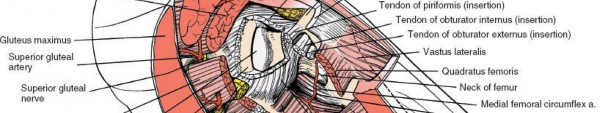
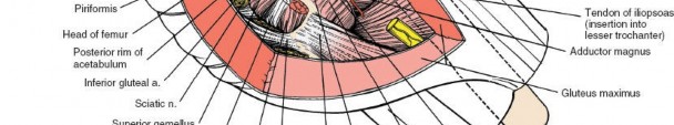
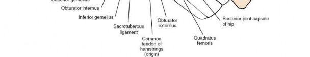 Figure 8-62 The gluteus minimus, piriformis, and short rotators have been resected to uncover the posterior aspect of the hip joint. Landmarks and Incision #### Landmarks
The
greater trochanter,
over which the skin incision is centered, is the easiest bony prominence to palpate around the hip. Its posterior aspect is relatively free of muscles; its anterior and lateral aspects are covered by the tensor fasciae latae and the gluteus medius and minimus muscles and are much less accessible.
-
The
gluteus medius
attaches by a broad insertion into its lateral aspect. Below this insertion, the bone is covered by the beginnings of the iliotibial tract. A bursa, occasionally a site of inflammation, lies between the tract and the bone over the relatively bare portion of the trochanter.
The bursa can be the site of bacterial infection (historically, most frequently tuberculosis). - The gluteus minimus is attached to the anterior aspect of the trochanter, where its tendon is divided in the anterolateral approach (see Figs. 8-61 and 8-62).
- The piriformis inserts via a tendon into the middle of the upper border of the greater trochanter. (see Figs. 8-61 and 8-62).
- Obturator externus tendon. Immediately below the insertion of the piriformis lies the trochanteric fossa, a deep pit that marks the attachment of the obturator externus tendon (see Figs. 8-61 and 8-62).
- The obturator internus tendon inserts with the two gemelli into the upper border of the trochanter, posterior to the insertion of the piriformis (see Figs. 8-61 and 8-62).
Incision
The upper part of the incision crosses the lines of cleavage of the skin at almost 90 degrees, but the resulting scar is always hidden by clothing. Most patients who undergo this approach are elderly and tend not to form exuberant scar tissue, and they heal with a fine line scar.
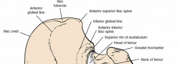
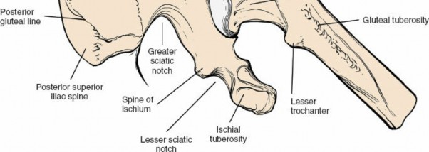 Figure 8-63 Osteology of the posterior aspect of the hip and pelvis. Superficial Surgical Dissection and Its Dangers Superficial dissection consists of cutting through the outer muscle layer by splitting the fibers of the gluteus maximus, the single largest muscle in the body. The fibers of the gluteus maximus are extremely coarse; they run obliquely downward and laterally across the buttock. The muscle’s innervation, the inferior gluteal nerve, emerges from the pelvis beneath the inferior border of the piriformis and almost immediately enters the muscle’s deep surface close to its medial border, its origin. From there, the nerve’s branches spread throughout the muscle. Splitting the gluteus maximus close to its lateral insertion does not denervate significant portions of the muscle, because its main nerve supply passes well medial to the most medial point of splitting (see Fig. 8-60).
The gluteus maximus is quiet during normal walking or standing still; it comes into play during stair climbing or standing up from a sitting position. (During normal walking, hip extension is primarily a function of the hamstrings rather than the gluteus maximus.)
Deep Surgical Dissection and Its Dangers
Deep dissection consists of incising some portion of the inner muscular layer (the short external rotators of the hip) to expose the posterior hip joint capsule (see Fig. 8-55). Five muscles form the inner layer: the piriformis, the superior gemellus, the tendon of the obturator internus, the inferior gemellus, and the quadratus femoris.
Recognizing the relationship of the piriformis to passing structures is the key to understanding the neurovascular anatomy of the area (see Fig. 8-61). All neurovascular structures that enter the buttock from the pelvis pass through the greater sciatic notch, either superior or inferior to the piriformis, which itself passes from the pelvis to the buttock through the notch.
The 10 critical neurovascular structures are as follows:
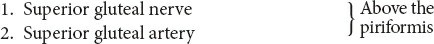
(These are the only structures to pass above the piriformis, hence, their name “superior.”)
The
superior gluteal nerve
emerges from the pelvis above the piriformis. It crosses behind the posterior border of the gluteus medius and runs in the space between the gluteus medius and the gluteus minimus, supplying both before sending fibers to the tensor fasciae latae.
The
superior gluteal artery,
the largest branch of the internal iliac artery, enters the buttock above the upper border of the piriformis and runs with its nerve, supplying the gluteus medius and gluteus minimus and sending a nutrient vessel to the ilium on the gluteal line. The nutrient vessel may bleed when a larger posterior iliac bone graft is taken. The superior gluteal artery also sends branches to the overlying gluteus maximus, forming part of the muscle’s dual arterial supply.
17 If you are using the anterior or posterior approaches to the acetabulum using a trochanteric osteotomy, the superior gluteal vessels must be intact in order to avoid muscle necrosis of the gluteus medius and minimus. This is because the origin and insertion of the muscles is detached in these approaches. If the acetabular fracture involves a displaced fracture of the greater sciatic notch, preoperative angiography is advised to ensure that the neurovascular pedicle to these structures is intact.
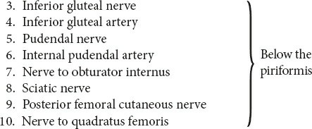
The
inferior gluteal nerve
reaches the buttock beneath the lower border of the piriformis. It enters the deep surface of the gluteus maximus almost immediately.
The
inferior gluteal artery,
which follows the inferior gluteal nerve, supplies the gluteus maximus. The branch it sends along the sciatic nerve was the original axial artery of the limb. In rare cases, this artery can serve
as one guide to the sciatic nerve during surgery. The inferior gluteal artery may also be torn in pelvic fractures, but not as frequently as the superior gluteal artery.
The
pudendal nerve
is not encountered during the posterior approach to the hip because its course in the buttock is very short. It turns around the sacrospinous ligament before entering the perineum.
The
internal pudendal artery_18 may be needed to ligate the parent trunk.
The _nerve to the obturator internus
enters its muscle almost as soon as it emerges from behind the inferior border of the piriformis. The nerve also supplies the superior gemellus.
The massive
sciatic nerve,
which is formed by roots from the lumbosacral plexus (L4, L5, S1, S2, S3), appears in the buttock from beneath the lower border of the piriformis, just lateral to the inferior gluteal and pudendal nerves and vessels. It is usually surrounded by fat and is often easier to feel than to see. It passes vertically down the buttock together with its artery, lying on the short external rotator muscles of the inner muscular sleeve, the obturator internus, the two gemelli, and the quadratus femoris. Farther distally, it passes deep (anterior) to the biceps femoris and disappears from view, lying on the adductor magnus.
The sciatic nerve is safe during posterior approaches as long as you are aware of its position. It can be injured if it is trapped in the posterior blade of the self-retaining retractor that holds the fascial edge. It also can be damaged if it is not protected during reduction of the prosthetic head into the acetabulum.
The tibial portion of the sciatic nerve supplies all the hamstring muscles except the short head of the biceps femoris and the extensor portion of the adductor magnus in the thigh. All its branches arise from the medial side of the nerve. Dissections around the sciatic nerve in the thigh therefore should remain on the lateral (safe) side, since the only branch coming off that side runs to the short head of the biceps, a muscle that causes few clinical problems if its nerve supply is damaged (see supply skin over the sole of the foot, the dorsum of
the foot (except for its medial side), and the calf and lateral side of the lower leg. Damage to the sciatic nerve at the level of the hip joint injures both tibial and common peroneal elements, resulting in a balanced flaccid paralysis below the knee, together with paralysis of the hamstring muscles. Complete sciatic nerve lesions are relatively rare; more often, the damage seems to affect either the tibial or the common peroneal components. Hence, neurologic findings may vary regardless of the level of the lesion.
Common peroneal nerve palsies do occur after posterior approaches to the hip. The question then arises as to whether the nerve was damaged at the operative site or whether it was compressed by external pressure on the nerve as it winds around the neck of the fibula. The differential diagnosis can be made by doing an electromyogram (EMG) of the short head of the biceps, the only muscle of the thigh that is supplied by the common peroneal division of the sciatic nerve. Lesions in the pelvis or at the level of the hip joint denervate this muscle. Lesions at the level of the fibular head leave it unaffected.
Only 20% of the cross section of the sciatic nerve at the hip joint is formed by nerve fibers. The remaining 80% is made up of connective tissue. Nerve repairs in this area are often unsuccessful, because bundle-to-bundle contact is difficult to achieve.
The
posterior femoral cutaneous nerve (posterior cutaneous nerve of the thigh)
supplies a large area of skin on the back of the thigh. The nerve actually lies on the sciatic nerve until the sciatic passes deep to the biceps femoris. Then the posterior femoral cutaneous nerve continues superficial to the hamstring muscles but deep to the fascia lata, sending out several cutaneous branches.
The
nerve to the quadratus femoris
emerges from the pelvis behind the sciatic nerve and runs with it on its deep surface as it crosses the tendon of the obturator internus and the two gemelli. The nerve then passes deep to the quadratus femoris before entering its anterior surface. It also gives off muscular branches to the inferior gemellus.
Obturator Internus
The obturator internus is one of the few muscles that make a right-angled turn; it curves round the lesser sciatic notch of the ischium. The muscle has a tricipital tendon that is reinforced at its insertion by the superior and inferior gemelli.
Quadratus Femoris
The quadratus femoris is quadrate, that is, four-sided. Its transversely
running fibers form a clear surgical landmark. The muscle has an excellent blood supply. At the lower border of its insertion lies the cruciate anastomosis, consisting of the ascending branch of the first perforating artery, the descending branch of the inferior gluteal artery, and transverse branches of the medial and lateral femoral circumflex arteries (see Fig. 8-61).
13 In this maneuver, the quadrate tubercle of the femur is detached, leaving the muscular insertion of the quadratus femoris intact. The resultant muscle-bone pedicle can be swung upward, and the bone can be inserted across the fracture site.
Medial Approach to the Hip
19 was originally designed for surgery on flexed, abducted, and externally rotated hips, the kinds of deformities caused by certain types of congenital dislocation of the hip.
The uses of the medial approach include the following:
2. Biopsy and treatment of tumors of the inferior portion of the femoral neck and medial aspect of proximal shaft
3. Psoas release
4. Obturator neurectomy
The upper part of the approach can be used for an obturator neurectomy. If a neurectomy is combined with an adductor release, the approach can be performed through a short transverse incision or a short longitudinal incision in the groin that permits division of the adductors close to their pelvic origin, an area where there is less bleeding.
Position of the Patient
Place the patient supine on the operating table with the affected hip flexed, abducted, and externally rotated. This may not be possible in cases with fixed deformity, when the position is often determined for you. The sole of the foot on the affected side should lie along the medial side of the
Fig. 8-64).
Landmarks and Incision
#### Landmarks
Palpate the
adductor longus
from the medial side of the thigh and follow it up to its origin at the pubis, in the angle between the pubic crest and the symphysis. The adductor longus is the only muscle of the adductor group that is easy to palpate.
With your finger anchored on the greater trochanter, move your thumb along the inguinal creases medially and obliquely downward until you can feel the
pubic tubercle.
They are at the same level as the top of the greater trochanter.
Incision
Fig. 8-65).
Internervous Plane
8-66).
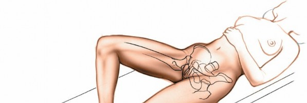
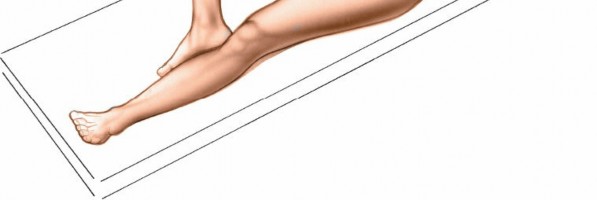 Figure 8-64 Position of the patient on the operating table for the medial approach to the hip.
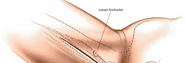
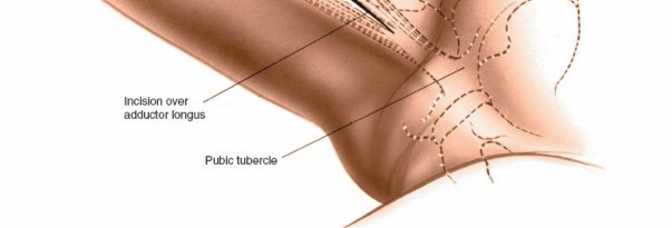 Figure 8-65 Incision for the medial approach to the hip.
More deeply, the plane of dissection lies between the adductor brevis and the adductor magnus. The adductor brevis is supplied by the anterior division of the obturator nerve. The adductor magnus has two nerve supplies: Its adductor portion is supplied by the posterior division of the obturator nerve, and its ischial portion is supplied by the tibial part of the sciatic nerve. The two muscles, therefore, form the boundaries of an internervous plane (see above and below the lesser trochanter to isolate the psoas tendon.
Figure 8-65 Incision for the medial approach to the hip.
More deeply, the plane of dissection lies between the adductor brevis and the adductor magnus. The adductor brevis is supplied by the anterior division of the obturator nerve. The adductor magnus has two nerve supplies: Its adductor portion is supplied by the posterior division of the obturator nerve, and its ischial portion is supplied by the tibial part of the sciatic nerve. The two muscles, therefore, form the boundaries of an internervous plane (see above and below the lesser trochanter to isolate the psoas tendon.
Dang
Nerves
The
anterior division of the obturator nerve
lies on top of the obturator externus and runs down the medial side of the thigh between the adductor longus and the adductor brevis, to which it is bound by a thin tissue. It supplies the adductor longus, the adductor brevis, and the gracilis in the thigh (Fig. 8-67B).
The
posterior division of the obturator nerve
8-67C).
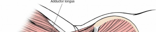

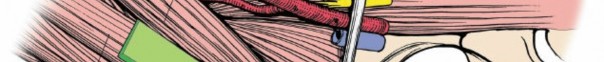
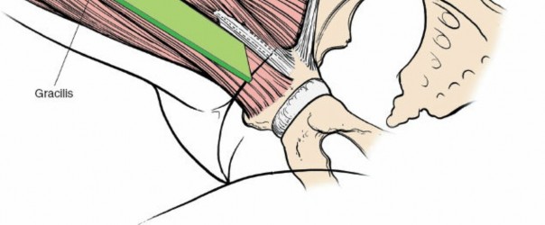 Figure 8-66 The intermuscular interval between the adductor longus and the gracilis is not an internervous plane because both muscles are innervated by the anterior division of the obturator nerve. The plane is safe, however, because the muscles receive their nerve supplies proximal to the dissection.
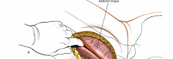
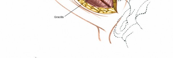
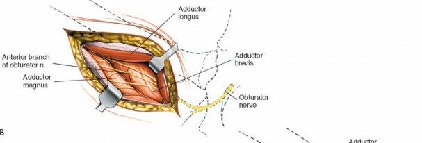
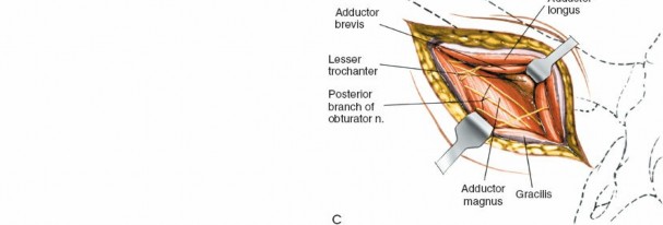 Figure 8-67 A: Develop the plane between the gracilis and the adductor longus. B: Retract the adductor longus and the gracilis to reveal the adductor brevis with the overlying anterior division of the obturator nerve. C: Retract the adductor brevis from the muscle belly of the adductor magnus to uncover the posterior division of the obturator nerve. Note the lesser trochanter in the depths of the wound.
Most of the time, the approach is designed specifically to cut these nerves to relieve muscular spasticity. If you are not using it for that purpose, avoid transecting them.
Vessels
The
medial femoral circumflex artery
(see Applied Surgical Anatomy of the Thigh in Chapter 98-69).
How to Enlarge the Approach
#### Local Measures
After reaching the femur and detaching the insertions of the psoas and iliacus, you can expose some 5 cm of femoral shaft distal to the lesser trochanter by blunt dissection.
Extensile Measures
This exposure is almost never enlarged by extensile measures.
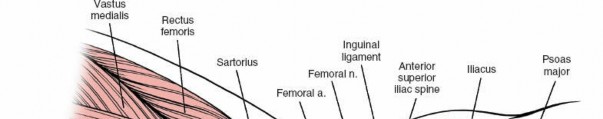


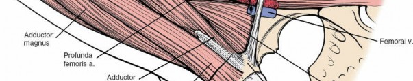
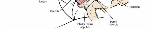 Figure 8-68 Anatomy of the medial approach to the hip. The thigh is abducted, slightly flexed, and externally rotated. The plane of the superficial dissection runs between the adductor longus and the gracilis.
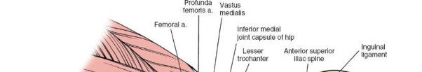
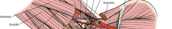
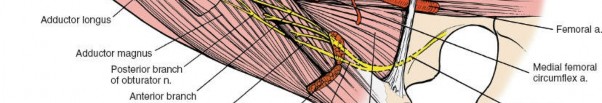
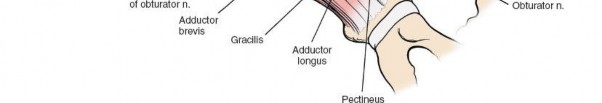 Figure 8-69 The deep muscular layer of the medial approach to the hip. The dissection lies between the adductor brevis and the adductor magnus. The gracilis, adductor longus, and sartorius have been resected to reveal the deeper structures of the medial aspect of the thigh. Note the relationships of the anterior and posterior divisions of the obturator nerve to the adductor longus and adductor brevis. Note the proximity of the medial femoral circumflex artery to the insertion of the psoas tendon.
Psoas Major.
Origin. Anterior surface of transverse processes and bodies of the lumbar vertebrae and corresponding intervertebral disks. Insertion. Lesser trochanter of femur. Action. Flexor of hip and flexor of lumbar spine when leg is fixed. Nerve supply. Segmental nerves from second and third lumbar roots.
Iliacus.
Origin. Upper two-thirds of iliac fossa, inner lip of iliac crest, anterior aspect of sacroiliac joint, and from the anterior sacroiliac and iliolumbar ligaments. Insertion. Lesser trochanter of femur by common tendon with psoas. Action. Flexor of hip. Tilts pelvis forward when leg is fixed. Nerve supply. Femoral nerve (L2-L4)._
Figure 8-69 The deep muscular layer of the medial approach to the hip. The dissection lies between the adductor brevis and the adductor magnus. The gracilis, adductor longus, and sartorius have been resected to reveal the deeper structures of the medial aspect of the thigh. Note the relationships of the anterior and posterior divisions of the obturator nerve to the adductor longus and adductor brevis. Note the proximity of the medial femoral circumflex artery to the insertion of the psoas tendon.
Psoas Major.
Origin. Anterior surface of transverse processes and bodies of the lumbar vertebrae and corresponding intervertebral disks. Insertion. Lesser trochanter of femur. Action. Flexor of hip and flexor of lumbar spine when leg is fixed. Nerve supply. Segmental nerves from second and third lumbar roots.
Iliacus.
Origin. Upper two-thirds of iliac fossa, inner lip of iliac crest, anterior aspect of sacroiliac joint, and from the anterior sacroiliac and iliolumbar ligaments. Insertion. Lesser trochanter of femur by common tendon with psoas. Action. Flexor of hip. Tilts pelvis forward when leg is fixed. Nerve supply. Femoral nerve (L2-L4)._
Applied Surgical Anatomy of the Medial Approach Overview The anatomy of this approach is the anatomy of the adductor compartment of the thigh. The adductors do not cover the hip joint, since they all originate below the level of the joint itself.
The adductor compartment of the thigh consists of three layers of muscles, with the two divisions of the obturator nerve running between each pair of layers. The superficial layer consists of the adductor longus and the gracilis; the middle layer, the adductor brevis; and the deep layer, the adductor magnus.
Landmarks and Incision
#### Landmarks
The
adductor longus
is the only muscle of the adductor group that is easily palpable at its tendinous origin. Its structure is considered in detail in the superficial surgical dissection.
The _pubic tubercle_Fig. 8-70).
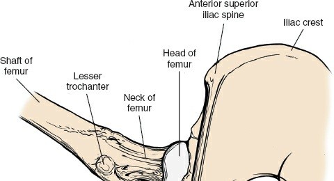
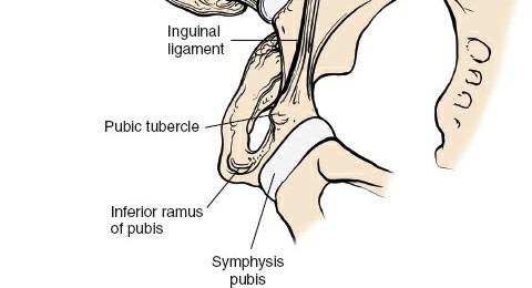 Figure 8-70 Osteology of the medial approach to the hip.
#### Incisions
Figure 8-70 Osteology of the medial approach to the hip.
#### Incisions
A longitudinal incision crosses the relaxed skin tension line obliquely, since they run down and medially cross the front of the thigh. A transverse insertion made in the groin, used for release of the adductors, is parallel with the lines and should heal with minimal scar formation. Meticulous closure of the deep fascia minimizes ugly depressed scars.
Superficial Surgical Dissection
The approach runs between the adductor longus and the gracilis. Both muscles are supplied by the anterior division of the obturator nerve, but the nerves enter them close to their pubic origins, leaving the intermuscular plane available for surgical use (see Fig. 8-68).
The
gracilis
is extremely long and thin, with long parallel-running fibers. Its aponeurotic origin, a thin sheet of tendinous fibers arising from the pubis, lies in an anteroposterior plane.
The
adductor longus
arises by a strong tendon which accounts for its involvement in the relatively high incidence of avulsion fractures. The origin may ossify in those who, like horseback riders, use their hip adductors excessively. This ossification is known as rider’s bone. Calcification and ossification are also seen in soccer players in whom the lesion may give rise to chronic pain and loss of function.
Given the size and relative “weakness” of these two muscles, it is difficult to understand why they were once given the name of
The
obturator nerve
is derived from anterior divisions of the L2-L4 nerve roots. The nerve divides in the obturator notch into
anterior
and
posterior
divisions. The anterior division passes over the upper border of the obturator externus and descends on the medial side of the thigh behind the adductor longus, on the anterior surface of the adductor brevis. It supplies sensory fibers to the hip joint.
The nerve is commonly transected in cases of adduction contractures of the hip caused by spasticity of the adductor muscles. To find it, define the interval between the adductor longus and the adductor brevis, the point at which the nerve is available for section. The nerve lies on the anterior surface of the adductor brevis, bound to it by a thin areolar covering. There, it may divide into three or even four bundles. Therefore, when performing an anterior obturator neurectomy, dissect the nerve bundles proximally as far as possible to avoid overlooking branches coming off higher up.
The anterior branch gives cutaneous distribution to the medial side of the knee, perhaps the reason hip pain is often referred to the knee, especially in cases of slipped upper femoral epiphysis. (A second pain commonly referred to the inside of the knee, originating in the ovary, is thought to be due to direct irritation of the obturator nerve by ovarian pathology, because the nerve runs adjacent to the ovary in the pelvis.)
Deep Surgical Dissection
The intermuscular interval used in deep surgical dissection lies between the adductor brevis and the adductor magnus (see Fig. 8-69).
The
adductor brevis
runs down the thigh sandwiched between the anterior and posterior divisions of the obturator nerve.
In certain animals, the
adductor magnus
inserts into the tibia. In humans, the superficial medial ligament of the knee is thought to have
arisen as the degenerative tendon of the adductor magnus and is sometimes referred to as the fourth hamstring muscle.
The
posterior division of the obturator nerve
runs distally on the surface of the adductor magnus supplying it. It gives off a fine terminal branch which runs with the femoral artery through the hiatus in the adductor magnus muscle to the popliteal fossa where it supplies part of the knee joint capsule. **
Scientific References
-
**
1. SMITH-PETERSEN MN: A new supra-articular subperiosteal approach to the hip joint. _Am J Orthop Surg._ 1917;15:592.
2. SMITH-PETERSEN MN: Approach to and exposure of the hip joint for mold arthroplasty. _J Bone Joint Surg Am._ 1949;31:40–46.
3. HONORTH MB: Congenital dislocation of the hip: technique of open reduction. _Ann Surg._ 1952;135:508.
4. CHARNLEY J: _Low Friction Arthroplasty of the Hip: Theory and Practice_. New York, NY: Springer-Verlag; 1979.
5. HARRIS WH: A new lateral approach to the hip joint. _J Bone Joint Surg Am._ 1967;49:891–898.
6. MüLLER ME: Total hip prosthesis. _Clin Orthop Relat Res._ 1970;72:46–68.
7. CJARNLEY J: Arthroplasty of the hip: a new operation. _Lancet._
1961;2:129.
8. EVANS P: The postural function of the iliotibial tract. _Ann R Coll Surg Engl._ 1979;61:271–280.
9. MOORE AT: The Moore self-locking vitallium prosthesis in fresh femoral neck fractures: a new low posterior approach (the southern exposure). In: _American Academy of Orthopaedic Surgeons: Instructional Course Lectures_. Vol. 16. St. Louis, MO: CV Mosby; 1959.
10. MOORE AT: The self-locking metal hip prosthesis. _J Bone Joint Surg Am._ 1957;39:811–812.
11. THOMPSON FR: Vitallium intramedullary hip prosthesis: preliminary report. _N Y State J Med._ 1958;52:3011–3020.
12. CHARNLEY J: Anchorage of the femoral head prosthesis to the shaft of the femur. _J Bone Joint Surg Br._ 1960;42:28–30.
13. MYERS MH, HARVEY JP, MOORE TM: Treatment of displaced
subcapital and transcervical fractures of the femoral neck by muscle pedicle–bone graft and internal fixation. _J Bone Joint Surg Am._
1973;55:257–274.
14. HENRY AK: _Extensile Exposure_. 3rd ed. Edinburgh: Churchill Livingstone; 1972.
15. MARCY GH, FLETCHER RS: Modification of the posterolateral approach to the hip for insertion of femoral head prosthesis. _J Bone Joint Surg Am._ 1954;36:142–143.
16. OSBOURE RP: The approach to the hip joint: a critical review and a suggested new route. _Br J Surg._ 1930;18:49–52.
17. NACHBUR B, MEYER RP, VRKKALA K ET AL: The mechanisms of severe arterial injury in surgery of the hip joint. _Clin Orthop Relat Res._ 1979;141:122–133.
18. RETZIUS AA: Ueber das ligamentum pelvioprostaticum oper den apparat, durch welchen die harnblase, die prostata an und der untern becken öffnung besestidt sind. _Arch Anat Physiol_. 1841;188–189.
19. LUDLOFF K: Zur blutigen einrenkung der angeborenen hüftluxation. _Z Orthop Chir._ 1908;22:272–276.
20. FERGUSON AB JR: Primary open reduction of congenital dislocation of the hip using a median adductor approach. _J Bone Joint Surg Am._ 1973;55:671–689.
21. LAST RJ: _Anatomy Regional and Applied_. New York, NY: Churchill Livingstone; 1978.
22. SWIONTKOWSKI MF: Intracapsular fractures of the hip. _J Bone Joint Surg AM._ 1994;76:129–138.
23. BOHLER N, HIPMAIR G: The minimal invasive surgery anterior approach with supine patient positioning: a step-wise introduction of technique. _Hip Int._ 2006;16 Suppl 4:48–53.
24. BABST D, STEPPACHER SD, GANZ R ET AL: The iliocapsularis muscle: an important stabilizer in the dysplastic hip. _Clin Orthop Relat Res_. 2011;469(6):1728–1734.
25. HARDINGE K: The direct lateral approach to the hip. _J Bone Joint Surg Br._ 1982;64:17–19.
26. GOTTSCHALK F, KOUROSH S, LEVEAU B: The functional anatomy of tensor fasciae latae and gluteus medius and minimus. _J Anat._ 1989;166:179–189.
Academic Citations & Sources
You Might Also Like