Anterior Thoracic Spine Approach: An Intraoperative Masterclass for Fellows
Key Takeaway
Master the anterior thoracic spine approach for complex pathologies. This immersive guide covers meticulous patient positioning, intricate surgical anatomy, and granular, real-time intraoperative steps. Fellows will learn critical dissection techniques, neurovascular protection, and strategies to avoid pitfalls, ensuring optimal patient outcomes and effective complication management.
Introduction and Epidemiology
The anterior approach can be used to access the thoracic spine for decompression, deformity correction, and stabilization. This approach allows for access to treat conditions such as intervertebral disc herniation, infection, tumor, and trauma. Historically, thoracic spinal pathologies were approached via posterior laminectomies; however, this often yielded catastrophic neurologic outcomes, particularly for anteriorly situated compressive lesions. The evolution of the transthoracic anterior approach revolutionized the surgical management of thoracic myelopathy and structural instability by providing direct visualization of the anterior column without necessitating manipulation of the spinal cord.

Thoracic disc herniations are relatively rare, comprising approximately 1% to 2% of all surgically treated intervertebral disc herniations. Despite their low incidence, they frequently present with profound myelopathy due to the narrow sagittal diameter of the thoracic spinal canal. Similarly, the thoracic spine is the most common site for spinal metastatic disease, with the anterior column being involved in the vast majority of cases. Traumatic injuries, specifically burst fractures resulting from axial loading and flexion forces, frequently compromise the anterior and middle columns, necessitating anterior column reconstruction to restore biomechanical integrity and decompress the neural elements.

The anterior thoracic approach provides unparalleled access from T4 to T12. Access to the upper thoracic spine (T1 to T3) typically requires specialized approaches, such as a trans-sternal or low cervical approach, while the thoracolumbar junction (T12 to L2) often necessitates a thoracoabdominal approach with diaphragmatic takedown. Mastery of the standard transthoracic approach is a fundamental requirement for the complex spine surgeon.

Surgical Anatomy and Biomechanics
The thoracic spinal cord may have a tenuous blood supply, particularly in patients with congenital anomalies and kyphosis. The midthoracic cord represents a watershed zone for vascularity. The artery of Adamkiewicz supplies the thoracic cord but can have a variable origin. Its origin is usually (80%) from the left side at the T10 level but can vary from T5 to L5.

Because of the thoracic spine's tenuous blood supply and the potential for anterior spinal artery compression caused by thoracic disc herniations, indirect approaches were developed, obviating the need for wide laminectomy and direct visualization of the cord. The goal of these techniques, including the posterolateral transpedicular and transthoracic anterior approaches, is to minimize direct cord manipulation via retraction and prevent subsequent microcontusion and cord ischemia.

Vascular and Visceral Considerations
The segmental arteries arise directly from the aorta and course horizontally across the midpoint of the vertebral bodies. When exposing the anterior column, these vessels must be identified, isolated, and ligated at the mid-vertebral level rather than near the neural foramen. Ligation at the mid-body preserves the collateral anastomotic network that supplies the neuroforaminal vessels, thereby mitigating the risk of spinal cord ischemia.

The position of the great vessels dictates the laterality of the approach. The descending thoracic aorta lies on the left anterolateral aspect of the spine, while the azygous vein and inferior vena cava are situated on the right. In the upper thoracic spine (T4 to T8), a right-sided approach is often preferred to avoid the aortic arch and the heart. In the lower thoracic spine (T9 to T12), a left-sided approach is generally favored to avoid liver retraction and because the aorta is more easily mobilized than the thin-walled inferior vena cava.

Neurologic and Lymphatic Structures
The sympathetic chain runs longitudinally along the heads of the ribs and the lateral aspect of the vertebral bodies. While mobilization is often required, excessive traction or bilateral injury can result in dysautonomia or altered temperature regulation in the lower extremities. The thoracic duct, responsible for lymphatic drainage, typically ascends on the right side of the lower thoracic spine before crossing to the left at approximately the T4 to T5 level. Iatrogenic injury to the thoracic duct results in a chylothorax, a severe complication requiring specialized dietary and surgical management.

Indications and Contraindications
Location of pathology is a key element to choosing the optimal approach to the thoracic spine. In terms of disc pathology, evidence has shown that more axially located disc herniations are better treated with an anterior approach, whereas a posterolateral approach is better suited for lateral or foraminal disc herniations.

Beyond disc herniations, the anterior approach is highly indicated for anterior column tumors requiring corpectomy, osteomyelitis or discitis requiring extensive debridement, and severe rigid kyphoscoliosis necessitating anterior release. Contraindications are largely related to the patient's cardiopulmonary reserve, as the procedure requires single-lung ventilation.
| Pathology Category | Operative Indications | Non Operative Indications |
|---|---|---|
| Thoracic Disc Herniation | Myelopathy, progressive neurologic deficit, intractable radiculopathy failing conservative care, giant calcified central discs | Incidental asymptomatic findings, isolated axial back pain, mild radiculopathy responsive to injections |
| Spinal Metastasis | Spinal Instability Neoplastic Score (SINS) > 12, radioresistant tumors with epidural compression, intractable pain | Highly radiosensitive tumors (e.g., myeloma, lymphoma) without instability, poor life expectancy (< 3 months) |
| Thoracic Trauma | Burst fractures with > 50% canal compromise and neurologic deficit, severe kyphotic deformity | Mechanically stable compression fractures, neurologically intact burst fractures without severe kyphosis |
| Spinal Infection | Epidural abscess with neurologic deficit, progressive deformity, failure of IV antibiotics, severe bony destruction | Early discitis without mechanical instability, neurologically intact patients responding to medical therapy |
| Spinal Deformity | Rigid Scheuermann kyphosis, severe rigid scoliosis requiring anterior release to achieve flexibility | Flexible deformities correctable via posterior-only approaches, mild curves not meeting surgical thresholds |
Pre Operative Planning and Patient Positioning
Radiographs of the thoracic spine and chest should be obtained to determine the level of surgery and help in “rib counting.” It is often helpful to obtain lumbar radiographs also to determine the number of lumbar segments below the most distal thoracic rib. Knowing this information preoperatively helps in counting “up” from the sacrum intraoperatively if needed.

In the absence of obvious bony pathology such as fractures, infections, or tumors, it is very easy to inadvertently localize the wrong level in the thoracic spine. The surgeon should be sure to have a strategy for intraoperative level identification based on careful scrutiny of radiographs and magnetic resonance imaging (MRI) or computed tomography (CT) scans before surgery, understanding that the quality of intraoperative fluoroscopy may not be optimal.
When obtaining an MRI to better understand the nature of the pathology in relation to the thoracic spinal cord, the surgeon should ask for a topogram to be performed so that there is no question as to the level or levels of involvement. On CT or MRI scans, the surgeon should pay close attention to the position of the aorta and inferior vena cava, especially on the axial cuts, as this may affect the side from which the spine is approached, especially if a corpectomy will be performed.

Anesthesia and Airway Management
Anesthesia considerations include the use of an oral gastric tube and double-lumen endotracheal tube, which allows for collapse of the ipsilateral lung. If the surgical site is T10 or caudal, selective deflation of the ipsilateral lung is usually not necessary. Additionally, a left-sided approach may prove more advantageous at the thoracolumbar junction, as the elevated right hemidiaphragm may prevent adequate exposure. If the surgical site is proximal to T10, selective deflation is helpful in keeping the lung out of the field, but it may lead to more postoperative issues with atelectasis. We routinely use neurologic monitoring when performing thoracic operations, specifically utilizing somatosensory evoked potentials (SSEPs) and motor evoked potentials (MEPs).

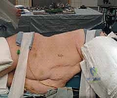
Patient Positioning Considerations
The patient should be in the lateral decubitus position with the arms in prayer position. The thorax vertex should be positioned over the break of the bed, all pressure points should be padded, pillows should be placed between legs and arms, and an axillary roll should be used to prevent compression of the axillary vessels. The operating surgeon typically stands behind the patient during the exposure. However, it may be helpful to transition to the anterior side of the table during the decompression and corpectomy phases to optimize the visual trajectory across the spinal canal and ensure complete removal of contralateral pathology.

Rigid fixation to the operating table is mandatory. The use of a beanbag or rigid lateral supports ensures the patient does not roll during table tilting. The bed is often flexed to open the intercostal spaces, facilitating easier access and reducing the need for aggressive rib retraction.

Detailed Surgical Approach and Technique
Incision and Superficial Dissection
The skin incision is planned directly over the rib that corresponds to the vertebral level of interest. Generally, to access the T8-T9 disc space, the 8th rib is resected. The incision follows the contour of the rib from the lateral border of the paraspinal musculature to the costochondral junction.

The superficial musculature is divided in line with the incision using electrocautery. The latissimus dorsi is transected, followed by the serratus anterior. In higher thoracic approaches, the lower fibers of the trapezius and the rhomboids may also require division. Meticulous hemostasis of the muscular layers is critical to prevent postoperative hematoma.

Rib Resection and Pleural Entry
Once the target rib is identified, the periosteum is incised longitudinally along the outer surface of the rib using electrocautery. A periosteal elevator is utilized to strip the periosteum superiorly and inferiorly. A Doyen elevator is then carefully passed subperiosteally around the deep surface of the rib, taking great care to avoid injury to the underlying parietal pleura and the neurovascular bundle situated in the subcostal groove at the inferior margin of the rib.

The rib is transected posteriorly near the costotransverse junction and anteriorly at the costochondral junction using rib shears. The resected rib is thoroughly cleaned of soft tissue and retained for use as autologous bone graft during the reconstruction phase. The parietal pleura is then incised through the bed of the resected rib to enter the thoracic cavity.

Intrathoracic Exposure and Retraction
Upon entering the pleural cavity, the ipsilateral lung is deflated by the anesthesia team. Lung retractors, protected by moist laparotomy sponges, are placed to gently displace the lung anteriorly and medially. Adhesions between the visceral and parietal pleura, if present, must be sharply divided.

The parietal pleura overlying the spine is visualized. The segmental vessels are identified crossing the mid-portion of the vertebral bodies. The pleura is incised longitudinally over the spine. The segmental arteries and veins at the levels of planned corpectomy or discectomy are isolated, ligated with silk ties or surgical clips, and divided. Bipolar electrocautery should be used judiciously near the neural foramina to avoid thermal injury to the nerve roots or spinal cord.

Decompression and Corpectomy
For a thoracic corpectomy, the discs adjacent to the affected vertebral body are excised first. An annulotomy is performed, and the disc material is removed using pituitary rongeurs and curettes. The vertebral body is then decorticated. Using a high-speed burr or an ultrasonic bone scalpel, a trough is created in the anterior two-thirds of the vertebral body.

The posterior wall of the vertebral body is thinned down to a cortical shell. This shell is then carefully fractured away from the underlying posterior longitudinal ligament (PLL) and dura using micro-curettes and Kerrison rongeurs. The epidural space is meticulously explored to ensure complete decompression of the spinal cord. If the pathology is a calcified disc herniation, the disc must be drilled down to a thin shell before being dissected off the dura to prevent dural tears or cord contusion.

Reconstruction and Fixation
Following adequate decompression, the endplates of the adjacent intact vertebral bodies are prepared by removing the cartilaginous endplates while preserving the subchondral bone to prevent graft subsidence. A structural graft (autograft, allograft, or an expandable titanium cage) is packed with the previously harvested local rib bone and inserted into the defect.

Anterior instrumentation is then applied to provide immediate biomechanical stability. An anterolateral plate or a screw-rod construct is utilized. Screws must be placed parallel to the endplates and directed slightly anteriorly to avoid breaching the spinal canal. Bicortical purchase is ideal for maximum pull-out strength, provided the contralateral great vessels are not at risk.

Closure
Prior to closure, the thoracic cavity is copiously irrigated. A chest tube (typically 28 to 32 French) is placed through a separate stab incision inferior to the primary wound and directed apically to drain air and fluid. The ribs are approximated using heavy pericostal sutures or specialized rib approximators. The parietal pleura and intercostal musculature are closed, followed by a meticulous layered closure of the serratus anterior, latissimus dorsi, subcutaneous tissue, and skin.

Complications and Management
The anterior thoracic approach is associated with a unique set of complications distinct from posterior spinal surgery. Pulmonary complications are the most frequent, necessitating aggressive preoperative optimization and postoperative respiratory therapy.

Vascular injuries, while rare, can be life-threatening. Meticulous dissection around the aorta and vena cava is paramount. Neurologic complications, particularly spinal cord ischemia, remain a catastrophic risk. Maintenance of mean arterial pressure (MAP) above 85 mmHg during decompression and the strict avoidance of excessive cord retraction are critical preventative measures.
| Complication | Estimated Incidence | Prevention and Salvage Strategies |
|---|---|---|
| Atelectasis and Pneumonia | 10% - 20% | Early mobilization, aggressive incentive spirometry, adequate pain control to allow deep breathing. Treat with targeted antibiotics and pulmonary toilet. |
| Intercostal Neuralgia | 5% - 15% | Avoid aggressive retraction of the intercostal nerve. Utilize intercostal nerve blocks intraoperatively. Manage postoperatively with gabapentinoids and NSAIDs. |
| Dural Tear and CSF Leak | 2% - 5% | Leave a thin shell of calcified disc or bone over the dura during drilling. Primary repair if possible; otherwise, use fascial grafts, fibrin glue, and lumbar drains. |
| Chylothorax | < 1% | Identify and ligate the thoracic duct if injured (usually right-sided approaches). Postoperative management includes a medium-chain triglyceride (MCT) diet, total parenteral nutrition, or surgical ligation. |
| Vascular Injury (Great Vessels) | < 1% | Careful preoperative imaging review. Use blunt retractors. Immediate packing and consultation with vascular surgery if major injury occurs. |
| Spinal Cord Ischemia | < 1% | Ligate segmental vessels at the mid-vertebral body. Maintain high MAPs. Utilize continuous SSEP and MEP monitoring. |

Post Operative Rehabilitation Protocols
Postoperative care is heavily focused on pulmonary recovery and early mobilization. The chest tube is placed on water seal and monitored for air leaks and fluid output. It is typically removed when drainage is less than 150 to 200 cc over a 24-hour period and no air leak is present, usually on postoperative day 2 or 3. A postoperative chest radiograph is mandatory immediately following chest tube removal to rule out a delayed pneumothorax.
Pain management is a significant challenge due to the thoracotomy incision. Multimodal analgesia is employed, often utilizing patient-controlled epidural analgesia (PCEA), intercostal nerve blocks, paracetamol, and gabapentinoids to minimize systemic opioid consumption, which can depress respiratory drive.
Patients are mobilized out of bed on postoperative day one. Depending on the bone quality and the rigidity of the internal fixation construct, a Thoracolumbosacral Orthosis (TLSO) may be prescribed for 6 to 12 weeks, though modern robust instrumentation often precludes the need for rigid bracing in patients with normal bone mineral density.

Summary of Key Literature and Guidelines
The efficacy and safety of the anterior thoracic approach have been well-documented in orthopedic and neurosurgical literature. Landmark studies by Bohlman and Zdeblick established the superiority of anterior decompression for central calcified thoracic disc herniations, demonstrating that laminectomy for these lesions carries an unacceptable risk of paraplegia.
For thoracic trauma, particularly burst fractures, the optimal approach remains a topic of debate. Wood et al. conducted randomized controlled trials comparing operative versus non-operative treatment for stable thoracolumbar burst fractures, highlighting that non-operative management is often sufficient for neurologically intact patients without severe kyphosis. However, when surgery is indicated for anterior column compromise and neurologic deficit, anterior corpectomy and plating have demonstrated excellent rates of neurologic recovery and deformity correction.
Furthermore, literature concerning the artery of Adamkiewicz emphasizes the importance of vascular preservation. Studies utilizing preoperative CT angiography to map the artery of Adamkiewicz have shown that while identifying the vessel is possible, the routine mid-vertebral ligation of segmental arteries remains a safe and reliable method to preserve spinal cord perfusion during anterior thoracic exposures. Continuous neuromonitoring remains the gold standard for intraoperative neural assessment, with guidelines strongly recommending its use in all anterior thoracic spinal reconstructions.
Clinical & Radiographic Imaging
You Might Also Like