Interdigital Neuroma: Comprehensive Pathoanatomy, Diagnosis, and Surgical Management
Key Takeaway
Interdigital neuroma, commonly known as Morton's neuroma, is a compressive neuropathy of the common digital nerve, most frequently affecting the third web space. This condition is characterized by perineural fibrosis rather than a true neoplastic process. Surgical management, typically involving neurectomy via a dorsal or plantar approach, is indicated following the exhaustion of conservative measures such as orthotics and corticosteroid injections.
INTERDIGITAL NEUROMA
HISTORICAL BACKGROUND AND EVOLUTION OF PATHOGENIC THEORIES
The clinical entity currently recognized as an interdigital neuroma has a rich historical background, characterized by evolving theories regarding its exact etiology. The condition was first described in the medical literature in 1845 by Durlacher, a chiropodist to the Queen of England. However, it was Thomas G. Morton in 1876 who expanded the clinical description, leading to the ubiquitous, albeit anatomically imprecise, eponym "Morton's toe" or "Morton's neuroma."
Morton originally postulated that the pathology resulted from the pinching of the common digital branch of the lateral plantar nerve to the fourth web space. He theorized that this nerve was compressed between the mobile fourth and fifth metatarsal heads. Despite his detailed clinical descriptions, Morton's specific anatomical theory was not widely accepted by his contemporaries or subsequent anatomists.
Over the ensuing decades, a multitude of alternative theories emerged to explain the pathogenesis of this debilitating forefoot pain:
* Ligamentous Laxity: Laxity of the transverse metatarsal ligament, allowing a break in the anterior arch with plantar displacement of the central metatarsal heads, thereby exerting pressure on the adjacent digital nerve.
* Joint Instability: Micro-instability or subluxation of the fourth metatarsophalangeal (MTP) joint.
* Pressure Neuralgia: The development of a "pressure neuralgia" resulting from repetitive mechanical loading on the nerve during the stance phase of weight-bearing.
* Arch Collapse: "Flattening or falling" of the transverse metatarsal arch, resulting in excessive, unmitigated pressure over the central metatarsal heads.
* Neoplastic and Vascular Theories: Early, largely discarded theories also included the presence of a true benign tumor involving the lateral-most branch of the medial plantar nerve, or lumen occlusion in the common digital artery adjacent to the nerve leading to ischemic neuropathy.
In 1940, Betts introduced a compelling anatomical theory. He believed that the singular anatomy of the fourth digital branch of the medial plantar nerve was the primary culprit. This branch emerges from beneath the medial side of the flexor digitorum brevis. While coursing obliquely across the plantar surface of the muscle, it frequently receives a communicating branch from the common digital branch of the lateral plantar nerve. This communicating branch emerges from the lateral side of the flexor digitorum brevis, crossing obliquely deep to the plantar aponeurosis to join the most lateral branch of the medial plantar nerve 2 to 3 cm before it bifurcates into the proper digital branches to the adjacent surfaces of the third and fourth toes.
Betts surmised that because of this additional communicating branch—which the other common digital nerves do not receive—the common digital nerve to the third web space is anatomically thicker. Consequently, it is more susceptible to being compressed against the unyielding deep transverse intermetatarsal ligament (DTML) located dorsal to it.
However, this theory was challenged by Levitsky et al., who conducted extensive cadaveric dissections. They found that this communicating branch was entirely absent in 73% of cadaveric feet. Furthermore, clinical data demonstrated that neuromas were identified in an almost equal distribution between the second and third web spaces, undermining the theory that a thickened third web space nerve is the sole pathogenic factor. Today, while none of the historical theories are universally accepted in isolation, the consensus points toward a multifactorial mechanical entrapment neuropathy.
PATHOANATOMY AND BIOMECHANICS
Despite the nomenclature, an interdigital neuroma is not a true neoplasm. Histological analysis consistently demonstrates perineural fibrosis, local vascular proliferation, endoneurial edema, and axonal degeneration. The presence of Renaut bodies (subperineurial whorls of loosely textured cells) is frequently observed, indicating chronic mechanical irritation and nerve entrapment.
The biomechanical etiology is best understood through the dynamic anatomy of the forefoot during the gait cycle. The common digital nerve courses plantar to the deep transverse intermetatarsal ligament (DTML). During the heel-off (terminal stance) phase of gait, the metatarsophalangeal joints are maximally dorsiflexed. This dorsiflexion tethers the common digital nerve tightly against the distal edge of the unyielding DTML.
Concurrently, the intermetatarsal bursa, which lies dorsal to the nerve and just proximal to the DTML, may become inflamed and hypertrophic due to repetitive mechanical shear. The combination of a hypertrophic bursa, the rigid DTML, and the ground reaction forces transmitted through the plantar fat pad creates a highly constrained space, leading to chronic compression, microtrauma, and subsequent perineural fibrosis of the nerve.
Clinical Pearl: The pathology is almost exclusively found in the second and third intermetatarsal spaces. The first and fourth spaces are rarely involved because they are not subjected to the same degree of mechanical tethering and lack the specific anatomical constraints of the central rays.
CLINICAL EVALUATION AND DIAGNOSIS
Patient Presentation
Patients typically present with severe, episodic, burning, or lancinating pain in the forefoot, most commonly localized to the third (and less frequently, the second) intermetatarsal space. The pain often radiates into the adjacent toes.
A hallmark symptom is the sensation of "walking on a marble" or having a "pebble in the shoe." Symptoms are classically exacerbated by weight-bearing, particularly in narrow-toed or high-heeled shoes, which compress the metatarsal heads and increase MTP joint dorsiflexion. Relief is almost universally obtained by removing the shoe and massaging the forefoot.
Physical Examination
A meticulous physical examination is paramount to differentiate an interdigital neuroma from other causes of metatarsalgia (e.g., MTP joint synovitis, Freiberg's infraction, stress fractures).
* Web Space Tenderness: Direct plantar palpation of the affected web space, just proximal to the metatarsal heads, elicits exquisite pain.
* Mulder's Click: This is the pathognomonic clinical test. The examiner grasps the first and fifth metatarsal heads and squeezes them together (compressing the transverse arch) while simultaneously applying dorsal pressure to the plantar aspect of the affected web space with the other hand. A palpable and often audible "click" (Mulder's sign) occurs as the enlarged neuroma and associated bursa are subluxated dorsally between the metatarsal heads.
* Sullivan's Sign: In chronic cases, the space-occupying nature of the neuroma and bursa may cause splaying of the adjacent toes when the patient stands.
* Sensory Deficit: Careful neurological examination may reveal diminished two-point discrimination or hyperesthesia on the opposing surfaces of the toes supplied by the affected nerve.
Imaging Studies
While the diagnosis is primarily clinical, imaging can be useful in equivocal cases or to rule out concomitant pathology.
* Radiographs: Weight-bearing anteroposterior, lateral, and oblique views of the foot are mandatory to rule out stress fractures, arthritis, or avascular necrosis (Freiberg's disease).
* Ultrasound: High-resolution ultrasonography is highly sensitive and specific. It typically reveals a well-demarcated, hypoechoic, ovoid mass in the intermetatarsal space. Dynamic ultrasound can visualize the neuroma subluxating during a Mulder's maneuver.
* Magnetic Resonance Imaging (MRI): MRI is reserved for complex or revision cases. A neuroma appears as a teardrop-shaped mass that is hypointense on T1-weighted images and variable on T2-weighted images, often demonstrating enhancement with gadolinium contrast.
NON-OPERATIVE MANAGEMENT
Conservative management should be exhausted before considering surgical intervention, as it yields satisfactory results in up to 30-40% of patients, particularly those with a short duration of symptoms.
- Footwear Modification: The cornerstone of conservative care. Patients must transition to shoes with a wide toe box to prevent lateral compression of the metatarsal heads, and a low heel to reduce MTP joint dorsiflexion and subsequent nerve tethering.
- Orthotics: Custom or over-the-counter orthotics incorporating a metatarsal pad are highly effective. The pad must be placed proximal to the metatarsal heads to elevate the transverse arch and splay the metatarsals, thereby decompressing the intermetatarsal space.
- Corticosteroid Injections: A localized injection of a corticosteroid and local anesthetic mixture into the dorsal web space can provide significant diagnostic information and therapeutic relief. It reduces the inflammation of the adjacent intermetatarsal bursa and the perineural edema.
- Alternative Injections: Sclerosing alcohol injections and radiofrequency ablation (RFA) have been described, though their use remains controversial due to variable success rates and the risk of extensive surrounding tissue necrosis.
SURGICAL INDICATIONS AND PREOPERATIVE PLANNING
Surgical intervention is indicated for patients who have persistent, debilitating symptoms despite a minimum of 3 to 6 months of dedicated non-operative management.
Preoperative planning must include a definitive localization of the symptomatic web space. Operating on the incorrect web space is a known, albeit preventable, complication. If the patient has bilateral symptoms or multiple neuromas in the same foot, careful consideration must be given to staging the procedures, as simultaneous adjacent neurectomies increase the risk of vascular compromise to the intervening toe.
SURGICAL TECHNIQUES
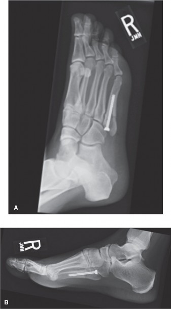
The two primary surgical approaches for the excision of an interdigital neuroma are the dorsal approach and the plantar approach. The dorsal approach is overwhelmingly preferred for primary excisions due to the avoidance of a potentially painful plantar scar. The plantar approach is generally reserved for revision surgery (stump neuroma) or when a concurrent plantar pathology requires addressing.
1. The Dorsal Approach (Primary Neurectomy)
Positioning and Preparation:
The patient is placed supine on the operating table. A small bump may be placed under the ipsilateral hip to maintain the foot in a neutral, upward-facing position. A calf or ankle tourniquet is applied to ensure a bloodless surgical field. Intravenous antibiotics are administered according to institutional protocols.
Incision and Superficial Dissection:
* A 3 to 4 cm longitudinal incision is made centered over the dorsal aspect of the affected intermetatarsal space, beginning at the web space and extending proximally.
* Blunt dissection is utilized through the subcutaneous tissues. Care must be taken to identify and retract any dorsal cutaneous nerve branches to prevent postoperative dorsal numbness or painful dorsal neuromas.
* The dorsal interosseous fascia is incised longitudinally.
Deep Dissection and Ligament Release:
* A self-retaining retractor (e.g., Weitlaner) or a lamina spreader is carefully inserted between the adjacent metatarsal heads to splay the intermetatarsal space.
* The intermetatarsal bursa is frequently encountered at this stage. It is often hypertrophic and should be excised to improve visualization and prevent postoperative fluid collection.
* The deep transverse intermetatarsal ligament (DTML) is identified as a taut, transverse band connecting the plantar aspects of the metatarsal heads.
* Using a Freer elevator to protect the underlying neurovascular bundle, the DTML is completely transected using a scalpel or dissecting scissors.
Surgical Warning: Complete transection of the DTML is the most critical step of the procedure. Failure to completely release this ligament is a primary cause of persistent postoperative pain and surgical failure.
Nerve Identification and Resection:
* Once the DTML is released, plantar pressure is applied to the foot by the assistant. This maneuver pushes the neuroma dorsally into the surgical field.
* The common digital nerve is identified. It will typically exhibit a fusiform swelling (the neuroma) just proximal to its bifurcation into the proper digital nerves.
* The nerve is traced distally, and the proper digital branches to the adjacent toes are identified and transected.
* The common digital nerve is then grasped with a hemostat and traced proximally.
* Traction is applied to the nerve, pulling it distally. The nerve is then sharply transected as far proximally as possible, deep within the intrinsic musculature of the foot (typically 2-3 cm proximal to the DTML).
Pitfall: The nerve must be resected proximal to the weight-bearing pad of the metatarsal heads. If the nerve is cut too distally, the resulting nerve stump will retract into the weight-bearing area, leading to a highly symptomatic and recalcitrant stump neuroma.
Closure:
* The tourniquet is deflated, and meticulous hemostasis is achieved using bipolar electrocautery.
* The DTML is not repaired.
* The skin is closed using interrupted non-absorbable sutures or a running subcuticular suture, depending on surgeon preference.
* A bulky, compressive soft dressing is applied.
2. The Plantar Approach (Revision Neurectomy)
The plantar approach provides direct, unparalleled access to the common digital nerve proximal to the metatarsal heads, making it the approach of choice for excising recurrent or stump neuromas.
Positioning and Incision:
* The patient is positioned supine with the knee flexed and the foot resting flat on the table, or prone, depending on surgeon preference.
* A longitudinal incision is made on the plantar aspect of the foot, centered over the affected intermetatarsal space. The incision begins just proximal to the web space and extends proximally for 3 to 4 cm.
* Note: Transverse plantar incisions have been described to follow resting skin tension lines, but longitudinal incisions provide superior proximal exposure for nerve tracking.
Dissection and Resection:
* The incision is carried through the thick plantar skin and subcutaneous fat pad.
* The plantar aponeurosis fibers are separated longitudinally.
* The common digital nerve is immediately visualized in the subcutaneous fat, plantar to the DTML.
* In a revision setting, the nerve stump is identified, often encased in dense scar tissue. The stump is dissected free, traced proximally into healthy, unscarred tissue, and sharply resected deep within the intrinsic muscles.
* Hemostasis is achieved, and the thick plantar skin is closed with deep, interrupted vertical mattress sutures (e.g., 3-0 nylon) to ensure robust approximation and minimize scar widening.
POSTOPERATIVE PROTOCOL AND REHABILITATION
Dorsal Approach:
* Weeks 0-2: The patient is allowed heel-weight-bearing or weight-bearing as tolerated in a rigid, hard-soled postoperative shoe. Elevation of the limb is strictly enforced to minimize edema and prevent hematoma formation.
* Week 2: The bulky dressing is removed, and sutures are extracted. The patient may transition to a wide, supportive athletic shoe.
* Weeks 3-6: Progressive return to normal activities and full weight-bearing. High-impact activities and tight footwear should be avoided until 6 weeks postoperatively.
Plantar Approach:
* Postoperative management is more conservative due to the risk of delayed healing and painful scarring of the plantar skin.
* Weeks 0-3: Strict non-weight-bearing or touch-down weight-bearing in a splint or controlled ankle motion (CAM) boot.
* Week 3: Suture removal. The plantar skin takes longer to heal; premature suture removal can lead to wound dehiscence.
* Weeks 4-8: Gradual transition to weight-bearing in a hard-soled shoe, followed by a supportive athletic shoe.
COMPLICATIONS AND MANAGEMENT
While neurectomy is generally highly successful (80-85% patient satisfaction), complications can occur and must be discussed during the informed consent process.
- Stump Neuroma: The most common cause of failed neuroma surgery. It occurs when the proximal nerve stump is left in a weight-bearing area or becomes tethered in scar tissue. Management requires revision surgery via a plantar approach to resect the stump further proximally, occasionally burying the new stump into the adjacent intrinsic musculature or bone.
- Web Space Numbness: Permanent numbness in the opposing halves of the toes is an expected outcome of the procedure, not a complication. However, patients must be explicitly counseled about this preoperatively to manage expectations.
- Complex Regional Pain Syndrome (CRPS): A rare but devastating complication characterized by disproportionate pain, autonomic dysfunction, and trophic changes. Early recognition and aggressive multidisciplinary management (gabapentinoids, sympathetic blocks, physical therapy) are essential.
- Hematoma and Infection: Minimized by meticulous hemostasis, appropriate use of electrocautery, strict postoperative elevation, and prophylactic antibiotics.
- Digital Ischemia: Extremely rare, usually associated with simultaneous adjacent web space neurectomies or aggressive dissection compromising the common digital artery.
In conclusion, the successful management of an interdigital neuroma relies on a profound understanding of forefoot pathoanatomy, a rigorous diagnostic workup, and meticulous surgical technique. Whether utilizing a dorsal or plantar approach, the principles of complete ligamentous release and adequate proximal nerve resection remain the cornerstones of achieving a pain-free, functional outcome for the patient.
You Might Also Like