ABOS Part I Orthopaedic Trauma Review: Monteggia, Humerus Fractures & Surgical Approaches | Part 22151
Key Takeaway
This ABOS Part I Orthopaedic Trauma Review module offers 22 advanced multiple-choice questions mirroring ABOS Part I and AAOS OITE exams. Derived from high-yield clinical cases, it covers Monteggia fracture-dislocations, proximal humerus, and humeral shaft fractures, including surgical approaches, neurovascular risks, and rehabilitation protocols for comprehensive exam preparation.
ABOS Part I Orthopaedic Trauma Review: Monteggia, Humerus Fractures & Surgical Approaches | Part 22151
A 45-year-old male presents to the emergency department after a high-energy fall onto an outstretched hand (FOOSH). Radiographs reveal a fracture of the ulnar diaphysis with anterior angulation and an associated anterior dislocation of the radial head. A thorough neurovascular exam is unremarkable. Based on the provided case information, what is the most likely Bado classification for this injury?
Correct Answer: C
The patient's presentation of an anterior dislocation of the radial head with an associated fracture of the ulnar diaphysis, typically angulated anteriorly, perfectly matches the description of a Bado Type I Monteggia fracture-dislocation. The case states that Type I is the most common, accounting for approximately 60% of adult cases, and typically results from a FOOSH with hyperpronation, leading to anterior radial head dislocation and anterior ulnar angulation.
Option A (Type II): This type involves a posterior or posterolateral dislocation of the radial head with a posteriorly angulated ulnar diaphysis fracture. This does not match the anterior dislocation described.
Option B (Type III): This type involves a lateral or anterolateral dislocation of the radial head with a fracture of the ulnar metaphysis (proximal to the coronoid). The patient's radial head dislocation is anterior, and the ulnar fracture is diaphyseal, not metaphyseal.
Option D (Type IV): This type involves an anterior dislocation of the radial head with fractures of both the radial and ulnar diaphyses at the same level. While the radial head dislocation is anterior, the vignette only mentions an ulnar fracture, not a concomitant radial shaft fracture at the same level.
Option E (Monteggia-equivalent): This term refers to injuries that functionally behave like a Monteggia but may not fit the classic Bado types (e.g., isolated radial head dislocation with an associated coronoid fracture). While important, the described injury clearly fits a specific Bado type.
A 38-year-old construction worker sustains a Bado Type II Monteggia fracture-dislocation after a fall from a height. During surgical planning, the orthopedic surgeon reviews the relevant anatomy. Which of the following structures is considered the primary stabilizer of the radial head within the radial notch of the ulna?
Correct Answer: C
The case explicitly states, 'The annular ligament is the primary stabilizer of the radial head within the radial notch of the ulna. It forms a fibrous ring encircling the radial head and neck, attaching to the anterior and posterior margins of the radial notch.' Its integrity is crucial for maintaining the proximal radioulnar joint (PRUJ) stability.
Option A (Medial Collateral Ligament - MCL): The MCL provides valgus stability to the elbow and is not the primary stabilizer of the radial head within the radial notch.
Option B (Lateral Ulnar Collateral Ligament - LUCL): The LUCL is part of the lateral collateral ligament complex and is critical for posterolateral rotatory stability of the elbow, but not the primary stabilizer of the radial head within the radial notch.
Option D (Interosseous Membrane - IOM): The IOM connects the ulna and radius, transmitting axial loads and contributing to overall forearm stability, but it is not the primary direct stabilizer of the radial head within the radial notch.
Option E (Biceps tendon): The biceps tendon primarily functions in elbow flexion and forearm supination. While it crosses the elbow joint, it does not directly stabilize the radial head within the radial notch.
A 28-year-old male presents with a Bado Type III Monteggia fracture-dislocation after a motorcycle accident. Pre-operative neurovascular examination reveals a partial wrist drop and inability to extend the metacarpophalangeal (MCP) joints of the fingers, with intact sensation. Which nerve is most likely injured in this patient, and why is it particularly vulnerable in this type of Monteggia injury?
Correct Answer: C
The clinical presentation of wrist drop and inability to extend the MCP joints of the digits and thumb, with intact sensation, is characteristic of a Posterior Interosseous Nerve (PIN) palsy. The case specifically highlights that the PIN, a deep branch of the radial nerve, passes through the supinator muscle and 'is particularly vulnerable to injury during Type III Monteggia fractures and during surgical approaches involving the lateral elbow or extensive dissection around the radial neck.' Type III Monteggia involves a lateral or anterolateral dislocation of the radial head and a ulnar metaphysis fracture, which can directly impact the PIN due to the significant displacement of the radial head and potential for traction or direct compression.
Option A (Ulnar nerve): While the ulnar nerve can be at risk, especially in Type II Monteggia with posterior displacement, its injury typically presents with numbness in the small finger and ulnar half of the ring finger, and weakness of intrinsic hand muscles, not a wrist drop.
Option B (Median nerve): The median nerve lies anteriorly and is less commonly injured in Monteggia fractures. Its injury would typically present with sensory loss in the thumb, index, middle, and radial half of the ring finger, and weakness of thumb opposition and finger flexion, not a wrist drop.
Option D (Radial sensory nerve): Injury to the radial sensory nerve would cause sensory deficits over the dorsum of the hand, but not motor deficits like wrist drop.
Option E (Anterior Interosseous Nerve - AIN): The AIN is a motor branch of the median nerve. Its injury would result in loss of flexion of the thumb IP joint and index finger DIP joint (OK sign deficit), not a wrist drop.
A 55-year-old female presents with a Bado Type I Monteggia fracture-dislocation. The surgical team is preparing for operative management. Based on the biomechanical principles outlined in the case, which of the following statements represents the cornerstone of successful treatment for this injury?
Correct Answer: C
The case explicitly states, 'The key biomechanical principle in treating Monteggia injuries is that the ulna dictates the stability of the entire forearm and the radial head. Anatomic reduction and rigid internal fixation of the ulna are prerequisites for successful radial head reduction and maintaining its concentric alignment with the capitellum.' This principle is reiterated multiple times throughout the text, emphasizing that restoring the ulna's length, rotation, and alignment is paramount.
Option A (Primary open reduction and repair of the annular ligament): While annular ligament repair may be necessary if the radial head remains unstable after ulnar fixation, it is not the primary cornerstone. The ulna's stability is the prerequisite.
Option B (Achieving concentric reduction of the radial head first): This is incorrect. The radial head's position is dictated by the ulna. Attempting to reduce the radial head first without addressing the ulnar fracture will likely be unsuccessful or unstable.
Option D (Application of an external fixator): While external fixation might be used temporarily in severe open fractures or for damage control, it is not the definitive treatment for adult Monteggia fracture-dislocations, which almost universally require open reduction and internal fixation (ORIF) of the ulna.
Option E (Early aggressive range of motion exercises): Early motion is important post-operatively, but it must be controlled and initiated *after* stable fixation of the ulna and concentric reduction of the radial head. Aggressive motion before definitive fixation would destabilize the injury.
A 62-year-old male presents with a complex Monteggia fracture-dislocation involving significant comminution of the ulnar shaft and suspected involvement of the radial head articular surface. The initial plain radiographs are difficult to interpret fully due to the comminution. Which advanced imaging modality is most highly recommended in this scenario to provide detailed information for surgical planning?
Correct Answer: C
The case states, 'Computed Tomography (CT) Scan: Highly recommended for complex Monteggia injuries. It provides invaluable detailed information on: Fracture morphology of the ulna (comminution, bone loss). Presence and extent of radial head or neck fractures (often occult on plain films). Coronoid process fractures (critical for elbow stability). Articular impaction or osteochondral lesions. Identifying intra-articular loose bodies or incarcerated soft tissues preventing reduction. 3D Reconstructions: Aid in surgical planning, especially for complex articular involvement.'
Option A (Magnetic Resonance Imaging - MRI): MRI is rarely indicated in acute settings unless suspicion for significant ligamentous injury (e.g., severe MCL/LCL tears) persists despite stable fixation of the ulna and radial head, or to evaluate annular ligament integrity more directly. It is not the primary modality for detailed bony fracture morphology.
Option B (Ultrasound): Ultrasound has limited utility in assessing complex bony fractures and articular surfaces in the acute setting of a Monteggia injury.
Option D (Bone scintigraphy): Bone scintigraphy (bone scan) is used to assess metabolic activity in bone, typically for stress fractures, infections, or tumors, not for acute fracture morphology or surgical planning.
Option E (Repeat plain radiographs with different views): While additional plain views can be helpful, they are often insufficient to fully delineate complex comminuted fractures or subtle articular involvement, which is precisely why CT is recommended.
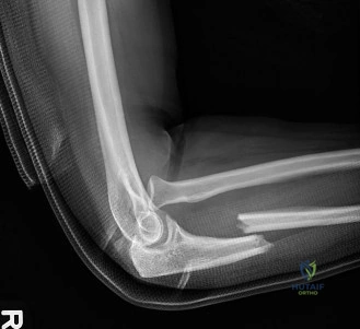
A 40-year-old male undergoes open reduction and internal fixation for a Bado Type I Monteggia fracture-dislocation. The surgeon has successfully achieved anatomical reduction and rigid internal fixation of the ulnar fracture using a locking compression plate, as depicted in the provided image. What is the immediate next critical step in the surgical procedure, according to the case's detailed surgical technique?
Correct Answer: C
The case outlines the step-by-step surgical technique, stating under 'Radial Head Assessment': 'Once the ulna is rigidly fixed, the radial head should spontaneously reduce into its concentric position relative to the capitellum. Confirm with fluoroscopy in AP and lateral views, ensuring the radial head bisects the capitellum in all positions of elbow flexion and forearm rotation.' This is the immediate and critical next step after ulnar fixation.
Option A (Perform a thorough neurovascular assessment): While a neurovascular assessment is crucial, it is typically performed at the end of the procedure (Final Stability Check) and also pre-operatively, but not as the *immediate* next step after ulnar fixation before confirming radial head reduction.
Option B (Initiate aggressive range of motion exercises): This is incorrect. Early aggressive motion is contraindicated at this stage and could destabilize the repair. Controlled motion begins post-operatively during rehabilitation.
Option D (Proceed with layered wound closure and apply a splint): Wound closure and splint application are final steps, performed only after all reductions and fixations are confirmed stable and neurovascular status is checked.
Option E (Directly repair the torn annular ligament): Annular ligament repair is only indicated if the radial head *does not* spontaneously reduce or remains unstable *after* anatomical ulnar fixation. It is not an automatic next step.
A 32-year-old male undergoes ORIF for a Bado Type I Monteggia fracture-dislocation. Post-operatively, despite what the surgeon believes was rigid internal fixation of the ulna, fluoroscopy reveals persistent subluxation of the radial head. Based on the case, what is the most common reason for persistent radial head dislocation or subluxation after ulnar fixation, and what is the appropriate next step?
Correct Answer: B
The case clearly states under 'Complications and Management' and 'Persistent Radial Head Dislocation/Subluxation': 'This is the most common and critical complication. It's almost always due to inadequate anatomical reduction or unstable fixation of the ulna (malreduction, malalignment, shortening, or rotation).' The management is to 're-establish anatomical reduction and rigid fixation of the ulna.'
Option A (An irreparable annular ligament tear; proceed with radial head replacement): While an annular ligament tear can contribute to instability, it is rarely the *primary* reason for persistent dislocation if the ulna is anatomically reduced. Radial head replacement is generally discouraged in acute Monteggia for unreconstructible radial head fractures, and even less so for an isolated annular ligament issue.
Option C (Interposition of the biceps tendon): While soft tissue interposition can occur, the case emphasizes that inadequate ulnar reduction is the *most common* reason. If soft tissue interposition is suspected after optimal ulnar fixation, open reduction of the radial head would be performed, but the primary focus remains the ulna.
Option D (Associated coronoid process fracture): Coronoid fractures can destabilize the elbow, but the most common reason for persistent radial head dislocation in a Monteggia is still inadequate ulnar reduction. If a coronoid fracture was significant enough to cause persistent instability, it should have been addressed during the initial fixation.
Option E (Posterior interosseous nerve impingement): PIN impingement causes neurological symptoms (wrist drop) but does not directly cause persistent radial head dislocation. Neurolysis would be for nerve recovery, not joint stability.
A 22-year-old male presents with a Bado Type IV Monteggia fracture-dislocation after a high-energy trauma. During the initial assessment, he is found to have a complete wrist drop and inability to extend his fingers at the MCP joints, with no sensory deficits. The surgical team plans for emergent ORIF. Based on the case, what is the most appropriate initial management strategy for this pre-existing neurological deficit?
Correct Answer: B
The case states under 'Complications and Management' regarding nerve injury: 'Pre-operative PIN palsy is typically observed as most are neurapraxic and resolve spontaneously over weeks to months. Post-operative PIN palsy requires immediate evaluation. If a complete palsy exists and there is suspicion of direct transection or impingement (e.g., by hardware), surgical exploration is warranted. Otherwise, observation, splinting, and electrophysiological studies guide management.'
Option A (Immediate surgical exploration and neurolysis): This is generally not the initial approach for a pre-operative PIN palsy unless there is strong evidence of direct transection or entrapment by a bone fragment that cannot be resolved by fracture reduction. Most pre-operative palsies are traction-related neurapraxias.
Option C (Administer high-dose corticosteroids): There is no evidence to support the use of corticosteroids for traumatic nerve palsies in this context.
Option D (Delay surgical fixation): Delaying definitive fixation of a Monteggia fracture-dislocation is associated with poorer outcomes and increased difficulty of reduction. The fracture should be fixed promptly, and the nerve managed concurrently.
Option E (Perform a tendon transfer immediately): Tendon transfers are reconstructive procedures for *permanent* nerve deficits and are performed much later, typically after 6-12 months of observation if no recovery occurs.
A 48-year-old male has undergone successful ORIF of a Bado Type I Monteggia fracture-dislocation. He is now 5 days post-operative. The surgeon emphasizes the importance of a structured rehabilitation protocol. According to the case, what is the primary goal and initial approach during the early post-operative phase (Phase 1)?
Correct Answer: C
The case describes the 'Phase Protection and Early Motion' (Phase 1) goals as: 'Reduce pain and swelling, protect surgical repair, initiate controlled early motion.' It further specifies for ROM: 'Begin with gentle active-assisted and passive range of motion (AAROM/PROM) within a safe arc determined by surgeon (e.g., 30-100 degrees flexion, neutral pronation/supination).'
Option A (Achieve full active range of motion (AROM) in all planes immediately): This is too aggressive and could jeopardize the surgical repair and radial head stability. Early motion must be controlled and within a safe arc.
Option B (Maintain strict immobilization for 6 weeks): The case states that 'The duration of strict immobilization is generally minimal, often just until initial pain and swelling subside (e.g., 3-7 days).' Prolonged immobilization is a known cause of stiffness.
Option D (Begin progressive resistive strengthening exercises): Strengthening exercises are initiated in a later phase (Phase 2: Progressive Motion and Light Strengthening), starting with isometrics, not immediately post-op.
Option E (Allow immediate weight-bearing): The case explicitly states 'Non-weight-bearing for the affected limb' during the early phase.
A 68-year-old male with a history of head trauma presents with a severe, comminuted Bado Type II Monteggia fracture-dislocation. Due to the high-energy nature of the injury and his medical history, the surgeon is concerned about the development of heterotopic ossification (HO). Which prophylactic measure is commonly considered to reduce the risk of HO in such high-risk patients?
Correct Answer: C
The case states under 'Complications and Management' for Heterotopic Ossification (HO): 'Prevention: Prophylactic NSAIDs (e.g., indomethacin) or low-dose radiation therapy (controversial in acute setting, typically reserved for high-risk cases) can be used.' The patient's history of head trauma is a known risk factor for HO.
Option A (Early aggressive, uncontrolled range of motion exercises): While early controlled motion is important, aggressive and uncontrolled motion can increase inflammation and potentially contribute to HO formation, not prevent it.
Option B (Prolonged immobilization): Prolonged immobilization is a risk factor for elbow stiffness and does not prevent HO; in fact, it can sometimes be associated with it.
Option D (High-dose systemic corticosteroids): Corticosteroids are not a standard prophylactic measure for HO in this context and carry significant side effects.
Option E (Immediate surgical excision of any suspected early ossification): Surgical excision of HO is typically performed only for symptomatic, mature HO after 6-12 months, not immediately post-injury or for early suspected ossification.
A 35-year-old male sustains a Monteggia fracture-dislocation with an associated severely comminuted radial head fracture that is deemed unreconstructible. The ulnar fracture has been anatomically reduced and rigidly fixed. Based on the current literature and guidelines discussed in the case, what is the generally discouraged approach for managing the unreconstructible radial head fracture in this acute setting, especially in a younger, active adult?
Correct Answer: B
The case states under 'Summary of Key Literature and Guidelines' and 'Radial Head Fractures': 'Unreconstructible radial head fractures in the context of an acute Monteggia are challenging. While historically radial head excision was considered, it is now generally discouraged in the acute setting due to the risk of severe valgus instability and proximal radial migration, especially in younger, active adults.'
Option A (Radial head arthroplasty): The case mentions that 'Radial head arthroplasty may be considered in selected cases to restore mechanical stability, particularly in older patients with low demand, but its role in *acute* Monteggia is not universally accepted compared to its use in terrible triad injuries.' While not universally accepted for acute Monteggia, it is a consideration for stability, unlike excision.
Option C (Open reduction of the radial head to clear any interposed soft tissue): This is a necessary step if the radial head cannot be reduced, regardless of whether it's fractured or not, to clear obstructions. This is not discouraged.
Option D (Annular ligament reconstruction): If the radial head remains unstable after ulnar fixation and any necessary radial head management, annular ligament reconstruction is a valid and often necessary step to maintain stability. This is not discouraged.
Option E (Temporary K-wire stabilization): Temporary K-wire stabilization can be considered in highly unstable cases to maintain reduction, although it limits early motion. This is a recognized technique, not generally discouraged.
A 68-year-old female presents with a displaced 3-part proximal humerus fracture following a fall. She is scheduled for open reduction and internal fixation via a deltopectoral approach. During the deep dissection, after identifying the cephalic vein and retracting the deltoid laterally and pectoralis major medially, the surgeon proceeds to manage the subscapularis to gain access to the humeral head. Which of the following neurovascular structures is at greatest risk during the inferior aspect of this exposure, particularly when mobilizing the deltoid or performing extensive inferior dissection?
Correct Answer: C
The axillary nerve is the neurovascular structure at greatest risk during the inferior aspect of a deltopectoral approach, especially when mobilizing the deltoid or performing extensive inferior dissection. The case content explicitly states: 'The axillary nerve typically exits the quadrilateral space and wraps around the surgical neck of the humerus, approximately 5-7 cm distal to the acromial edge. It innervates the deltoid and teres minor muscles. During a deltopectoral approach, careful dissection in the inferior aspect of the exposure, especially when mobilizing the deltoid, is crucial to protect this nerve.'
Option A (Musculocutaneous nerve): While the musculocutaneous nerve is in the vicinity, piercing the coracobrachialis, it is generally protected by staying lateral to the conjoined tendon and is more at risk with excessive medial retraction of the biceps/coracobrachialis, not primarily with inferior deltoid mobilization.
Option B (Radial nerve): The radial nerve courses in the spiral groove posteriorly and is primarily at risk during posterior or anterolateral approaches to the humeral shaft, particularly in its distal two-thirds, not typically during the inferior aspect of a deltopectoral approach for the proximal humerus.
Option D (Median nerve): The median nerve lies medially within the neurovascular bundle alongside the brachial artery. It is generally well-protected during anterior approaches by staying lateral to the neurovascular bundle.
Option E (Brachial artery): The brachial artery also lies medially with the median nerve. While any major vessel can be injured, the axillary nerve is specifically highlighted as being at risk with inferior deltoid mobilization in this approach due to its anatomical course around the surgical neck.
A 42-year-old male sustains a comminuted, displaced mid-shaft humeral fracture after a high-energy motor vehicle accident. Pre-operative imaging reveals significant anterior comminution and a suspected associated brachial artery injury. The patient also presents with a complete radial nerve palsy. Based on the case content, which of the following is the most appropriate indication for an anterior approach to the humeral shaft in this specific scenario?
Correct Answer: C
The case content explicitly lists 'Fractures associated with vascular injury requiring exploration and repair' and 'Open fractures with anterior soft tissue involvement' as indications for an anterior approach to the humeral shaft. In this scenario, the suspected brachial artery injury is a critical indication that necessitates an anterior approach for direct exploration and repair. The open fracture further supports this choice.
Option A (The presence of a complete radial nerve palsy, as it mandates primary nerve exploration via an anterior approach): While radial nerve palsy can be an indication for primary nerve exploration, and an anterior approach can facilitate this (especially with a lateral extension), the case states that 'a posterior approach may offer better direct visualization of the nerve.' The primary driver for the anterior approach in this specific vignette is the vascular injury and open fracture, not solely the radial nerve palsy.
Option B (The comminuted nature of the fracture, which is best addressed with an anterior locking plate for superior stability): While anterior locking plates are effective for comminuted fractures, the comminution itself does not exclusively dictate an anterior approach over other approaches (e.g., anterolateral or posterior) unless other specific factors are present.
Option D (The patient's young age and high functional demand, favoring early operative intervention via the most direct route): While young, active patients often benefit from operative management, this is a general indication for surgery, not a specific reason to choose an anterior approach over others in the context of the given associated injuries.
Option E (The ability to avoid extensive dissection of the radial nerve, as it lies posterior to the brachialis in the distal two-thirds of the humerus): This is a true statement about the anterior approach's advantage in protecting the radial nerve, but it's a benefit of the approach, not the primary indication for choosing it in the presence of a vascular injury and open fracture. The need for vascular exploration overrides this consideration as the primary determinant.
A 55-year-old male undergoes open reduction and internal fixation of a displaced 4-part proximal humerus fracture using a locking plate via a deltopectoral approach. Post-operatively, radiographs show satisfactory reduction and hardware placement. However, at the 6-month follow-up, the patient develops increasing shoulder pain and crepitus, and repeat radiographs reveal superior migration of the humeral head with articular penetration by several locking screws. Based on the case content, which of the following is the most likely biomechanical factor contributing to this complication?
Correct Answer: E
The case content, under 'Biomechanics of Fixation' and 'Summary of Key Literature and Guidelines,' emphasizes the importance of 'multi-directional locking screws are critical to capture fragments and resist varus collapse' and 'adequate number and placement of locking screws (especially in the inferomedial quadrant for calcar support)' to minimize complications like screw cutout and avascular necrosis. Superior migration of the humeral head with articular penetration by screws (screw cutout) is a classic complication of varus collapse, which often occurs due to inadequate inferomedial calcar support, particularly in osteoporotic bone or comminuted fractures.
Option A (Insufficient plate length, leading to inadequate working length and stress concentration): While insufficient plate length can contribute to hardware failure, it's less directly linked to superior screw cutout in the humeral head compared to the specific issue of calcar support.
Option B (Excessive number of distal cortical screws, causing stress shielding of the fracture site): Stress shielding can be a concern, but it's not the primary mechanism for superior screw cutout in the humeral head. It's more related to diaphyseal healing.
Option C (Lack of multi-directional locking screws, failing to capture fragments and resist varus collapse): While multi-directional screws are important, the specific issue of 'inferomedial calcar support' is a more precise biomechanical factor directly addressing varus collapse, which leads to screw cutout. Multi-directional screws help, but if the inferomedial support is poor, varus collapse can still occur.
Option D (Plate positioning too distal to the bicipital groove, resulting in poor screw trajectory into the humeral head): The case states 'Position the plate laterally, usually 5-8 mm posterior to the bicipital groove, to avoid impingement and ensure optimal screw trajectory into the humeral head.' While incorrect plate positioning can lead to issues, positioning too distal might affect overall stability but is not the most direct cause of superior screw cutout due to varus collapse. Plate prominence (too proud) is more related to impingement.
A 30-year-old male presents with a displaced spiral fracture of the distal third of the humeral shaft. He is scheduled for open reduction and internal fixation via an anterolateral approach. During the deep dissection, the surgeon carefully identifies the interval between the biceps and brachialis muscles. As the dissection proceeds distally to expose the fracture, which critical neurovascular structure must be meticulously identified and protected as it pierces the lateral intermuscular septum to enter the anterior compartment?
Correct Answer: D
The case content explicitly states: 'The radial nerve is the most frequently injured nerve in humeral shaft fractures. It courses in the spiral groove posteriorly, crossing from medial to lateral. Approximately 10-14 cm proximal to the lateral epicondyle, it pierces the lateral intermuscular septum to enter the anterior compartment, lying between the brachialis and brachioradialis muscles in the distal arm. For direct anterior or anterolateral approaches to the shaft, the radial nerve is generally posterior to the plane of dissection (i.e., posterior to the brachialis). However, extensive distal anterior dissection or inadvertent posterior extension of the plane places the nerve at risk.'
Option A (Musculocutaneous nerve): The musculocutaneous nerve pierces the coracobrachialis more proximally and lies between the biceps and brachialis. While important, it is not the nerve that pierces the lateral intermuscular septum in the distal arm.
Option B (Median nerve): The median nerve lies medially within the neurovascular bundle and does not pierce the lateral intermuscular septum.
Option C (Ulnar nerve): The ulnar nerve also lies medially and then courses posteriorly around the medial epicondyle distally; it does not pierce the lateral intermuscular septum.
Option E (Brachial artery): The brachial artery lies medially with the median nerve and does not pierce the lateral intermuscular septum.
A 72-year-old female with osteoporosis presents with a displaced 2-part surgical neck fracture of the humerus. She is scheduled for ORIF via a deltopectoral approach. During pre-operative planning, the surgeon is considering the optimal plate placement and screw trajectory. According to the case content and current literature, which of the following statements regarding plate application for proximal humerus fractures is most accurate?
Correct Answer: C
The case content, under 'Biomechanics of Fixation' and 'Summary of Key Literature and Guidelines,' states: 'Locking plates are particularly beneficial in osteoporotic bone or comminuted fractures, providing angular stability independent of bone-plate interface compression.' It further emphasizes: 'For proximal humerus fractures, multi-directional locking screws are critical to capture fragments and resist varus collapse.' And 'Literature consistently highlights the importance of anatomical reduction, adequate number and placement of locking screws (especially in the inferomedial quadrant for calcar support), avoidance of plate prominence, and careful protection of the axillary nerve.'
Option A (The plate should be positioned directly over the bicipital groove to maximize screw purchase into the humeral head): The case states: 'Position the plate laterally, usually 5-8 mm posterior to the bicipital groove, to avoid impingement and ensure optimal screw trajectory into the humeral head.' Positioning directly over the bicipital groove is incorrect and can lead to impingement or damage to the biceps tendon.
Option B (The plate should be flush with the humeral head to prevent acromial impingement): The case states: 'Ensure the plate is proud of the humeral head by approximately 5 mm to prevent acromial impingement.' A plate that is flush or recessed can lead to impingement. Being slightly proud helps prevent this.
Option D (Conventional compression plates are preferred over locking plates in osteoporotic bone due to better bone-plate interface compression): This is incorrect. The case states: 'Locking plates are particularly beneficial in osteoporotic bone or comminuted fractures, providing angular stability independent of bone-plate interface compression.' Conventional plates rely on compression, which is poor in osteoporotic bone.
Option E (Screw length should be maximized to ensure bicortical purchase in the humeral head for optimal stability): This is incorrect and dangerous. The case states: 'Ensure screws do not penetrate the articular surface – check with fluoroscopy.' Bicortical purchase in the humeral head would mean penetrating the articular surface, leading to joint damage and pain. Screws in the humeral head should be unicortical but long enough to provide good purchase without articular penetration.
A 28-year-old professional athlete undergoes ORIF of a displaced proximal humerus fracture via a deltopectoral approach. Post-operatively, he is placed in a sling. At the 3-week follow-up, he reports mild pain but is eager to begin aggressive rehabilitation. Based on the provided rehabilitation protocol, which of the following activities is most appropriate for this patient during the immediate post-operative phase (0-6 weeks)?
Correct Answer: C
The case content, under 'Phase 1 Immediate Post-operative Phase 0-6 Weeks' for Proximal Humerus (Deltopectoral Approach), states: 'Passive Range of Motion (PROM): Pendulum exercises (gentle, gravity-assisted distraction and rotation), supine passive external rotation (to 0-30 degrees), passive forward flexion (up to 90-120 degrees depending on stability), passive internal rotation.' It also lists 'No active shoulder abduction or external rotation against resistance' as a precaution.
Option A (Active shoulder abduction against light resistance with a Theraband): This is explicitly contraindicated in Phase 1. 'No active shoulder abduction or external rotation against resistance' is a precaution.
Option B (Active-assistive range of motion (AAROM) for shoulder flexion up to 150 degrees): AAROM is typically initiated in Phase 2 (6-12 weeks). While PROM for flexion up to 90-120 degrees is allowed, 150 degrees is likely too aggressive for the immediate phase, and AAROM is not the primary focus yet.
Option D (Full active range of motion (AROM) exercises for the shoulder to prevent stiffness): AROM is generally initiated in Phase 2. Phase 1 focuses on PROM to protect the healing fracture.
Option E (Weight-bearing through the affected arm to promote early bone healing): This is explicitly contraindicated. 'No weight-bearing through the arm' is a precaution in Phase 1.
A 60-year-old male presents with a non-union of the mid-shaft humerus, previously treated non-operatively. He is scheduled for revision ORIF via an anterior approach. During the deep dissection, the surgeon identifies the biceps brachii and brachialis muscles. To access the humeral shaft, the surgeon plans to split the brachialis muscle longitudinally in its distal portion. Which nerve is primarily responsible for innervating the brachialis muscle and must be protected during this maneuver?
Correct Answer: C
The case content, under 'Neurovascular Anatomy,' states: 'Musculocutaneous Nerve: Arising from the lateral cord of the brachial plexus, it pierces the coracobrachialis muscle to lie between the biceps and brachialis. It innervates these three muscles (biceps, brachialis, coracobrachialis) and continues as the lateral antebrachial cutaneous nerve. Excessive retraction of the biceps or coracobrachialis can risk traction injury.'
Option A (Axillary nerve): The axillary nerve innervates the deltoid and teres minor and is primarily at risk around the surgical neck, not typically during dissection of the brachialis.
Option B (Radial nerve): The radial nerve innervates the triceps and muscles of the posterior forearm. While it lies posterior to the brachialis in the distal arm, it does not innervate the brachialis itself.
Option D (Ulnar nerve): The ulnar nerve innervates some forearm flexors and intrinsic hand muscles and is located medially, not associated with the brachialis muscle's innervation.
Option E (Median nerve): The median nerve innervates most forearm flexors and some intrinsic hand muscles and is located medially, not associated with the brachialis muscle's innervation.
A 35-year-old male presents with a complex, comminuted 4-part proximal humerus fracture. Pre-operative planning is underway for ORIF via a deltopectoral approach. The surgeon is particularly concerned about understanding the precise fracture morphology and fragment orientation to plan screw trajectories and avoid articular penetration. Which imaging modality is explicitly highlighted in the case as indispensable for this purpose?
Correct Answer: D
The case content, under 'Pre-Operative Planning' and 'Imaging Review,' states: 'Computed Tomography (CT) Scan: Indispensable for complex fractures, especially 3- and 4-part proximal humerus fractures, humeral head splits, comminuted shaft fractures, non-unions, or tumors. 3D reconstructions aid in understanding fracture morphology, fragment orientation, and surgical approach planning.'
Option A (Standard anteroposterior and lateral plain radiographs): While essential, plain radiographs provide 2D views and may not fully elucidate complex 3D fracture morphology and fragment orientation, especially for 4-part fractures.
Option B (Scapular Y view radiographs): This is part of the trauma series for the shoulder and is critical for characterization, but like other plain films, it lacks the 3D detail of a CT scan.
Option C (Magnetic Resonance Imaging (MRI)): MRI is useful for assessing soft tissue injuries, rotator cuff integrity, or characterizing tumors, but it is not typically the primary modality for detailed bone fracture morphology and fragment orientation, especially when compared to CT with 3D reconstructions.
Option E (Angiography): Angiography is indicated if vascular injury is suspected, not primarily for detailed fracture morphology.
A 50-year-old male undergoes ORIF of a displaced 3-part proximal humerus fracture. During the deltopectoral approach, the subscapularis muscle is managed to gain access to the humeral head. Which of the following is the most common and recommended method for managing the subscapularis tendon to expose the humeral head in this approach, as described in the case?
Correct Answer: C
The case content, under 'Detailed Surgical Approach and Technique' and 'Subscapularis Management' for the Deltopectoral Approach, states: 'For direct access to the humeral head, the subscapularis tendon is either detached from the lesser tuberosity (often with a cuff sparing lesser tuberosity osteotomy) or split vertically in the direction of its fibers. If detached, repair is paramount.'
Option A (Complete tenotomy of the subscapularis tendon at its insertion, followed by repair): While detachment is mentioned, the phrase 'often with a cuff sparing lesser tuberosity osteotomy' implies a more controlled detachment that preserves the tendon's integrity for repair, rather than a simple tenotomy which might be less favorable for healing.
Option B (Vertical splitting of the subscapularis muscle in the direction of its fibers): This is listed as an alternative method, but the primary method often described for direct access to the humeral head, especially for complex fractures, is detachment from the lesser tuberosity, often with an osteotomy.
Option D (Retraction of the subscapularis medially without any incision or detachment): For direct access to the humeral head and fracture fragments, simple retraction is usually insufficient, especially for complex fractures requiring extensive exposure.
Option E (Resection of a portion of the subscapularis muscle to improve visualization): Resection of muscle is generally avoided to preserve function and is not a standard technique for exposing the humeral head in this approach.
A 48-year-old male undergoes ORIF of a comminuted humeral shaft fracture via an anterolateral approach. Post-operatively, he develops a new complete radial nerve palsy. The surgeon decides to observe the patient for initial recovery. Based on the case content, what is the typical expected recovery period for most radial nerve palsies following humeral shaft fracture fixation?
Correct Answer: B
The case content, under 'Complications and Management' and 'Nerve Injury,' states for Radial Nerve injury: 'Observation (most recover within 3-6 months), exploration if no recovery, nerve grafting, tendon transfer.'
Option A (Within 1-2 weeks): This is typically too short for significant nerve recovery, especially for a complete palsy.
Option C (Within 9-12 months): While some nerve recovery can continue beyond 6 months, the majority of spontaneous recoveries for radial nerve palsies associated with humeral shaft fractures occur within the 3-6 month window, after which exploration might be considered if no signs of recovery are present.
Option D (Recovery is rare and usually requires immediate surgical exploration): This is incorrect. The case states 'most recover' with observation, indicating that spontaneous recovery is common.
Option E (Recovery is highly unpredictable and rarely occurs spontaneously): This is incorrect. The case indicates that spontaneous recovery is common and predictable within a certain timeframe.
A 78-year-old female with severe osteoporosis presents with a highly comminuted 4-part proximal humerus fracture. She is a low-demand patient with multiple comorbidities. The orthopedic team is debating between operative (ORIF with locking plate via deltopectoral approach) and non-operative management. Based on the 'Summary of Key Literature and Guidelines' in the case, particularly the findings of the PROFHER trial, which of the following statements best reflects the current understanding regarding the superiority of ORIF for such a patient?
Correct Answer: C
The case content, under 'Summary of Key Literature and Guidelines' and 'Proximal Humeral Fractures,' states: 'The PROFHER trial (Prospective Randomised Orthopaedic Fracture Trial in the Elderly with a Humeral fracture) by Rangan et al. (2015), a landmark multicenter randomized controlled trial, found no significant difference in patient-reported outcomes (Oxford Shoulder Score) between operative (ORIF with locking plate) and non-operative management for displaced proximal humeral fractures in patients over 16 years. This study emphasized the importance of patient selection and the potential for good outcomes with non-operative care in many cases.'
Option A (ORIF with locking plates consistently demonstrates superior functional outcomes and lower complication rates compared to non-operative management in elderly, osteoporotic patients): This is incorrect. The PROFHER trial specifically challenged this notion, finding no significant difference in patient-reported outcomes, and complication rates for ORIF remain a concern.
Option B (The PROFHER trial concluded that ORIF with locking plates is the gold standard for all displaced proximal humeral fractures, regardless of patient age or bone quality): This is incorrect. The PROFHER trial's findings suggested the opposite, questioning the universal superiority of ORIF and highlighting the role of non-operative management.
Option D (Locking plate technology has eliminated the challenges of fixation in osteoporotic bone, making ORIF universally superior for complex proximal humerus fractures): While locking plates improved fixation stability in osteoporotic bone, they have not eliminated all challenges, and complications like screw cutout and AVN remain. The PROFHER trial's findings further temper the idea of universal superiority.
Option E (Non-operative management is only suitable for minimally displaced fractures, and all 4-part fractures in the elderly require ORIF for acceptable outcomes): This is incorrect. The PROFHER trial included displaced fractures and found no significant difference, suggesting that non-operative management can yield good outcomes even for displaced fractures in selected elderly patients, challenging the notion that all complex fractures require ORIF.
A 29-year-old male presents with a Bado Type III Monteggia fracture-dislocation. Which of the following best describes the classical presentation of a Bado Type III injury?
Assuming this represents a Bado II Monteggia equivalent with an associated comminuted radial head fracture, what is the most appropriate sequence of surgical management?
An 8-year-old boy presents after a fall. Radiographs demonstrate a fracture of the ulnar diaphysis and a displaced fracture of the radial neck, but the radiocapitellar articulation remains intact without true dislocation. Which of the following best describes this injury pattern?
None
