Ellis-Van Creveld's Syndrome: Uncover Key Orthopedic & Oral Signs
Key Takeaway
This topic focuses on Ellis-Van Creveld's Syndrome: Uncover Key Orthopedic & Oral Signs, Ellis-van Creveld's syndrome is an autosomal recessive genetic disorder characterized by disproportionate dwarfism, short limbs, and polydactyly. Patients typically present with congenital heart disease, often a ventricular septal defect, and various oral findings such as fused lips to the gingiva, abnormally shaped or missing teeth. This syndrome affects enchondral ossification maturation.
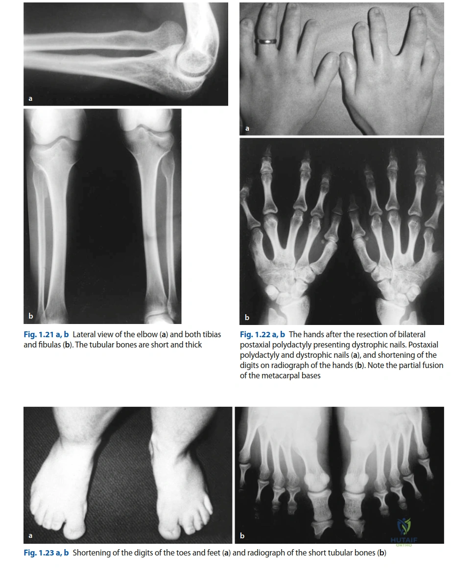
You are reviewing a 4-year-old child with a known diagnosis of Ellis-Van Creveld (EVC) syndrome. The parents are concerned about the child's gait and "knock-knee" appearance. You observe the patient and order the following radiograph.
Describe the key orthopedic findings and outline your systematic approach to evaluating this child for potential surgical intervention.
Candidate: "The radiograph shows mesomelic shortening of the long bones and postaxial polydactyly. For the genu valgum, I would assess the mechanical axis on long-leg standing films, screen for hip dysplasia, and ensure cardiac clearance before considering any correction, such as guided growth or osteotomy."
Candidates often jump immediately to describing the deformity or surgical options without acknowledging the multi-system nature of EVC. Failing to mention mandatory cardiac evaluation (ASD/single atrium) or the potential for respiratory compromise due to thoracic hypoplasia is a critical failure at the FRCS level.
A high-scoring answer follows a structured 'Patient-System-Deformity' framework: 1. Pathology: Identify mesomelic/acromelic dwarfism and characteristic polydactyly. 2. Systemic Risk: Emphasize the mandatory multidisciplinary team (MDT) approach, specifically citing cardiology clearance for ASD/atrial defects and anesthetic consultation for narrow-thorax respiratory risks. 3. Deformity Assessment: Propose full-length standing films (scanograms) to quantify mechanical axis deviation. 4. Surgical Strategy: Discuss 'Guided Growth' (hemiepiphysiodesis) as the first-line, less invasive option before considering formal osteotomies, while noting the role of bone quality and joint stability.
The pediatric team consults you regarding a neonate with EVC syndrome. They are concerned about feeding difficulties and potential aspiration. They present this image of the oral cavity:
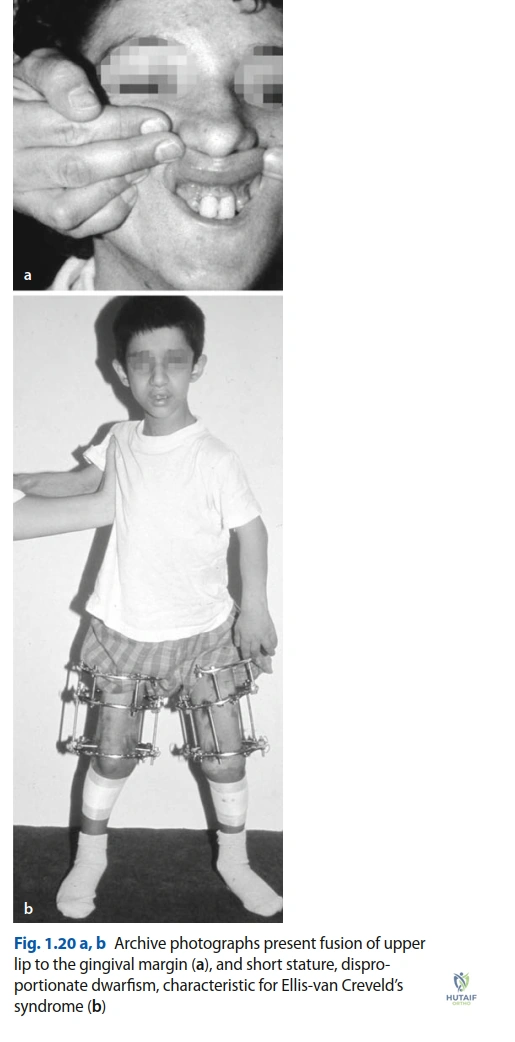
What are the primary oral-orthopedic surgical concerns in the neonatal period, and what is your priority in management?
Candidate: "The image shows natal teeth, gingival hyperplasia, and potentially alveolar-lip synechiae. My priority is to prevent aspiration of the mobile natal teeth and address the synechiae if they prevent adequate latching for breastfeeding."
Candidates often focus solely on the teeth. A failing answer ignores the functional impact on feeding and speech (synechiae/frenula) or fails to mention that neonatal extraction is a minor procedure requiring controlled conditions to avoid secondary hemorrhage or injury to developing tooth buds.
The Gold Standard identifies the "triad of oral management": 1. Safety: Evaluate natal/neonatal teeth for mobility and aspiration risk (Riga-Fede disease prevention). 2. Function: Assess alveolar-lip synechiae and frenula. If they prevent seal/latch, surgical release (frenulectomy/synechiae release) is indicated to restore feeding. 3. Coordination: Emphasize that these are procedures for a pediatric specialist. Mention that while minor, anesthesia requires a pediatric team familiar with the EVC airway, as micrognathia and narrow thoracic cage are frequent comorbidities that complicate safe sedation.
