First Metatarsophalangeal Joint Arthrodesis: Dorsal Plate and Compression Screw Fixation
Key Takeaway
Arthrodesis of the first metatarsophalangeal (MTP) joint using a low-profile contoured dorsal plate and interfragmentary compression screw is the gold standard for end-stage hallux rigidus and severe deformity. This technique provides rigid biomechanical stability, allowing for immediate postoperative weight-bearing. Precise joint preparation with spherical reamers and meticulous multiplanar alignment—specifically 15 to 25 degrees of dorsiflexion—are critical to ensuring optimal functional outcomes and preventing adjacent joint arthritis.
INTRODUCTION TO FIRST METATARSOPHALANGEAL JOINT ARTHRODESIS
Arthrodesis of the first metatarsophalangeal (MTP) joint remains the gold standard and most reliable surgical intervention for end-stage pathology of the first ray. Originally described in the late 19th century, the procedure has undergone significant evolutionary refinements in both joint preparation and fixation biomechanics. The contemporary technique, popularized by Kumar, Pradhan, and Rosenfeld, utilizes a low-profile contoured dorsal titanium plate combined with an interfragmentary compression screw.
This construct provides unparalleled biomechanical rigidity, effectively neutralizing the immense bending moments experienced across the first ray during the terminal stance phase of gait. By achieving absolute stability, this technique facilitates high union rates, predictable deformity correction, and the distinct advantage of immediate postoperative weight-bearing.
INDICATIONS AND CONTRAINDICATIONS
Primary Indications
The decision to proceed with a first MTP joint arthrodesis is based on a combination of clinical symptomatology, radiographic findings, and patient functional demands. Primary indications include:
* End-Stage Hallux Rigidus: Coughlin and Shurnas Grade III or IV osteoarthritis where joint-sparing procedures (e.g., cheilectomy) are insufficient.
* Severe Hallux Valgus: Deformities with an intermetatarsal angle (IMA) exceeding 20 degrees, or hallux valgus associated with severe degenerative joint disease.
* Inflammatory Arthropathies: Rheumatoid arthritis or gouty arthropathy resulting in joint destruction and severe multiplanar deformity.
* Neuromuscular Disorders: Conditions such as cerebral palsy or Charcot-Marie-Tooth disease where dynamic muscle imbalances necessitate rigid stabilization.
* Salvage Procedures: Failed prior forefoot surgeries, including failed Keller resection arthroplasties, failed silastic or metallic joint replacements, or recurrent hallux valgus.
Contraindications
- Active Infection: Local or systemic infection must be eradicated prior to arthrodesis.
- Severe Peripheral Vascular Disease: Inadequate perfusion compromises soft tissue healing and bone consolidation.
- Inadequate Soft Tissue Envelope: Severe dorsal scarring or compromised skin that cannot safely cover the hardware.
- Adjacent Joint Arthritis (Relative): Severe interphalangeal (IP) joint arthritis may be a relative contraindication, as an MTP fusion will increase stress on the IP joint.
BIOMECHANICAL RATIONALE
The first MTP joint is subjected to forces exceeding 100% of body weight during the push-off phase of the gait cycle. The windlass mechanism, driven by the plantar fascia, exerts a powerful plantar-flexing force on the proximal phalanx, creating a dorsal tension gap at the MTP joint.
💡 Biomechanical Pearl
A standalone interfragmentary screw provides excellent compression but is highly susceptible to bending and torsional failure. The addition of a dorsal plate acts as a tension band. By placing the plate on the dorsal (tension) side of the joint, the plantar-flexing forces of gait are converted into compressive forces across the plantar aspect of the arthrodesis site, drastically reducing the risk of nonunion and hardware failure.
PREOPERATIVE PLANNING
Meticulous preoperative planning is essential for achieving optimal alignment and restoring the weight-bearing mechanics of the forefoot.
* Clinical Assessment: Evaluate the vascular status, soft tissue envelope, and the presence of any adjacent joint hypermobility or arthritis (specifically the first tarsometatarsal and interphalangeal joints).
* Radiographic Evaluation: Standard weight-bearing anteroposterior (AP), lateral, and sesamoid axial views are mandatory. Assess bone stock, the presence of subchondral cysts, and the degree of deformity.
* Templating: Anticipate the size of the dorsal plate and the trajectory of the interfragmentary screw. If significant shortening is anticipated, plan for structural bone grafting.
SURGICAL ANATOMY AND EXPOSURE
Patient Positioning and Anesthesia
The procedure is typically performed under regional anesthesia (ankle block or popliteal block) combined with monitored anesthesia care or general anesthesia. The patient is positioned supine on the operating table. A calf or thigh tourniquet is applied to ensure a bloodless surgical field.
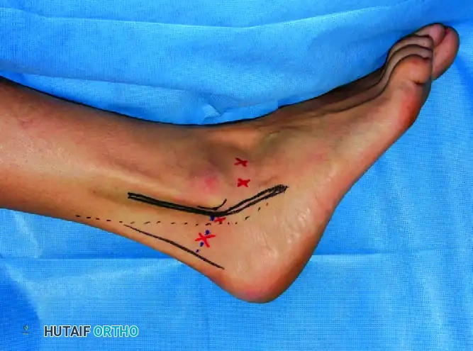
The Dorsal Approach
- Incision: A dorsal longitudinal incision is made, typically 5 to 7 cm in length, centered over the first MTP joint. If this is a revision case, utilize the previous incision to minimize the risk of skin necrosis, extending it as necessary.
- Superficial Dissection: Deepen the incision through the subcutaneous tissues. Meticulous hemostasis is achieved. Identify and protect the dorsomedial cutaneous nerve (a branch of the superficial peroneal nerve) and the proper digital nerve.
- Deep Dissection: Identify the extensor hallucis longus (EHL) tendon. The joint capsule is exposed and divided longitudinally, strictly medial to the EHL tendon. The EHL is then retracted laterally using a blunt retractor.
- Capsular Reflection: Reflect the capsule and collateral ligaments sharply from the metatarsal head and the base of the proximal phalanx.
- Plantar Mobilization: It is critical to mobilize the flexor hallucis longus (FHL) inferiorly. Release the plantar plate and sesamoid complex from the base of the proximal phalanx to allow for adequate exposure and to prevent tethering, which can restrict optimal positioning.
⚠️ Surgical Warning
Failure to adequately release the plantar structures and mobilize the FHL will result in a plantar-flexion contracture of the proximal phalanx, making it nearly impossible to achieve the required 15 to 25 degrees of dorsiflexion during the alignment phase.
JOINT PREPARATION: THE SPHERICAL REAMER TECHNIQUE
Historically, flat planar cuts were used for joint preparation. However, flat cuts result in significant shortening of the first ray and offer no ability to adjust alignment once the cuts are made. The contemporary standard utilizes spherical (cup and cone) reamers.
- Osteophyte Excision: Remove any prominent dorsal, medial, or lateral osteophytes (cheilectomy) using a rongeur or sagittal saw to clearly define the true articular margins.
- Metatarsal Preparation: A guide pin is placed centrally into the metatarsal head, aligned with the anatomical axis of the first metatarsal. A concave spherical reamer is passed over the pin to denude the cartilage down to bleeding subchondral bone.
- Phalangeal Preparation: A guide pin is placed centrally into the base of the proximal phalanx. A convex spherical reamer is used to prepare the phalangeal base.
- Preservation of Bone Stock: Reaming should be performed judiciously. The goal is to remove cartilage and expose healthy, bleeding cancellous bone while preserving maximum bone length.
💡 Clinical Pearl
Always save the autologous bone graft generated within the flutes of the spherical reamers. This highly osteogenic morselized graft is invaluable for packing into subchondral cysts or minor local defects prior to final fixation.
POSITIONING AND ALIGNMENT: THE CRITICAL STEP
The success of a first MTP arthrodesis hinges entirely on achieving perfect multiplanar alignment. Malpositioning is the leading cause of postoperative dissatisfaction, transfer metatarsalgia, and adjacent joint arthritis.
Appose the prepared concave-convex joint surfaces. The spherical nature of the preparation allows for infinite adjustments in all three planes without losing bony contact.
1. Sagittal Plane Alignment (Dorsiflexion)
- The MTP joint must be positioned in 15 to 25 degrees of dorsiflexion relative to the longitudinal axis of the first metatarsal.
- Clinical Simulation: Use a flat tray or a sterile rigid board pressed against the plantar aspect of the foot to simulate weight-bearing.
- Heel Clearance: When the foot is loaded on the flat tray, position the hallux to allow a heel clearance of approximately 1 inch (2.5 cm). This equates to roughly 10 to 15 degrees of dorsiflexion relative to the floor, ensuring the patient can roll over the hallux during the terminal stance phase of gait without vaulting.
2. Coronal Plane Alignment (Valgus)
- Determine the valgus alignment by comparing it to the opposite hallux and the adjacent second toe.
- The hallux should be positioned in 10 to 15 degrees of valgus. It should rest parallel to the second toe without impinging upon it, preventing the development of an interdigital heloma (corn).
3. Axial Plane Alignment (Rotation)
- Keep the rotation strictly neutral.
- Use the nail plate as a visual guide; it should face directly dorsal, matching the rotational profile of the lesser toes. Pronation of the hallux is poorly tolerated and leads to painful medial callosities.
Once optimal alignment is achieved in all three planes, temporarily secure the reduction with one or two crossed Kirschner wires (K-wires) driven from the proximal phalanx into the metatarsal head.
DEFINITIVE FIXATION TECHNIQUE
Interfragmentary Compression Screw
- Confirm alignment clinically and fluoroscopically.
- Place a guide wire for a cannulated interfragmentary compression screw (typically 3.5 mm or 4.0 mm). The trajectory is usually from the medial-distal aspect of the proximal phalanx, directed proximal-lateral into the metatarsal head. Alternatively, a dorsal-distal to plantar-proximal trajectory can be used, provided it does not interfere with the planned plate placement.
- Measure, drill, and insert the compression screw. Ensure the screw threads fully bypass the arthrodesis site to achieve true interfragmentary compression.
Low-Profile Contoured Dorsal Plate
- Preparation: Remove any remaining dorsal bony prominences that might prevent the plate from sitting flush against the bone.
- Plate Selection: Select a precontoured titanium alloy low-profile dorsal plate. These plates are anatomically designed with a built-in valgus and dorsiflexion angle (often available in 0, 5, or 10 degrees of built-in dorsiflexion to match the patient's anatomy).
- Application: Apply the plate dorsally over the MTP joint.
- Fixation: Secure the plate using standard non-locking or locking screws. Typically, the proximal (metatarsal) screws are placed first, followed by the distal (phalangeal) screws. If the plate features a compression slot, utilize it to achieve secondary dynamic compression across the joint before filling the remaining locking holes.
⚠️ Surgical Pitfall
Do not rely solely on the plate for compression. The interfragmentary lag screw is the primary engine for coaptation. The dorsal plate functions primarily as a neutralization and tension-band device.
BONE GRAFTING AND CLOSURE
- Grafting: Inspect the arthrodesis site. Fill any minor local defects, gaps, or subchondral cysts with the autologous bone graft previously harvested from the spherical reamers.
- Irrigation: Thoroughly irrigate the wound to remove any remaining bone debris.
- Layered Closure:
- Close the joint capsule and extensor retinaculum over the plate using absorbable sutures (e.g., 2-0 Vicryl) to provide a soft tissue buffer between the hardware and the skin.
- Close the subcutaneous tissue with inverted interrupted sutures.
- Close the skin with a running subcuticular suture or non-absorbable nylon, depending on surgeon preference and skin tension.
- Dressing: Apply a sterile, non-adherent dressing followed by a robust, well-padded compression bandage to minimize postoperative edema.
POSTOPERATIVE CARE AND REHABILITATION
The rigid biomechanical stability afforded by the dorsal plate and compression screw construct allows for accelerated rehabilitation compared to traditional K-wire fixation.
- Immediate Postoperative Phase (Weeks 0-2): Patients are allowed full weight-bearing immediately in a stiff-soled, flat postoperative shoe or a controlled ankle motion (CAM) boot. Crutches or a walker may be used for balance and comfort as needed. Elevation is strictly enforced to control swelling.
- First Follow-up (Week 2): The compression bandage is removed. The wound is inspected, and sutures are removed if the incision is fully healed. Radiographs are obtained to confirm hardware position and maintenance of alignment. The patient is transitioned back into the stiff-soled postoperative shoe.
- Intermediate Phase (Weeks 2-6): Weight-bearing continues in the stiff-soled shoe. Patients are encouraged to perform active range of motion exercises of the ankle and interphalangeal joints to prevent stiffness.
- Second Follow-up (Week 6): Clinical evaluation and weight-bearing radiographs are performed to assess for trabecular bridging across the arthrodesis site. If clinical and radiographic signs of union are present, the wearing of normal, comfortable, wide-toed shoes is permitted. High-impact activities and running are typically restricted until 10 to 12 weeks postoperatively.
COMPLICATIONS AND MANAGEMENT
While highly successful, first MTP arthrodesis carries potential complications that the orthopedic surgeon must be prepared to manage.
1. Nonunion
Nonunion occurs in approximately 5% to 10% of cases. Risk factors include smoking, poor bone stock, inadequate fixation, and failure to meticulously prepare the joint surfaces.
* Management: Asymptomatic nonunions (fibrous unions) require no intervention. Symptomatic nonunions require revision surgery, which typically involves hardware removal, aggressive debridement of the nonunion site, structural or cancellous autografting (often from the proximal tibia or iliac crest), and rigid refixation.
2. Malunion
Malunion is the most functionally devastating complication.
* Excessive Plantarflexion: Leads to severe stress on the interphalangeal joint, vaulting during gait, and dorsal impingement.
* Excessive Dorsiflexion: Results in transfer metatarsalgia to the lesser metatarsal heads and a "cocked-up" toe that rubs against footwear.
* Management: Symptomatic malunions necessitate a corrective closing or opening wedge osteotomy through the fusion mass, followed by refixation.
3. Hardware Irritation
Due to the paucity of dorsal soft tissue over the first MTP joint, the dorsal plate may become prominent and irritate the skin or the EHL tendon.
* Management: Once solid bony fusion is confirmed radiographically (typically after 6 to 12 months), the hardware can be safely removed in a minor outpatient procedure.
4. Adjacent Joint Arthritis
Over time, the loss of motion at the MTP joint increases kinematic stress on the interphalangeal joint distally and the first tarsometatarsal joint proximally, potentially leading to secondary osteoarthritis. Proper sagittal alignment (15-25 degrees of dorsiflexion) is the most effective way to mitigate this risk.
CONCLUSION
Arthrodesis of the first metatarsophalangeal joint utilizing a low-profile contoured dorsal plate and an interfragmentary compression screw represents the pinnacle of surgical management for severe first ray pathology. By adhering to strict principles of joint preparation with spherical reamers, executing precise multiplanar alignment, and applying rigid tension-band biomechanics, orthopedic surgeons can reliably achieve high union rates, restore pain-free ambulation, and significantly improve patient quality of life.
You Might Also Like