Tibial Intramedullary Nail (Reamed / Unreamed): A Comprehensive Patient Guide
Welcome to this in-depth guide on Tibial Intramedullary Nailing, a highly effective and widely used surgical procedure for stabilizing fractures of the tibia (shin bone). As an expert Medical SEO Copywriter and Orthopedic Specialist, our goal is to provide you with authoritative, comprehensive, yet easy-to-understand information about this critical orthopedic intervention. This content is for patient education only and should not be considered medical advice. Always consult with a qualified healthcare professional for diagnosis and treatment.
1. Comprehensive Introduction & Overview
The tibia, or shin bone, is the larger of the two bones in the lower leg and is crucial for weight-bearing and movement. Fractures of the tibia can result from high-impact trauma, falls, or even underlying medical conditions. When a tibia fracture occurs, precise and stable fixation is often necessary to ensure proper healing and restoration of function.
Tibial intramedullary nailing is a surgical technique that involves inserting a specially designed metal rod (the nail) into the medullary canal (the hollow center) of the tibia. This nail acts as an internal splint, stabilizing the bone fragments and promoting healing. The procedure is considered the gold standard for many types of tibia shaft fractures due to its biomechanical advantages, early mobilization potential, and high success rates.
A key distinction within this procedure lies in whether the medullary canal is "reamed" or "unreamed" prior to nail insertion. Both techniques have their specific indications, advantages, and potential drawbacks, which we will explore in detail. Understanding these differences can help patients appreciate the nuanced decisions made by their orthopedic surgeon.
2. Deep-dive into Technical Specifications & Mechanisms
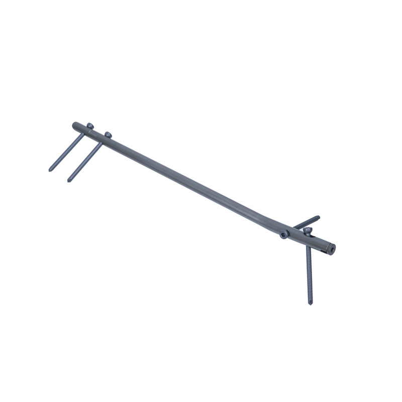
Design and Materials of the Tibial Intramedullary Nail
Tibial intramedullary nails are sophisticated orthopedic implants designed for strength, biocompatibility, and optimal biomechanical performance.
- Materials:
- Most nails are made from high-grade titanium alloys (e.g., Ti-6Al-4V) or stainless steel alloys. These materials are chosen for their excellent strength-to-weight ratio, corrosion resistance within the body, and biocompatibility, meaning they are well-tolerated by human tissues.
- Shape and Structure:
- Hollow Cylindrical Design: The nail is typically hollow, allowing for easier insertion over a guide wire and potentially accommodating future removal or bone grafting if necessary.
- Anatomical Curvature: Nails are pre-contoured to match the natural anterior bow of the tibia, ensuring a snug fit and optimal alignment.
- Proximal and Distal Locking Holes: These holes allow for the insertion of locking screws, which pass through the nail and into the bone fragments. These screws prevent rotational instability and maintain the length of the fractured bone, crucial for healing.
- End Caps: Some nails feature an end cap that can be inserted into the proximal end of the nail after insertion. This cap prevents tissue irritation from the nail's end and can sometimes offer additional stability.
- Associated Instrumentation: The procedure requires a specialized set of instruments, including guide wires, reamers (for reamed technique), drills, targeting devices for locking screws, and insertion handles.
Reamed Nailing vs. Unreamed Nailing: Mechanisms and Biomechanics
The choice between reamed and unreamed nailing is a critical surgical decision influenced by fracture characteristics, patient factors, and surgeon preference.
Reamed Intramedullary Nailing
- Mechanism: In reamed nailing, a series of progressively larger reamers are used to enlarge the medullary canal of the tibia. This process creates a uniform channel that is slightly larger than the chosen nail's diameter.
- Advantages:
- Improved Bone-Nail Contact: Reaming allows for the insertion of a larger diameter nail, which fills the medullary canal more completely. This leads to increased surface contact between the nail and the bone, enhancing primary stability and load sharing.
- Greater Biomechanical Stability: A larger diameter nail provides superior bending and torsional stiffness, contributing to a more stable construct and potentially faster healing.
- Enhanced Load Sharing: The tight fit distributes stress more effectively across the bone-nail interface, reducing stress shielding and promoting bone remodeling.
- Reduced Risk of Fatigue Failure: The larger diameter and tighter fit reduce the stress on the nail itself, potentially lowering the risk of hardware fatigue and breakage.
- Disadvantages:
- Disruption of Endosteal Blood Supply: The reaming process can damage the blood vessels lining the inside of the bone (endosteum), which are crucial for bone healing. However, this disruption is often temporary, and the blood supply typically recovers.
- Increased Intramedullary Pressure and Heat Generation: Reaming generates heat and increases pressure within the canal, which can potentially lead to bone necrosis or fat emboli (small fat particles entering the bloodstream).
- Potential for Complications: A slightly higher theoretical risk of fat embolism syndrome, though clinically significant cases are rare.
Unreamed Intramedullary Nailing
- Mechanism: In unreamed nailing, the intramedullary nail is inserted directly into the medullary canal without prior reaming. The nail diameter is typically chosen to be slightly smaller than the narrowest part of the unreamed canal.
- Advantages:
- Preservation of Endosteal Blood Supply: By avoiding reaming, the delicate endosteal blood supply is largely preserved, which theoretically could promote faster healing, especially in cases with compromised vascularity.
- Reduced Risk of Fat Embolism: Lower intramedullary pressure during insertion minimizes the release of fat globules into the bloodstream.
- Less Invasive: The procedure can be slightly less traumatic to the bone marrow and surrounding tissues.
- Shorter Operative Time: Eliminating the reaming steps can reduce the overall surgery duration.
- Disadvantages:
- Smaller Diameter Nail: The nail must be small enough to fit into the unreamed canal, which means a smaller diameter compared to reamed nails.
- Reduced Bone-Nail Contact: Less contact between the nail and the inner cortex of the bone leads to less primary stability and load sharing.
- Lower Biomechanical Stability: A smaller diameter nail offers less bending and torsional stiffness, potentially making the construct less stable and increasing the risk of delayed union or non-union, especially in comminuted fractures.
- Increased Risk of Nail Fatigue: The smaller nail diameter might experience higher stresses, potentially increasing the risk of hardware fatigue failure over time.
Biomechanics in Fracture Fixation
Regardless of the reaming technique, the fundamental biomechanical principles of intramedullary nailing are crucial for successful healing:
- Load Sharing: The nail shares axial compressive loads with the bone, preventing excessive stress shielding (where the implant carries too much load, preventing the bone from bearing weight and stimulating healing).
- Rotational Stability: Locking screws inserted proximally and distally prevent the bone fragments from rotating relative to each other, which is vital for bone healing.
- Bending Stiffness: The nail resists bending forces, maintaining the alignment of the fractured bone. This stiffness is directly proportional to the nail's diameter and material properties.
- Torsional Stiffness: The resistance to twisting forces, primarily provided by the locking screws and the nail's fit within the canal.
3. Extensive Clinical Indications & Usage
Tibial intramedullary nailing is a versatile procedure with a broad range of clinical applications for various types of tibia fractures.
Primary Clinical Indications
- Diaphyseal Tibia Fractures: These are fractures occurring in the shaft (middle section) of the tibia. Both open (skin broken) and closed (skin intact) diaphyseal fractures are common indications.
- Transverse fractures: Straight across the bone.
- Oblique fractures: Diagonal across the bone.
- Spiral fractures: Twisting pattern.
- Comminuted fractures: Multiple bone fragments.
- Segmental Tibia Fractures: Fractures where the bone is broken in two or more separate places along its length. Nailing can stabilize all segments simultaneously.
- Proximal and Distal Metaphyseal Tibia Fractures: Fractures near the knee or ankle joint that extend into the diaphysis. Modern nails with advanced locking options can effectively stabilize these fractures while preserving joint function.
- Pathological Fractures: Fractures occurring through bone weakened by conditions like tumors or osteoporosis. Nailing provides immediate stability and pain relief.
- Non-unions and Malunions: Cases where a previous fracture has failed to heal (non-union) or has healed in an incorrect alignment (malunion). Nailing can be used in revision surgery to promote healing and correct deformity.
- Impending Fractures: Prophylactic nailing in cases where a bone lesion (e.g., metastatic tumor) is at high risk of fracturing.
Detailed Surgical Procedure (Simplified for Patients)
While the specifics vary, a general outline of the tibial intramedullary nailing procedure includes:
- Pre-operative Planning:
- Thorough clinical examination, X-rays, and often CT scans to assess the fracture pattern, bone quality, and determine appropriate nail length and diameter.
- Antibiotics are typically administered before incision to minimize infection risk.
- Anesthesia: The patient receives either general anesthesia (fully asleep) or regional anesthesia (e.g., spinal block) with sedation.
- Patient Positioning: The patient is typically positioned supine (on their back) on a specialized operating table that allows for traction and fluoroscopic (real-time X-ray) imaging.
- Incision and Entry Point:
- A small incision (approximately 2-5 cm) is made at the front of the knee, just below the kneecap.
- A precise entry point into the medullary canal is created, usually through the proximal tibia, ensuring proper alignment with the bone's axis.
- Fracture Reduction: The orthopedic surgeon carefully manipulates the leg to align the fractured bone fragments using traction and external maneuvers, guided by fluoroscopy.
- Medullary Canal Preparation:
- Reamed Technique: If chosen, progressively larger flexible reamers are passed through the entry point and down the medullary canal, gradually widening it to the desired diameter.
- Unreamed Technique: No reaming is performed; the canal is left in its natural state.
- Nail Insertion:
- A guide wire is typically inserted first, crossing the fracture site and extending down the canal.
- The intramedullary nail is then carefully advanced over the guide wire through the entry point and across the fracture site, until its distal tip is positioned correctly near the ankle.
- Locking Screw Insertion:
- Proximal Locking: Screws are inserted through the proximal locking holes of the nail and into the bone, typically using a specialized jig or freehand technique with fluoroscopic guidance. These prevent rotation and maintain length.
- Distal Locking: Similar to proximal locking, screws are inserted through the distal holes, often requiring more precise fluoroscopic targeting due to the bone's anatomy. These also provide rotational and axial stability.
- Wound Closure: After confirming optimal nail position and locking screw placement with fluoroscopy, the incision is thoroughly irrigated, and the layers of tissue and skin are closed with sutures or staples. A sterile dressing is applied.
Post-operative Care and Rehabilitation
Successful patient outcomes heavily rely on diligent post-operative care and rehabilitation.
- Pain Management: Medications are prescribed to manage post-surgical pain.
- Wound Care: Instructions will be provided for keeping the incision clean and dry to prevent infection.
- Weight-Bearing Progression:
- Initial weight-bearing status varies based on fracture type, stability of fixation, and surgeon's preference. It can range from immediate full weight-bearing to partial or non-weight-bearing for several weeks.
- Gradual progression is guided by clinical and radiographic healing.
- Physical Therapy: Crucial for regaining strength, range of motion, and function. Exercises focus on knee and ankle mobility, muscle strengthening, and gait training.
- Follow-up Appointments: Regular visits with the orthopedic surgeon are necessary to monitor healing progress with X-rays and adjust the rehabilitation plan.
4. Risks, Side Effects, or Contraindications
While tibial intramedullary nailing is generally safe and highly effective, like any surgical procedure, it carries potential risks and complications.
General Surgical Risks
- Infection: At the surgical site or deeper within the bone (osteomyelitis).
- Bleeding: Excessive blood loss during or after surgery.
- Nerve or Vascular Damage: Injury to surrounding nerves or blood vessels.
- Anesthesia Risks: Reactions to anesthesia, respiratory problems, etc.
- Blood Clots (DVT/PE): Deep vein thrombosis (DVT) in the leg or pulmonary embolism (PE) if a clot travels to the lungs.
Specific Risks Associated with Tibial Nailing
- Non-union: The fracture fails to heal completely, requiring further intervention.
- Malunion: The fracture heals in an incorrect position or alignment, potentially leading to limb deformity or functional impairment.
- Hardware Failure: The nail or locking screws can break, especially if healing is delayed or if the patient places excessive stress on the implant too early.
- Pain at the Entry Site: Pain or irritation around the knee, particularly from the proximal end of the nail, which may necessitate hardware removal.
- Fat Embolism Syndrome: Although rare, reaming can theoretically increase the risk of fat globules entering the bloodstream, potentially affecting the lungs or brain.
- Compartment Syndrome: A dangerous condition where swelling within the muscle compartments of the lower leg increases pressure, potentially leading to muscle and nerve damage.
- Refracture: The bone may re-fracture after hardware removal, particularly if removed too early or if bone quality is poor.
- Delayed Union: The fracture takes longer than expected to heal.
- Nail Protrusion/Irritation: The nail or screws may protrude slightly, causing soft tissue irritation.
Contraindications
Certain conditions may make tibial intramedullary nailing unsuitable or increase its risks:
- Active Infection: Especially osteomyelitis, which must be treated before implanting hardware.
- Severe Open Fractures with Extensive Soft Tissue Damage: Initial management may focus on wound debridement and external fixation before considering nailing.
- Significant Bone Loss or Bone Defects: May require alternative fixation methods or bone grafting.
- Extensive Articular Involvement: Fractures extending significantly into the knee or ankle joint may be better managed with plates and screws to achieve anatomical reduction of the joint surface.
- Severe Osteoporosis: While not an absolute contraindication, very poor bone quality can compromise screw purchase and nail stability.
- Patient Unable to Comply with Post-operative Care: Compliance with weight-bearing restrictions and physical therapy is crucial.
5. Expert Tips from Dr. Mohammed Hutaif
"As an orthopedic specialist, I've seen firsthand the life-changing impact of well-executed tibial intramedullary nailing. Here are my key insights for patients undergoing or considering this procedure:
- Early Diagnosis and Proper Planning are Paramount: A thorough pre-operative assessment, including detailed imaging, is critical. This allows us to choose the correct nail size, length, and whether a reamed or unreamed technique is most appropriate for your specific fracture pattern and bone quality. Don't hesitate to ask your surgeon about their rationale.
- Patient Compliance with Physical Therapy is Non-Negotiable: Surgery is only half the battle. Your commitment to the prescribed physical therapy regimen is the single most important factor in regaining full function. Skipping sessions or not performing exercises diligently can significantly delay your recovery and impact your long-term outcome.
- Understand the Reamed vs. Unreamed Choice: While your surgeon will make the ultimate decision, understanding the principles behind reamed (stronger construct, larger nail) versus unreamed (preserves blood supply, less invasive) can empower you. Ask why a specific technique was chosen for your case.
- Manage Your Expectations for Recovery: Healing takes time. While some patients may bear weight relatively quickly, full bone healing can take several months. It's a marathon, not a sprint. Be patient with your body and celebrate small victories in your recovery journey.
- Prioritize Follow-up Appointments: Regular follow-up visits with your surgeon are crucial. We monitor your healing progress with X-rays, assess your rehabilitation, and address any concerns. These appointments help us detect potential issues early and adjust your treatment plan as needed.
- Nutrition and Lifestyle Matter: Support your bone healing by maintaining a healthy diet rich in calcium and Vitamin D. Avoid smoking, as it significantly impairs bone healing and increases complication rates. Limit alcohol consumption.
- Don't Hesitate to Communicate: If you experience new pain, swelling, redness, or any other concerning symptoms, contact your surgical team immediately. Early intervention can prevent minor issues from becoming major complications."
6. Massive FAQ Section
Q1: What exactly is a tibial intramedullary nail?
A1: A tibial intramedullary nail is a strong, hollow metal rod (usually titanium or stainless steel) that an orthopedic surgeon inserts into the hollow center (medullary canal) of your shin bone (tibia). It acts as an internal splint to stabilize a broken tibia, allowing it to heal properly.
Q2: What is the main difference between reamed and unreamed nailing?
A2: The main difference lies in how the medullary canal is prepared.
* Reamed Nailing: The surgeon uses special instruments (reamers) to gradually widen the bone's hollow canal before inserting the nail. This allows for a larger, stronger nail and a tighter fit, offering more stability.
* Unreamed Nailing: The nail is inserted directly into the bone's natural canal without prior widening. This technique is less invasive and preserves more of the bone's internal blood supply, but typically uses a smaller diameter nail, which might offer less inherent stability.
Q3: How long does the surgery typically take?
A3: The surgery itself usually takes between 1 to 3 hours, depending on the complexity of the fracture and whether it's an open or closed procedure. This estimate does not include preparation and recovery time in the operating room.
Q4: How long is the full recovery period after tibial nailing?
A4: Full recovery can vary significantly, but generally ranges from 3 to 6 months for bone healing, and up to a year or more to regain full strength and return to pre-injury activity levels. Early weight-bearing and consistent physical therapy play a huge role in accelerating recovery.
Q5: Will I need to have the tibial nail removed?
A5: Not always. In many cases, the nail can remain in place indefinitely, causing no issues. However, removal might be recommended if you experience pain, irritation (especially around the knee), infection, or if the implant interferes with future joint replacement surgery. Removal is typically performed at least 12-18 months after the initial surgery, once the bone has fully healed.
Q6: What are the most common potential complications?
A6: While generally safe, potential complications include infection, non-union (fracture not healing), malunion (healing in poor alignment), pain at the knee entry site, hardware failure, and nerve or blood vessel damage. Your surgeon will discuss these risks with you.
Q7: Can I put weight on my leg immediately after surgery?
A7: It depends on the type and stability of your fracture, as well as your surgeon's preference. Some patients may be allowed immediate weight-bearing as tolerated, while others may need to remain non-weight-bearing or partially weight-bearing for several weeks. Always follow your surgeon's specific instructions.
Q8: What kind of pain can I expect after the surgery?
A8: You can expect moderate to severe pain initially, which will be managed with prescribed pain medication. As you recover, the pain should gradually decrease. Some patients experience localized pain around the knee (entry site) or at the fracture site for several months.
Q9: How can I optimize my recovery and bone healing?
A9: To optimize recovery:
* Strictly follow your surgeon's weight-bearing and physical therapy instructions.
* Maintain a healthy diet rich in calcium, Vitamin D, and protein.
* Avoid smoking and excessive alcohol consumption, as these hinder bone healing.
* Keep your incision site clean and dry.
* Attend all follow-up appointments.
Q10: When can I return to sports or strenuous activities?
A10: Return to sports or strenuous activities is highly individualized. It typically requires complete bone healing, restoration of strength and range of motion, and clearance from your orthopedic surgeon and physical therapist. This can often take 6 to 12 months, or even longer for high-impact sports.
Q11: Are there any alternatives to tibial intramedullary nailing for tibia fractures?
A11: Yes, alternatives exist depending on the fracture type and location. These may include:
* Casting or Bracing: For stable, non-displaced fractures.
* External Fixation: For severe open fractures or highly comminuted fractures, often as a temporary measure.
* Plate and Screw Fixation: For fractures extending into the joint (intra-articular fractures) or very distal/proximal shaft fractures.
Q12: What materials are the nails typically made from?
A12: Tibial intramedullary nails are primarily made from biocompatible materials like high-grade titanium alloys or stainless steel alloys. These materials are chosen for their strength, durability, and ability to be safely implanted in the human body long-term.