The Straight Osteotome (Exostosis Chisel): An In-Depth Orthopedic Guide
The Straight Osteotome, often referred to as an "Exostosis Chisel," is an indispensable instrument in the orthopedic surgeon's toolkit. Renowned for its precision and effectiveness in bone removal and reshaping, this specialized surgical tool plays a pivotal role in a myriad of procedures aimed at restoring function, alleviating pain, and improving patient quality of life. This comprehensive guide delves into every facet of the Straight Osteotome, from its intricate design and material science to its critical role in surgical applications, meticulous maintenance, and profound impact on patient outcomes.
1. Comprehensive Introduction & Overview
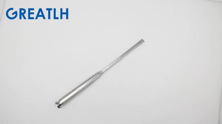
A Straight Osteotome is a hand-held surgical instrument characterized by a straight, sharp cutting edge at its tip, extending from a sturdy shaft to an ergonomic handle. Its primary function is the controlled removal of bone tissue, particularly exostoses (bone spurs), osteophytes, and other bony prominences that can cause pain, impingement, or restrict movement. Unlike a gouge, which has a curved cutting edge, the straight osteotome provides a flat, precise cut, making it ideal for shaving off unwanted bone or performing controlled osteotomies.
Historically, bone chisels and osteotomes have been fundamental to bone surgery for centuries, evolving from rudimentary tools to the sophisticated, high-precision instruments used today. Modern orthopedic surgery demands tools that offer both durability and fine control, and the Straight Osteotome perfectly embodies these requirements. Its design facilitates the precise application of force, typically via a surgical mallet, to achieve clean and accurate bone resections with minimal collateral tissue damage.
This guide will provide a deep dive into:
* The meticulous design and material science behind the Straight Osteotome.
* Its detailed surgical and clinical applications across various orthopedic specialties.
* Best practices for fitting and usage to optimize surgical outcomes.
* Rigorous maintenance and sterilization protocols essential for patient safety and instrument longevity.
* The biomechanical principles governing its effectiveness.
* The direct improvements in patient outcomes attributable to its judicious use.
2. Deep-Dive into Technical Specifications & Mechanisms
The efficacy of a Straight Osteotome is a direct result of its sophisticated design and the advanced materials used in its construction. Understanding these technical aspects is crucial for appreciating its role in orthopedic surgery.
2.1 Design & Ergonomics
The Straight Osteotome is typically composed of three primary sections:
* Handle: Designed for optimal grip and comfort, often featuring a textured or knurled surface to prevent slippage during use. Handles can vary in diameter and length to accommodate different hand sizes and surgical approaches. The proximal end is usually flat or slightly rounded to receive mallet strikes effectively without causing discomfort to the surgeon's hand.
* Shaft: A robust, straight shaft connects the handle to the blade. Its length varies, allowing access to superficial or deeper anatomical sites. The shaft's rigidity is paramount to ensure efficient transmission of force from the mallet to the cutting edge without bending or absorbing impact energy.
* Blade/Tip: This is the working end of the instrument, featuring a finely honed, straight cutting edge. Blades come in various widths (e.g., 5mm, 10mm, 15mm, 20mm, 25mm) to suit different surgical requirements and the size of the bone to be resected. The bevel of the blade can be single or double-sided, influencing the cutting action and the angle of bone removal. A sharp, durable edge is critical for clean bone cuts, minimizing bone trauma and promoting faster healing.
Table: Common Straight Osteotome Design Variations
| Feature | Description | Surgical Impact |
|---|---|---|
| Handle | Ergonomic, textured, various diameters. | Enhanced grip, reduced surgeon fatigue, better control. |
| Shaft | Straight, rigid, varying lengths (e.g., 10cm, 15cm, 20cm). | Access to superficial/deep sites, efficient force transmission. |
| Blade Width | 5mm to 25mm, graduated markings often present. | Precision in bone removal, adaptable to lesion size. |
| Blade Bevel | Single bevel (more aggressive cut) or double bevel (more balanced cut). | Influences cutting angle, depth, and control. |
| Overall Balance | Weight distribution optimized for stability. | Improved maneuverability and reduced tremor. |
2.2 Materials
The choice of material for a surgical osteotome is critical for its performance, durability, and biocompatibility.
* High-Grade Surgical Stainless Steel: This is the most common material, typically alloys like 420 or 440A stainless steel. These alloys offer an excellent balance of hardness, corrosion resistance, and edge retention. The steel undergoes specific heat treatment processes to achieve optimal hardness for maintaining a sharp edge while resisting fracture.
* Titanium: Used for specific applications, titanium osteotomes are lighter, non-magnetic (MRI compatible), and highly biocompatible, making them suitable for patients with nickel allergies or procedures requiring MRI follow-up. However, titanium is generally softer than surgical steel, potentially leading to faster dulling of the edge.
* Surface Coatings: Some osteotomes may feature advanced surface coatings, such as tungsten carbide, particularly on the cutting edge, to enhance hardness, improve edge retention, and extend instrument life. Anti-glare finishes are also common to reduce reflections in the surgical field.
2.3 Biomechanics & Mechanism of Action
The Straight Osteotome operates on the principle of controlled mechanical force application.
* Force Transmission: When struck by a surgical mallet, the impact force is efficiently transmitted along the rigid shaft to the sharp cutting edge. The surgeon guides the osteotome, determining the angle and depth of the cut.
* Shear Force: The sharp edge applies concentrated shear force to the bone tissue. This force, combined with the impact, causes the bone to cleave or shave off in a controlled manner. A dull instrument requires significantly more force, increasing the risk of uncontrolled bone fracture or slippage.
* Precision: The straight design allows for linear, controlled bone removal, which is crucial for achieving precise resections and maintaining anatomical integrity of adjacent structures. The varying blade widths enable surgeons to select the appropriate tool for the specific size of the exostosis or bone segment.
* Ergonomics: An ergonomically designed handle and balanced instrument reduce surgeon fatigue, allowing for greater control and precision, especially during lengthy procedures. This directly translates to more accurate cuts and better patient outcomes.
3. Extensive Clinical Indications & Usage
The Straight Osteotome is a versatile instrument with a broad range of applications in orthopedic surgery. Its primary role is in the removal of unwanted bony tissue and precise bone reshaping.
3.1 Surgical Applications
- Exostosis and Osteophyte Removal: This is the most common indication.
- Calcaneal Spurs (Heel Spurs): Removal of bony growths on the calcaneus that cause plantar fasciitis or Achilles tendinopathy.
- Haglund's Deformity: Resection of the posterosuperior calcaneal prominence that impinges on the Achilles tendon.
- Dorsal Midfoot Exostosis (Tarsal Boss): Removal of bony prominences on the dorsal aspect of the foot, often causing pain and difficulty with shoewear.
- Joint Osteophytes: Removal of bone spurs around joints (e.g., knee, hip, shoulder, spine) that cause impingement, pain, or restrict range of motion in arthritic conditions.
- Osteochondromas: Resection of sessile or pedunculated benign bone tumors, particularly when causing symptoms.
- Minor Bone Reshaping and Osteotomies:
- Bunionectomy: While often performed with power burrs, osteotomes can be used for precise removal of the medial eminence or for specific osteotomies.
- Joint Debridement: Preparing joint surfaces for arthroplasty by removing small osteophytes or irregular bone fragments.
- Spinal Fusion Preparation: Removing small bony prominences or preparing vertebral endplates for graft placement in spinal fusion procedures.
- Bone Graft Harvesting: In some cases, small cortical bone grafts can be carefully harvested using an osteotome.
- Trauma Surgery:
- Fracture Debridement: Removing small, loose bone fragments from a fracture site to prepare for reduction and fixation.
- Preparing Bone Ends: Shaping bone ends to achieve optimal alignment and contact for internal fixation.
3.2 Fitting & Usage Instructions
Proper technique is paramount to maximize the effectiveness of the Straight Osteotome and minimize surgical risks.
Pre-operative:
1. Instrument Inspection: Before sterilization, and again before use, meticulously inspect the osteotome for any signs of damage, dullness, nicks on the blade, or corrosion. A dull or damaged instrument can lead to uncontrolled cuts and increased bone trauma.
2. Size Selection: Choose the appropriate blade width based on the size of the exostosis or the bone segment to be resected, and the anatomical constraints of the surgical site.
3. Patient Positioning: Ensure the patient is correctly positioned to allow optimal access to the surgical site and to maintain stability throughout the procedure.
Intra-operative:
1. Grip and Hand Position: Hold the osteotome firmly with a two-hand technique if possible. One hand guides the blade precisely, while the other provides counter-pressure or helps stabilize the instrument. The guiding hand should be positioned close to the working end for maximum control.
2. Controlled Mallet Strikes: The osteotome is typically struck with a surgical mallet. Strikes should be firm but controlled, rhythmic taps, rather than forceful blows. The force should be sufficient to cut through the bone but not so excessive as to cause uncontrolled fracture or slippage.
3. Angle of Attack: Position the blade at the desired angle relative to the bone surface. For exostosis removal, aim to shave off the prominence smoothly, following the natural contour of the bone.
4. Soft Tissue Protection: Always use appropriate retractors to protect surrounding soft tissues, nerves, and blood vessels from the osteotome and mallet strikes.
5. Clearance and Irrigation: Regularly clear bone debris from the surgical field. Irrigation with saline helps to cool the bone, prevent thermal necrosis, and improve visualization.
6. Progressive Removal: For larger exostoses, remove bone in small, controlled increments rather than attempting a single, large resection.
Post-operative:
1. Careful Removal: Once the desired bone removal is achieved, carefully withdraw the osteotome from the surgical site.
2. Initial Cleaning: Perform initial cleaning to remove gross contaminants before sending for sterilization.
4. Maintenance & Sterilization Protocols
Maintaining surgical instruments, especially those with sharp edges like osteotomes, is critical for patient safety, infection control, and instrument longevity.
4.1 Cleaning
- Immediate Post-Use: Remove gross contaminants (blood, tissue, bone fragments) immediately after surgery by wiping with a moist cloth or sponge. This prevents organic material from drying and hardening, which makes cleaning more difficult.
- Manual Cleaning: If automated cleaning is not immediately available, instruments should be manually cleaned using a soft brush and an enzymatic cleaner. Pay close attention to the blade and any textured areas.
- Automated Cleaning:
- Ultrasonic Bath: Place instruments in an ultrasonic cleaner with appropriate cleaning solution. Ultrasonic waves create cavitation bubbles that dislodge microscopic debris.
- Washer-Disinfector: A preferred method, these machines clean and disinfect instruments automatically, following validated cycles.
- Rinsing: Thoroughly rinse all instruments with distilled or deionized water to remove all cleaning solution residues, which can cause staining or corrosion during sterilization.
4.2 Inspection
After cleaning and before sterilization, each osteotome must be meticulously inspected:
* Sharpness: Check the cutting edge for sharpness. A dull edge can be identified by visual inspection or by gently testing on a specialized sharpness indicator (never on skin or glove).
* Damage: Look for nicks, dents, bends, cracks, or signs of corrosion (pitting, rust). Damaged instruments should be repaired or removed from circulation.
* Cleanliness: Ensure no residual debris remains on the instrument.
4.3 Sterilization
Steam Sterilization (Autoclaving): This is the most common and effective method for heat-stable instruments like stainless steel osteotomes.
* Packaging: Instruments must be properly packaged in sterilization pouches or rigid sterilization containers that allow steam penetration while maintaining sterility after the cycle.
* Parameters: Follow validated parameters for temperature, pressure, and exposure time (e.g., 132°C (270°F) for 4 minutes for wrapped instruments, or 121°C (250°F) for 15-30 minutes).
* Drying: Ensure instruments are thoroughly dried within the sterilizer to prevent water spots and microbial growth.
Other Methods (Less Common for Osteotomes):
* Flash Sterilization: Used only in emergency situations when there's insufficient time for standard sterilization. Instruments are unwrapped and immediately used. This method carries higher risks and should be minimized.
* Ethylene Oxide (EtO): For heat-sensitive instruments, but rarely used for osteotomes. Requires aeration time.
* Hydrogen Peroxide Gas Plasma: Another low-temperature sterilization method, but typically for more delicate, heat-sensitive instruments.
4.4 Storage
Sterilized instruments must be stored in a dry, clean, and protected environment to maintain their sterility until use. Packaging should be intact and undamaged.
5. Biomechanics and Patient Outcome Improvements
The precise biomechanical interaction of the Straight Osteotome with bone tissue directly translates into significant improvements in patient outcomes.
5.1 Biomechanical Advantages
- Controlled Bone Removal: The sharp, straight edge allows for the precise removal of bone in predictable planes. This minimizes the risk of inadvertently damaging healthy bone or adjacent soft tissues.
- Reduced Thermal Necrosis: Unlike high-speed burrs that generate significant heat, the percussive action of an osteotome generates less heat, reducing the risk of thermal necrosis to the bone cells, which can impair healing.
- Clean Cut Surfaces: A sharp osteotome creates clean, smooth bone surfaces. These surfaces are more conducive to healing and less likely to cause irritation to surrounding soft tissues compared to ragged edges left by less precise instruments.
- Optimal Force Application: The design facilitates the efficient transmission of force from the mallet, allowing the surgeon to achieve desired bone cuts with minimal effort, thereby reducing surgeon fatigue and improving surgical accuracy.
5.2 Patient Outcome Improvements
The judicious use of the Straight Osteotome contributes to a cascade of positive patient outcomes:
- Reduced Pain and Discomfort: By precisely removing impinging exostoses or osteophytes, the osteotome directly addresses the source of pain, nerve compression, or joint friction, leading to significant post-operative pain relief.
- Improved Mobility and Function: Removal of bony obstructions restores normal joint mechanics, increases range of motion, and improves overall limb function, allowing patients to return to daily activities and even sports.
- Faster Recovery and Rehabilitation: Clean surgical margins and minimal collateral tissue damage promote faster healing of both bone and soft tissues. This can shorten recovery times and accelerate the rehabilitation process.
- Reduced Risk of Complications:
- Lower Infection Risk: Proper sterilization and clean cuts reduce the risk of surgical site infections.
- Less Post-operative Swelling and Bruising: Precise bone removal and reduced tissue trauma contribute to less inflammation.
- Minimized Scar Tissue Formation: Cleaner cuts and less tissue disruption can lead to less exuberant scar tissue formation.
- Long-term Durability of Results: By addressing the mechanical source of the problem, the Straight Osteotome helps achieve durable long-term results, preventing recurrence of symptoms related to bony impingement.
- Enhanced Quality of Life: Ultimately, these improvements culminate in a significantly enhanced quality of life for patients, allowing them to live more active, pain-free lives.
6. Risks, Side Effects, or Contraindications
While the Straight Osteotome is a powerful and precise tool, its use is not without potential risks, side effects, and contraindications. Surgeons must be acutely aware of these to ensure patient safety.
6.1 Risks
- Iatrogenic Fracture: The primary risk is unintended fracture of the bone being operated on, or adjacent bones, due to excessive force, incorrect angle of attack, or a dull instrument. This is particularly relevant in osteoporotic bone.
- Soft Tissue Damage: Nerves, blood vessels, tendons, ligaments, and muscle tissue can be inadvertently damaged if not properly retracted and protected during the procedure. This can lead to numbness, weakness, bleeding, or functional deficits.
- Incomplete Bone Removal: If the exostosis is not fully removed, symptoms may persist or recur, necessitating revision surgery.
- Excessive Bone Removal: Removing too much healthy bone can compromise structural integrity, weaken the bone, or alter joint mechanics in an undesirable way.
- Infection: As with any surgical instrument, inadequate sterilization or breaks in sterile technique can lead to surgical site infections.
- Instrument Failure: A defective or improperly maintained osteotome can break during surgery, potentially leaving fragments in the wound, requiring further intervention.
- Thermal Injury: While less common than with power burrs, repetitive or forceful strikes in a confined area without adequate irrigation can still generate localized heat, leading to bone necrosis.
6.2 Side Effects
- Post-operative Pain, Swelling, and Bruising: These are common and expected side effects of any bone surgery, typically managed with analgesics, RICE (Rest, Ice, Compression, Elevation), and physical therapy.
- Scar Tissue Formation: The surgical incision will result in scar tissue.
- Numbness or Weakness: If a nerve is bruised or damaged during the procedure, temporary or permanent numbness, tingling, or muscle weakness can occur.
6.3 Contraindications
- Severe Osteopenia/Osteoporosis: In patients with significantly compromised bone density, the risk of iatrogenic fracture with an osteotome is substantially higher. Alternative methods like power burrs or specialized cutting instruments may be preferred.
- Active Infection: Performing surgery through an area of active infection is contraindicated, as it can spread the infection.
- Poor Patient General Health: Patients with significant comorbidities that increase surgical risk (e.g., uncontrolled diabetes, severe cardiovascular disease) may not be suitable candidates for elective osteotome procedures.
- Unsuitable Anatomical Location: In areas where critical neurovascular structures are in extremely close proximity and cannot be adequately protected, or where precise control is exceptionally difficult, other surgical approaches or instruments might be safer.
- Surgeon Inexperience: Procedures involving osteotomes require significant skill and experience. Inexperienced surgeons may have a higher risk of complications.
7. Massive FAQ Section
Here are frequently asked questions about the Straight Osteotome (Exostosis Chisel):
Q1: What is the primary purpose of a Straight Osteotome (Exostosis Chisel)?
A1: The Straight Osteotome is primarily used in orthopedic surgery for the precise and controlled removal of unwanted bony prominences, such as exostoses (bone spurs), osteophytes, and for minor bone reshaping or osteotomies.
Q2: What materials are Straight Osteotomes typically made from?
A2: Most Straight Osteotomes are crafted from high-grade surgical stainless steel (e.g., 420 or 440A) for optimal hardness, edge retention, and corrosion resistance. Some are made from titanium, which is lighter, non-magnetic, and highly biocompatible.
Q3: How do you ensure the sharpness of an osteotome?
A3: The sharpness of an osteotome is critical. It's ensured through meticulous manufacturing processes, proper heat treatment of the steel, and rigorous inspection before and after each sterilization cycle. Dull instruments must be re-sharpened by specialized technicians or replaced.
Q4: What are the different sizes available for Straight Osteotomes?
A4: Straight Osteotomes come with various blade widths, commonly ranging from 5mm up to 25mm or more, in increments of 5mm or 10mm. This allows surgeons to select the appropriate size for the specific bone structure and lesion.
Q5: Can osteotomes be used for delicate bone work?
A5: Yes, when used with a precise, controlled technique and a light mallet, smaller Straight Osteotomes are excellent for delicate bone work where controlled, linear bone removal is required, such as in hand or foot surgery.
Q6: What are the key steps in sterilizing a Straight Osteotome?
A6: Key steps include immediate post-use cleaning to remove gross contaminants, manual or automated cleaning (ultrasonic bath/washer-disinfector), thorough rinsing, meticulous inspection, proper packaging, and then steam sterilization (autoclaving) according to validated parameters.
Q7: How does the design of the handle impact surgical use?
A7: The ergonomic design of the handle, often featuring a textured or knurled surface, provides a secure, non-slip grip. This reduces surgeon fatigue, enhances control, and allows for more precise force transmission, directly impacting surgical accuracy and patient safety.
Q8: What are the potential risks associated with using an osteotome?
A8: Potential risks include iatrogenic bone fracture, damage to surrounding soft tissues (nerves, vessels, tendons), incomplete or excessive bone removal, surgical site infection, and instrument failure if not properly maintained.
Q9: How does proper osteotome use improve patient outcomes?
A9: Proper use of a Straight Osteotome leads to precise bone removal, reduced tissue trauma, cleaner surgical margins, and less heat generation compared to some other methods. This translates to reduced post-operative pain, improved mobility, faster recovery, and a lower risk of complications, ultimately enhancing the patient's quality of life.
Q10: Is there a difference between an osteotome and a chisel?
A10: While often used interchangeably, in surgical contexts, an osteotome traditionally has a beveled cutting edge on both sides (double bevel), designed for cutting bone, whereas a chisel typically has a single-beveled edge, more for scraping or shaping. However, the term "exostosis chisel" often refers to an osteotome used for removing bony growths, and many "osteotomes" are available with single or double bevels depending on the manufacturer and intended use.
Q11: How often should an osteotome be replaced?
A11: An osteotome should be replaced when it can no longer be effectively sharpened, shows signs of significant wear, corrosion, or structural damage (e.g., nicks, cracks) that could compromise its integrity or sterility. Regular inspection is key to determining replacement needs.
Q12: Are Straight Osteotomes MRI compatible?
A12: Most Straight Osteotomes made from high-grade surgical stainless steel are generally considered safe for use in an MRI environment, but it's crucial to verify with the manufacturer, especially for specific alloys. Titanium osteotomes are inherently non-magnetic and are fully MRI compatible.