Understanding Small Joint Fusion Plates for Finger Arthrodesis (PIP/DIP)
1. Comprehensive Introduction & Overview
The intricate mechanics of our hands and fingers allow for a remarkable range of motion, essential for daily tasks. However, conditions like severe arthritis, trauma, or deformity can severely compromise the function and cause debilitating pain in the small joints of the fingers. When conservative treatments fail to provide relief, surgical intervention becomes a vital option. Among these, arthrodesis, or joint fusion, stands as a reliable procedure, particularly for the Proximal Interphalangeal (PIP) and Distal Interphalangeal (DIP) joints of the fingers.
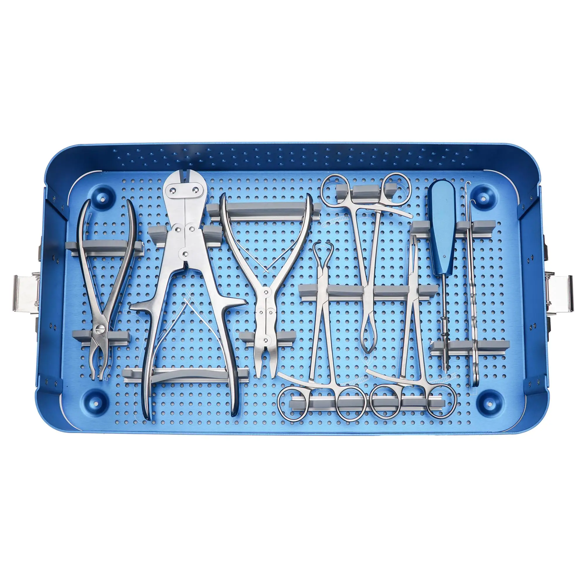
A "Small Joint Fusion Plate (PIP/DIP)" is a specialized orthopedic implant designed to facilitate and stabilize this fusion process. It acts as an internal splint, holding the two bones forming the joint firmly together, allowing them to grow into a single, solid bone. While fusion eliminates movement at the treated joint, it effectively eradicates pain, corrects deformity, and provides a stable, strong finger for gripping and pinching, significantly improving a patient's quality of life. This comprehensive guide will delve into every aspect of these crucial implants, from their innovative design to their profound impact on patient outcomes.
2. Deep-Dive into Technical Specifications / Mechanisms
Small joint fusion plates are marvels of orthopedic engineering, meticulously designed to meet the unique biomechanical demands of the fingers.
Design & Materials
The effectiveness of these plates lies in their precise design and the advanced materials used in their manufacture.
- Low-Profile and Anatomical Contouring: These plates are incredibly thin and contoured to match the natural curvature of the phalangeal bones. This low-profile design minimizes soft tissue irritation and palpability under the skin, which is crucial in the shallow anatomy of the fingers. Anatomical pre-contouring ensures optimal fit and reduces the need for extensive intraoperative bending, preserving the material's integrity.
- Materials:
- Titanium Alloys (e.g., Ti-6Al-4V ELI): The predominant material choice due to its exceptional biocompatibility, high strength-to-weight ratio, and excellent corrosion resistance. Titanium is non-ferromagnetic, allowing for safe MRI scans post-surgery. Its osseointegrative properties also promote bone growth onto the plate surface.
- Stainless Steel (e.g., 316L): While less common than titanium for primary implants due to potential MRI interference and lower osseointegration, it offers good mechanical properties and cost-effectiveness.
- Screw Holes and Fixation:
- Locking Screws: A hallmark of modern plating systems. Locking screws thread into the plate itself, creating a fixed-angle construct. This "internal fixator" concept provides angular stability, which is highly beneficial in osteopenic bone or comminuted fractures, preventing screw pull-out and maintaining reduction.
- Non-Locking (Compression) Screws: These screws pull the bone fragments towards the plate, generating compression at the fusion site. Many systems offer a combination of locking and non-locking holes, allowing the surgeon to achieve both compression across the joint and rigid fixation.
- Variable Angle Locking Screws: Some advanced systems feature variable angle locking holes, allowing the surgeon to insert screws at a range of angles while still achieving locking fixation. This flexibility is invaluable for navigating complex bone anatomy or avoiding critical structures.
- Screw Sizes: Typically very small, ranging from 1.0mm to 2.0mm in diameter, with specific lengths tailored to the phalanges.
- Plate Size and Geometry: Plates are available in various lengths and widths, specifically scaled for the PIP and DIP joints. Their geometry often includes a central aperture for bone grafting if needed, and tapered ends to minimize stress risers.
Biomechanics of Fusion Plates
The mechanical principles behind small joint fusion plates are critical for successful arthrodesis.
- Rigid Internal Fixation: The primary goal is to provide absolute stability at the fusion site. By rigidly immobilizing the joint, micromotion is minimized, which is essential for primary bone healing (direct bone formation without callus).
- Compression at the Fusion Site: Achieving interfragmentary compression across the joint surfaces is paramount. This compression reduces the gap between the bones, increases the surface area for healing, and stimulates osteogenesis according to Wolff's Law, which states that bone adapts to the loads it is subjected to. Many plates are designed with dynamic compression features or are applied with specific techniques to achieve this.
- Load Sharing: While the plate provides rigid fixation, it ideally should not completely shield the bone from physiological loads. "Load sharing" means the plate and bone both bear some of the stress, promoting bone healing and preventing stress shielding, which can lead to bone atrophy. The stiffness and placement of the plate are optimized for this balance.
- Optimizing Fusion Rates: By providing stable fixation and optimal compression, these plates significantly enhance the chances of a successful bony union (fusion). A high fusion rate is directly linked to patient satisfaction and long-term functional improvement.
3. Extensive Clinical Indications & Usage
Small joint fusion plates are employed in a variety of clinical scenarios where preserving motion is either impossible, undesirable, or has failed.
Indications for PIP/DIP Fusion
- Severe Osteoarthritis (OA): Degenerative joint disease leading to cartilage loss, bone-on-bone friction, pain, and deformity. Fusion eliminates the painful joint surfaces.
- Rheumatoid Arthritis (RA) and Psoriatic Arthritis: Inflammatory arthropathies causing significant joint destruction, instability, and severe deformity (e.g., swan-neck or boutonnière deformities). Fusion provides stability and pain relief.
- Post-Traumatic Arthritis: Arthritis developing after a fracture or severe injury to the joint, leading to chronic pain and stiffness.
- Failed Arthroplasty (Joint Replacement): If a previous joint replacement in the finger fails due to loosening, infection, or persistent pain, fusion is often the salvage procedure of choice.
- Unreconstructable Fractures: Severe, comminuted fractures of the phalanges involving the joint surface that cannot be adequately repaired to restore joint function.
- Chronic Instability or Deformity: Long-standing ligamentous laxity or deformities that cause pain and impair hand function.
- Neuropathic Joints (Charcot Arthropathy): Conditions where nerve damage leads to progressive joint destruction. Fusion provides stability in these often unstable and destructive joints.
- Infection: In cases of chronic osteomyelitis or septic arthritis affecting the joint, after thorough debridement and antibiotic treatment, fusion can be used to stabilize the residual bony defect and achieve eradication of infection.
- Tumor Resection: Following the removal of benign or malignant tumors involving the joint, fusion can reconstruct the defect and provide stability.
Surgical Procedure Overview (Patient-Friendly)
The procedure for small joint fusion is typically performed on an outpatient basis.
- Anesthesia: Usually regional anesthesia (e.g., an axillary block) combined with sedation, or sometimes general anesthesia.
- Incision: A small incision is made, typically on the dorsal (back) aspect of the finger, directly over the affected joint.
- Joint Preparation: The surgeon carefully exposes the joint. The remaining articular cartilage and any diseased bone are meticulously removed from the ends of the two bones forming the joint. The bone ends are then shaped to ensure a good fit and maximum surface contact, often by creating interlocking surfaces (e.g., fishmouth or chevron cuts) to enhance stability and promote fusion.
- Plate Placement and Screw Fixation: The small fusion plate is then positioned precisely over the prepared joint. Temporary pins may be used to hold the bones in the desired position (typically 10-20 degrees of flexion for PIP, slight flexion for DIP). Screws are then inserted through the plate into the bone on either side of the fusion site, securing the plate and compressing the joint surfaces.
- Wound Closure: Once the plate is securely in place and adequate compression is achieved, the incision is closed with sutures. A sterile dressing is applied.
Post-Operative Care & Rehabilitation
Recovery is a crucial phase for successful fusion.
- Immobilization: A splint or cast will be applied immediately after surgery to protect the fusion site and prevent unwanted motion. This is typically worn for 4-8 weeks, depending on the surgeon's preference and bone healing progress.
- Pain Management: Oral pain medications will be prescribed to manage post-operative discomfort.
- Wound Care: Instructions for keeping the surgical site clean and dry will be provided. Stitches are usually removed after 10-14 days.
- Physical Therapy: While the fused joint itself will not move, physical therapy is often recommended to maintain motion in the adjacent, unfused joints (e.g., the MCP joint for a PIP fusion, or the PIP joint for a DIP fusion). Gentle exercises for the wrist and elbow may also be initiated. The therapist will also guide patients on scar management and swelling reduction.
- Return to Activities: Light activities can resume as pain allows and with surgical clearance. Heavy gripping, lifting, or impact activities should be avoided for several months until radiographic evidence of solid fusion is confirmed. Full recovery and bone consolidation can take 3-6 months or longer.
4. Risks, Side Effects, or Contraindications
While highly effective, small joint fusion surgery, like any surgical procedure, carries potential risks and has specific contraindications.
General Surgical Risks
- Infection: Although precautions are taken, there is always a risk of infection at the surgical site.
- Bleeding: Minor bleeding is common; excessive bleeding is rare.
- Nerve Damage: Temporary or permanent numbness, tingling, or weakness due to nerve irritation or damage.
- Anesthetic Risks: Adverse reactions to anesthesia.
Specific Risks Associated with Finger Fusion
- Non-Union (Failure to Fuse): The most significant complication, where the bones fail to grow together. This can lead to persistent pain and may require revision surgery. Factors contributing to non-union include poor bone quality, inadequate fixation, infection, or patient non-compliance.
- Mal-Union: The bones fuse in an undesirable position, leading to a crooked finger or impaired function.
- Hardware Failure: Although rare with modern plates, screws or the plate itself can break, especially if fusion is delayed or if the finger is subjected to excessive stress too early.
- Hardware Irritation: The plate and screws can sometimes be palpable or cause tenderness, particularly in the thin skin over the fingers. This may necessitate hardware removal once fusion is complete, typically 6-12 months post-surgery.
- Persistent Pain: Even with successful fusion, some patients may experience residual pain, often due to adjacent joint arthritis or nerve irritation.
- Stiffness in Adjacent Joints: While the fused joint loses motion, there can be a compensatory increase in stress on adjacent joints, sometimes leading to stiffness or pain in those joints.
- Complex Regional Pain Syndrome (CRPS): A rare but severe chronic pain condition that can affect the hand and arm after trauma or surgery.
Contraindications
- Active Infection: Surgery is generally contraindicated in the presence of an active infection in or around the joint until it is fully eradicated.
- Insufficient Bone Stock: Severe bone loss or very poor bone quality (e.g., extreme osteoporosis) may make it difficult to achieve stable fixation, increasing the risk of hardware pull-out or non-union.
- Poor Vascularity: Compromised blood supply to the finger can impair bone healing.
- Uncontrolled Systemic Diseases: Conditions like uncontrolled diabetes, severe peripheral vascular disease, or severe autoimmune disorders can increase surgical risks and impair healing.
- Patient Non-Compliance: Patients who are unwilling or unable to adhere to post-operative instructions (e.g., immobilization, activity restrictions) may have a higher risk of complications.
5. Expert Tips from Dr. Mohammed Hutaif
As an orthopedic specialist, Dr. Mohammed Hutaif emphasizes several key aspects for optimal outcomes in small joint fusion.
- Patient Selection is Paramount: "Not every painful finger joint needs fusion. Careful evaluation of the patient's functional demands, overall hand health, and realistic expectations is crucial. For some, maintaining a degree of motion with an arthroplasty might be better, while for others, a stable, pain-free fused joint is superior, especially in the DIP joint or for patients engaged in heavy manual labor."
- Meticulous Pre-operative Planning: "Detailed imaging, including high-quality X-rays and sometimes CT scans, allows for precise templating of plate size and screw trajectories. Understanding the patient's anatomy and planning the optimal fusion angle are critical for long-term function. We aim for a functional angle that allows for effective gripping without impeding adjacent joints."
- Precision in Surgical Technique: "The key to successful fusion lies in meticulous joint preparation. Thorough removal of all cartilage and sclerotic bone, followed by careful contouring of the bone ends, maximizes the surface area for bone-to-bone contact. Achieving robust compression across the fusion site is equally vital; it's the engine for bony union. Using appropriate instrumentation and ensuring proper screw engagement are non-negotiable."
- Early, Appropriate Post-operative Care: "While rigid immobilization is essential for the fused joint, neglecting the rest of the hand can lead to stiffness. Early, gentle range-of-motion exercises for the adjacent, unfused joints, supervised by a hand therapist, are crucial to prevent generalized hand stiffness. Adherence to wound care and activity restrictions is also vital for preventing complications."
- Realistic Patient Expectations: "It's imperative to educate patients thoroughly about what to expect. They need to understand that the fused joint will no longer bend, but that this loss of motion is exchanged for pain relief and stability. Discussing the recovery timeline, potential limitations, and the possibility of hardware irritation helps manage expectations and improves satisfaction."
- Long-Term Benefits of Fusion: "When performed correctly and for the right indications, finger joint fusion provides a durable, pain-free solution. It allows patients to regain strong pinch and grip, significantly improving their ability to perform daily activities and often restoring a functional hand that was previously compromised by severe pain and deformity."
6. Massive FAQ Section
Here are some frequently asked questions regarding small joint fusion plates for PIP/DIP joints:
Q1: What is the main goal of finger joint fusion (arthrodesis)?
A1: The primary goal of finger joint fusion is to eliminate pain, correct deformity, and provide a stable, strong joint. By fusing the bones together, movement at that specific joint is removed, but the debilitating pain caused by conditions like severe arthritis is eradicated, leading to improved overall hand function for gripping and pinching.
Q2: How long does the small joint fusion surgery typically take?
A2: The surgical procedure itself is relatively quick, typically lasting between 45 minutes to 1.5 hours per joint, depending on the complexity of the case and the surgeon's experience. This time does not include pre-operative preparation or post-operative recovery room time.
Q3: What materials are the fusion plates made from, and are they safe?
A3: Most modern small joint fusion plates are made from medical-grade titanium alloys. These materials are highly biocompatible, meaning they are well-tolerated by the body and rarely cause allergic reactions. Titanium is also non-magnetic, allowing for safe MRI scans if needed in the future.
Q4: Will my finger be completely straight after fusion, or will it be slightly bent?
A4: The surgeon will typically fuse the joint in a slightly flexed (bent) position, rather than completely straight. This "functional position" is carefully chosen to optimize the hand's ability to grip objects and perform daily tasks, balancing strength with comfort and preventing interference with adjacent fingers. The exact angle depends on the specific joint (PIP or DIP) and the patient's individual needs.
Q5: What is the typical recovery period after small joint fusion?
A5: Initial recovery involves wearing a splint or cast for 4-8 weeks to allow the bones to begin fusing. During this time, pain and swelling will gradually subside. Full bony union can take 3-6 months or even longer. Return to light activities usually occurs after 2-3 months, with full unrestricted activity often delayed until 6 months, pending radiographic confirmation of solid fusion.
Q6: Will I need physical therapy after finger fusion surgery?
A6: Yes, physical therapy or hand therapy is often a crucial part of the recovery process. While the fused joint itself won't move, therapy focuses on maintaining strength and mobility in the adjacent, unfused joints, reducing swelling, managing scar tissue, and guiding you through a safe return to activities.
Q7: Can the small joint fusion plate be removed later?
A7: In most cases, the fusion plate is designed to be a permanent implant and is not routinely removed. However, if the hardware causes irritation, pain, or becomes infected after the bones have successfully fused, a second, simpler surgical procedure can be performed to remove the plate and screws.
Q8: What are the signs of a successful fusion (bony union)?
A8: Clinically, a successful fusion is indicated by the absence of pain at the joint and the lack of motion when the joint is stressed. Radiographically, X-rays will show solid bone bridging across the fusion site, with the bone ends completely grown together. This typically takes several months to be clearly visible.
Q9: What activities can I expect to do with a fused finger joint?
A9: A successfully fused finger joint will be pain-free and stable, allowing for strong gripping, pinching, and pushing activities. Patients often report significant improvement in their ability to perform daily tasks, hobbies, and even some work-related functions that were previously limited by pain or instability. Fine motor skills might be subtly altered, but the overall functional gain is substantial.
Q10: Is fusion generally better than joint replacement for PIP/DIP joints?
A10: The choice between fusion and joint replacement depends on several factors, including the specific joint, the patient's age, activity level, bone quality, and the underlying condition. Fusion is often preferred for DIP joints, for patients engaged in heavy manual labor, or in cases of severe bone loss/instability, as it offers a very stable, durable, and pain-free outcome. Joint replacement (arthroplasty) aims to preserve some motion but may have a higher risk of loosening or failure over time, especially with high demands. Your surgeon will discuss the best option for your individual case.
Q11: What happens if the fusion doesn't heal (non-union)?
A11: Non-union occurs when the bones fail to grow together. This can lead to persistent pain and instability. If a non-union is confirmed, further treatment options may include prolonged immobilization, bone graft procedures, or revision surgery with new fixation to encourage fusion.
Q12: How do I prepare for small joint fusion surgery?
A12: Preparation typically involves a thorough medical evaluation, including blood tests and imaging. You'll need to discuss all medications with your doctor, especially blood thinners, which may need to be stopped before surgery. You should also arrange for transportation home and assistance during the initial recovery period, as your hand will be immobilized. Following all pre-operative instructions from your surgical team is vital.
Disclaimer: This information is for educational purposes only and is not intended as medical advice. Always consult with a qualified healthcare professional for diagnosis and treatment of any medical condition.