Proximal Tibia Lateral Locking Plate (Golf Club Plate): A Comprehensive Patient Guide
1. Introduction & Overview
Welcome to this comprehensive guide on the Proximal Tibia Lateral Locking Plate, often referred to by its distinctive shape as the "Golf Club Plate." This advanced orthopedic implant plays a critical role in treating complex fractures of the proximal tibia, the upper part of the shin bone located just below the knee joint. These fractures, particularly those involving the knee's articular surface (tibial plateau fractures), can significantly impair knee function and mobility if not treated effectively.
The primary goal of using a Proximal Tibia Lateral Locking Plate is to provide stable internal fixation, allowing the fractured bone fragments to heal in their correct anatomical position. This stability is crucial for restoring the knee's alignment, joint congruence, and ultimately, the patient's ability to bear weight and regain mobility. Its unique design and sophisticated locking mechanism represent a significant advancement in orthopedic trauma surgery, offering improved outcomes for many patients.
2. Deep-dive into Technical Specifications / Mechanisms
The effectiveness of the Proximal Tibia Lateral Locking Plate stems from its innovative design, material science, and biomechanical principles.
Design and Materials
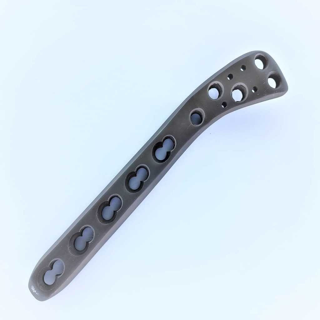
The "Golf Club Plate" gets its name from its characteristic shape: a broad, contoured head designed to conform to the anatomy of the lateral tibial plateau, transitioning into a narrower shaft that extends down the tibial diaphysis.
- Anatomical Contouring: The plate is pre-contoured to match the natural curvature of the lateral proximal tibia, minimizing the need for intraoperative bending and ensuring an optimal fit. This reduces surgical time and potential stress on the bone.
- Material Composition:
- Titanium Alloy (most common): Known for its excellent biocompatibility (meaning it's well-tolerated by the body), high strength-to-weight ratio, and resistance to corrosion. Titanium is also non-ferromagnetic, allowing for MRI compatibility in most cases.
- Stainless Steel: Another common material, offering good strength and cost-effectiveness. However, it may have limited MRI compatibility compared to titanium.
- Locking Mechanism: This is the defining feature of locking plates.
- Threaded Screw Holes: The plate features screw holes with internal threads that engage with corresponding threads on the head of the locking screws.
- Fixed-Angle Construct: When locking screws are fully inserted, they "lock" into the plate, creating a fixed-angle construct. This means the screws and plate act as a single, rigid unit, providing angular stability. This is paramount, especially in comminuted (fragmented) fractures or in bones with poor quality (osteoporosis).
- Screw Types:
- Locking Screws: Provide angular stability, preventing screw pull-out and maintaining reduction even in challenging bone.
- Non-Locking (Cortical/Lag) Screws: Can be used for initial fracture compression or to draw fragments together before locking screws are applied.
- Combination Holes: Many modern plates feature "combi-holes" that allow the surgeon to choose between inserting a locking screw or a non-locking screw for compression within the same hole, offering greater surgical flexibility.
- Low-Profile Design: The plate is designed to be as thin as possible while maintaining strength, reducing the risk of soft tissue irritation after implantation.
Biomechanics
The biomechanical advantages of the Golf Club Plate are significant, particularly in the context of complex tibial plateau fractures.
- Fixed-Angle Stability: Unlike conventional plates where screw purchase depends heavily on bone density, locking plates achieve stability by creating a fixed-angle construct. This distributes stress over a larger area, reducing the risk of screws loosening or pulling out, especially in osteoporotic bone.
- Load Sharing vs. Load Bearing: While the plate provides strong support, it's designed to promote "load sharing" with the healing bone, rather than solely "load bearing." This allows physiological stresses to stimulate bone healing (Wolff's Law) and reduces the risk of stress shielding, where the bone beneath the plate becomes weaker due to lack of stress.
- Resistance to Forces: The fixed-angle construct provides superior resistance to:
- Shear Forces: Forces that tend to slide bone fragments past each other.
- Bending Forces: Forces that try to bend the bone.
- Torsional Forces: Twisting forces.
- Minimally Invasive Potential: The robust stability offered by locking plates can sometimes facilitate minimally invasive surgical techniques, potentially leading to smaller incisions, less soft tissue disruption, and faster recovery.
3. Extensive Clinical Indications & Usage
The Proximal Tibia Lateral Locking Plate is indicated for a wide range of fractures affecting the proximal tibia, particularly those involving the articular surface.
Specific Clinical Indications
This plate is a preferred choice for:
- Tibial Plateau Fractures: These are fractures of the upper part of the shin bone that involves the knee joint. They are classified using systems like the Schatzker classification:
- Schatzker Type II (Lateral Split Depression): A split fracture of the lateral condyle with associated depression.
- Schatzker Type III (Lateral Depression): A pure depression fracture of the lateral condyle.
- Schatzker Type IV (Medial Condyle Fracture): While primarily a lateral plate, it can be used in conjunction with a medial plate for bicondylar fractures or for specific lateral components of type IV.
- Schatzker Type VI (Bicondylar with Diaphyseal Disruption): Complex fractures involving both condyles and extending into the diaphysis, often requiring dual plating.
- Metaphyseal Fractures: Fractures of the wider part of the bone near the joint, just below the tibial plateau.
- Fractures with Comminution: When the bone is broken into multiple fragments, the fixed-angle stability of the locking plate is invaluable.
- Osteoporotic Bone: In elderly patients with weakened bones, locking plates provide much better purchase and stability than traditional screws.
- Peri-prosthetic Fractures: Fractures occurring around an existing knee replacement implant.
- Non-union or Malunion Correction: In some cases, the plate may be used in revision surgeries to correct previously failed healing or malalignment.
Detailed Surgical Application (Simplified for Patients)
The surgical procedure involves careful planning and execution to ensure optimal outcomes.
- Pre-operative Planning:
- Imaging: X-rays, CT scans (often with 3D reconstructions), and sometimes MRI are used to thoroughly assess the fracture pattern, displacement, and soft tissue status.
- Templating: The surgeon uses digital or physical templates to plan the plate size, length, and screw trajectory.
- Patient Positioning: The patient is typically positioned supine (on their back) on an operating table, often with the knee slightly flexed to allow for optimal access.
- Incision and Exposure: An incision is made on the lateral side of the knee, carefully dissecting through layers of soft tissue to expose the fractured bone while preserving vital structures like nerves and blood vessels.
- Fracture Reduction: This is the most critical step, where the surgeon meticulously manipulates the bone fragments to restore the anatomical alignment of the tibia and the congruence of the articular surface. This may involve elevation of depressed fragments, use of bone grafts, and temporary fixation with K-wires or clamps.
- Plate Application: The Golf Club Plate is carefully positioned on the lateral aspect of the proximal tibia, ensuring it fits snugly against the bone and covers the fracture site appropriately.
- Screw Insertion:
- Initially, non-locking screws might be used to achieve compression across simple fracture lines or to temporarily hold the plate in place.
- Then, locking screws are strategically inserted through the threaded holes, engaging the bone fragments and locking into the plate. The number and trajectory of screws depend on the fracture pattern and bone quality.
- Fluoroscopy (real-time X-ray imaging) is used throughout the process to confirm proper plate placement, fracture reduction, and screw positioning.
- Wound Closure: Once stable fixation is achieved, the surgical site is irrigated, and the incision is closed layer by layer. A drain may be placed temporarily to prevent fluid accumulation.
Fitting/Usage Instructions (Post-operative Care for Patients)
Successful recovery heavily relies on adherence to post-operative instructions.
- Weight-Bearing Restrictions: This is crucial. Your surgeon will provide specific instructions, which may range from non-weight-bearing (no weight on the leg) for several weeks to touch-down weight-bearing (light foot contact) or partial weight-bearing, gradually progressing to full weight-bearing as healing progresses.
- Pain Management: Medications will be prescribed to manage post-operative pain. Follow the dosage instructions carefully.
- Wound Care: Keep the incision clean and dry. Follow instructions for dressing changes and watch for signs of infection (redness, swelling, warmth, pus).
- Physical Therapy (Rehabilitation): This is paramount for regaining knee motion, strength, and function. A structured physical therapy program will typically begin soon after surgery, focusing on:
- Early, controlled range of motion exercises (often with continuous passive motion (CPM) machines).
- Strengthening exercises for the quadriceps and hamstrings.
- Balance and proprioception training.
- Gait training (learning to walk again with proper form).
- Monitoring for Complications: Be vigilant for any unusual symptoms and report them to your doctor immediately.
4. Risks, Side Effects, or Contraindications
While the Proximal Tibia Lateral Locking Plate offers excellent outcomes, like any surgical procedure, it carries potential risks and contraindications.
Potential Risks and Side Effects
| Risk/Side Effect | Description |
|---|---|
| Infection | Can occur at the surgical site, ranging from superficial to deep, potentially requiring further surgery. |
| Non-union/Malunion | The fracture may fail to heal (non-union) or heal in an incorrect position (malunion). |
| Neurovascular Injury | Damage to nerves or blood vessels around the knee during surgery. |
| Hardware Irritation/Pain | The plate or screws may become prominent or cause discomfort, sometimes requiring removal after healing. |
| Loss of Reduction | The fracture fragments may shift after initial fixation, especially with premature weight-bearing. |
| Compartment Syndrome | A rare but serious condition where swelling within a muscle compartment compromises blood flow. |
| Deep Vein Thrombosis (DVT) | Blood clots forming in the deep veins, potentially leading to pulmonary embolism. |
| Arthrofibrosis/Stiffness | Scar tissue formation leading to restricted knee motion. |
| Post-traumatic Arthritis | Long-term risk, especially if the articular surface damage was severe or reduction was imperfect. |
| Hardware Failure | The plate or screws can break, although rare with proper technique and patient compliance. |
Contraindications
- Active Infection: Presence of an active infection at the surgical site is an absolute contraindication, as it significantly increases the risk of implant infection.
- Severe Soft Tissue Compromise: Poor skin condition, severe open wounds, or compromised blood supply around the fracture may necessitate delaying surgery or using alternative fixation methods.
- Patient Inability/Unwillingness to Comply: Patients who cannot or will not adhere to post-operative weight-bearing restrictions and physical therapy regimens may be at higher risk for complications.
- Certain Medical Comorbidities: Uncontrolled diabetes, severe peripheral vascular disease, or other medical conditions that significantly increase surgical risk may be contraindications.
- Insufficient Bone Stock: Extremely comminuted fractures with very small fragments that cannot hold screws effectively.
5. Expert Tips from Dr. Mohammed Hutaif
"As an orthopedic specialist, my primary focus when treating complex proximal tibia fractures with a Lateral Locking Plate is to ensure optimal functional recovery for my patients. Here are my key recommendations:
- Precision is Paramount: The most crucial step is achieving an accurate anatomical reduction of the fracture, especially restoring the joint surface. Even minor incongruities can lead to long-term issues like post-traumatic arthritis.
- Gentle Soft Tissue Handling: Minimizing soft tissue disruption during surgery is vital to preserve blood supply to the bone and prevent complications like infection and delayed healing.
- Strategic Screw Placement: It's not just about inserting screws; it's about placing them strategically to maximize stability and support the articular fragments effectively. Fluoroscopy is indispensable here.
- Personalized Rehabilitation: Every patient's recovery journey is unique. A tailored physical therapy program, initiated early and progressed cautiously, is essential for regaining strength, range of motion, and function. Adherence to weight-bearing protocols is non-negotiable.
- Patient Education and Compliance: I always spend considerable time educating my patients about their specific fracture, the surgical procedure, and the critical role they play in their own recovery. Understanding the 'why' behind each instruction significantly improves compliance and, ultimately, outcomes.
- Nutritional Support: Adequate nutrition, particularly protein and vitamin D, can play a supportive role in bone healing."
6. Massive FAQ Section
Q1: What is a Proximal Tibia Lateral Locking Plate, and why is it called a "Golf Club Plate"?
A1: It's a specialized orthopedic implant used to stabilize fractures of the upper part of the shin bone (proximal tibia), particularly those involving the knee joint. It's called a "Golf Club Plate" due to its distinctive shape, which resembles a golf club, with a broad head conforming to the lateral side of the tibia just below the knee, and a narrower shaft extending down the bone.
Q2: What types of fractures does the Golf Club Plate typically treat?
A2: It's primarily used for complex fractures of the proximal tibia, especially those involving the lateral tibial plateau (the weight-bearing surface of the knee joint). This includes various Schatzker classification types (e.g., Type II, III, IV, VI) and fractures with significant comminution or in osteoporotic bone.
Q3: How does a locking plate differ from a traditional plate?
A3: The key difference lies in the screw-plate interface. In a locking plate, screws thread directly into the plate, creating a fixed-angle construct that acts as a single, rigid unit. This provides angular stability independent of bone quality. Traditional plates rely on compression between the screw head and the plate, and friction between the plate and bone, which can be less stable in fragmented or osteoporotic bone.
Q4: Is the Proximal Tibia Lateral Locking Plate a permanent implant?
A4: In most cases, yes, the plate is intended to be a permanent implant. It can remain in the body indefinitely if it's not causing any problems. However, if it causes irritation, pain, or infection after the bone has fully healed (typically 1-2 years post-surgery), it can be surgically removed.
Q5: What are the main benefits of using this plate for my fracture?
A5: The main benefits include superior stability, especially in complex or osteoporotic fractures, reduced risk of screw pull-out, improved ability to restore anatomical alignment, and potential for earlier, controlled rehabilitation. This often leads to better functional outcomes and a lower risk of re-operation compared to older fixation methods.
Q6: How long is the recovery period after surgery with a Golf Club Plate?
A6: Recovery varies significantly based on the fracture's severity, individual healing capacity, and adherence to rehabilitation. Generally, initial bone healing takes 6-12 weeks, during which weight-bearing is restricted. Full recovery, including regaining strength and range of motion, can take 6 months to over a year. Your surgeon and physical therapist will guide your specific timeline.
Q7: Will I need physical therapy after the surgery?
A7: Absolutely. Physical therapy is a crucial component of recovery. It helps regain knee motion, strength, balance, and the ability to walk normally. Starting early with controlled exercises and progressing under the guidance of a therapist is vital for an optimal outcome.
Q8: Can I undergo an MRI scan with a titanium Proximal Tibia Lateral Locking Plate?
A8: Yes, medical-grade titanium alloy implants are generally considered MRI-safe. However, the implant may cause some artifact (distortion) on the MRI images, especially close to the plate itself. Always inform the MRI technician and your doctor about your implant before any scan. Stainless steel plates may have more restrictions.
Q9: What are the potential complications I should be aware of?
A9: Potential complications include infection, non-union (fracture not healing), malunion (healing in a poor position), nerve or blood vessel damage, hardware irritation, loss of reduction, blood clots (DVT), stiffness, and post-traumatic arthritis. Your surgeon will discuss these risks with you.
Q10: When can I return to sports or strenuous activities?
A10: Returning to sports or strenuous activities depends entirely on your healing progress, bone consolidation, and regaining full strength and stability. This typically takes a minimum of 6-12 months, and often longer for high-impact sports. Your surgeon will clear you for specific activities based on your recovery milestones and follow-up imaging.
Q11: How is the surgery performed?
A11: The surgery involves making an incision on the side of the knee, carefully exposing the fracture. The surgeon then meticulously realigns the bone fragments (reduction) and secures them in place with the Golf Club Plate and specialized screws. Real-time X-rays (fluoroscopy) are used to ensure precise placement.
Q12: What should I expect immediately after the surgery?
A12: Immediately after surgery, you will experience pain, which will be managed with medication. Your leg will likely be in a brace or splint, and you will have weight-bearing restrictions. You will begin gentle range-of-motion exercises as instructed by your physical therapist, often within days of the procedure.
This guide is for informational purposes only and does not constitute medical advice. Always consult with a qualified healthcare professional for diagnosis and treatment of any medical condition.