Understanding the Polyaxial Pedicle Screw System (5.5mm/6.0mm/7.5mm): A Comprehensive Patient Guide
1. Comprehensive Introduction & Overview
Welcome to this in-depth guide on the Polyaxial Pedicle Screw System, a cornerstone technology in modern spinal surgery. As an essential component in spinal fusion procedures, this advanced implant system plays a critical role in stabilizing the spine, correcting deformities, and facilitating the permanent joining (fusion) of vertebral segments. This guide is designed to provide patients with a clear, authoritative, and comprehensive understanding of this vital orthopedic instrument.
The human spine is a complex structure, and when compromised by injury, disease, or deformity, it can lead to significant pain and disability. Spinal fusion surgery aims to alleviate these issues by permanently connecting two or more vertebrae, eliminating motion between them. To achieve successful fusion, the spine needs robust internal support, and this is where pedicle screw systems come into play.
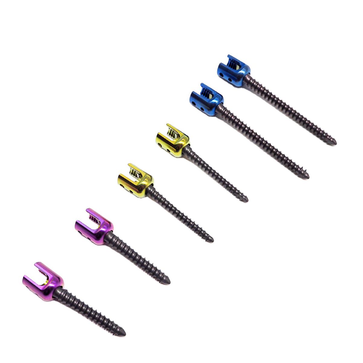
The Polyaxial Pedicle Screw System stands out due to its innovative design, particularly its "polyaxial" (multi-directional) head. This unique feature allows the screw head to articulate and angle in multiple directions relative to the screw body, offering surgeons unparalleled flexibility during rod placement. This flexibility is crucial for adapting to individual patient anatomy, especially in complex spinal deformities. Available in various diameters, such as 5.5mm, 6.0mm, and 7.5mm, these screws can be precisely matched to the specific pedicle size and bone quality of different vertebral segments, ranging from the thoracic (mid-back) to the lumbar (lower back) and sacral regions.
This guide will delve into the technical aspects of its design, its detailed clinical applications, the biomechanical principles behind its effectiveness, and how it contributes to improved patient outcomes. While this information is for educational purposes, it is not medical advice. Always consult with a qualified medical professional, such as Dr. Mohammed Hutaif, for personalized guidance regarding your specific condition.
2. Deep-dive into Technical Specifications / Mechanisms
The efficacy of the Polyaxial Pedicle Screw System lies in its sophisticated design and the advanced materials used in its construction. Understanding these elements helps appreciate why it's a preferred choice for spinal stabilization.
Design and Materials
The system typically comprises three main components: the pedicle screw, the polyaxial head (or "tulip"), and the locking cap (or "set screw"), all interconnected by a spinal rod.
- Pedicle Screw Body:
- Shape and Threading: The screw body is threaded, designed to anchor firmly into the vertebral pedicle (a strong bony projection from the back of the vertebra). The threads are optimized for bone purchase and resistance to pull-out.
- Diameters: Commonly available in diameters like 5.5mm, 6.0mm, and 7.5mm.
- 5.5mm/6.0mm: Often used in the thoracic and smaller lumbar pedicles, or in cases of osteopenic bone where a smaller diameter might be preferred to avoid pedicle fracture.
- 7.5mm: Typically reserved for larger lumbar and sacral pedicles, which can accommodate a larger screw for increased strength and stability, especially in high-stress areas or in patients with good bone quality.
- Lengths: Screws also come in various lengths (e.g., 30mm to 60mm), chosen based on the depth of the pedicle and vertebral body.
- Materials: Primarily constructed from medical-grade titanium or titanium alloys (e.g., Ti-6Al-4V). These materials are chosen for their:
- Biocompatibility: They are well-tolerated by the human body, minimizing adverse reactions.
- High Strength-to-Weight Ratio: Providing robust support without excessive bulk.
- Corrosion Resistance: Ensuring longevity within the body.
- MRI Compatibility: Generally safe for MRI scans, though some image artifacting may occur.
- Polyaxial Head (Receiver/Tulip):
- Articulation: This is the defining feature. The screw head is a spherical or semi-spherical component that allows the spinal rod to be inserted at various angles relative to the screw body. This angulation typically ranges from 30 to 60 degrees in all directions (conical angulation).
- Benefits of Polyaxiality:
- Easier Rod Placement: Significantly simplifies the connection of the spinal rod to multiple screws, especially in cases of spinal deformity where pedicles may not be perfectly aligned.
- Reduced Stress: Minimizes stress on the screws and vertebrae during rod placement, as the surgeon doesn't need to over-manipulate the bone to align the screw heads.
- Accommodates Anatomical Variation: Allows for precise contouring of the rod to the patient's natural or corrected spinal curvature.
- Locking Cap (Set Screw):
- Once the spinal rod is positioned within the polyaxial head, a locking cap is threaded down into the head. This cap applies pressure, "locking" the rod firmly in place and simultaneously fixing the polyaxial head's angulation, creating a rigid construct.
- Material: Usually titanium alloy.
- Spinal Rod:
- Connects multiple pedicle screws, forming the structural backbone of the fusion construct.
- Material: Typically medical-grade titanium or cobalt-chromium alloys, chosen for their strength and fatigue resistance.
Biomechanics
The biomechanical advantages of the Polyaxial Pedicle Screw System are fundamental to its success in spinal fusion:
- Load Sharing and Distribution: The system creates a rigid framework that effectively shares and distributes mechanical loads across the fused segments, reducing stress on individual vertebrae and implants.
- Rigid Fixation: Once the locking caps are tightened, the entire construct becomes rigid, preventing motion at the fusion site. This stability is paramount for promoting bone fusion.
- Deformity Correction: The polyaxial nature allows surgeons to apply powerful corrective forces (compression, distraction, derotation) to reduce spinal deformities like scoliosis or kyphosis, achieving optimal alignment before final locking.
- Enhanced Fusion Environment: By immobilizing the spinal segments, the system provides a stable biological environment for bone graft material to heal and integrate, leading to a solid bony fusion (arthrodesis).
- Reduced Risk of Pseudarthrosis: The superior stability offered by polyaxial screws helps reduce the incidence of pseudarthrosis (failed fusion), which is a common complication in spinal surgery.
- Adaptability: The ability to choose different screw diameters (5.5mm, 6.0mm, 7.5mm) allows for optimal purchase in varying bone qualities and pedicle sizes, maximizing biomechanical stability for each patient.
3. Extensive Clinical Indications & Usage
The Polyaxial Pedicle Screw System is a versatile tool used in a broad spectrum of spinal conditions requiring stabilization and fusion.
Clinical Indications for Use
Patients who may benefit from spinal fusion with a Polyaxial Pedicle Screw System include those suffering from:
- Spinal Deformities:
- Scoliosis: Lateral curvature of the spine, both adolescent idiopathic and adult degenerative. The polyaxial heads are particularly useful for navigating the complex rotational deformities.
- Kyphosis: Excessive forward curvature (e.g., Scheuermann's kyphosis, post-traumatic kyphosis, or iatrogenic kyphosis).
- Spondylolisthesis: Forward slippage of one vertebra over another, often causing nerve compression and instability.
- Spinal Instability:
- Degenerative Disc Disease: Severe wear and tear of spinal discs leading to instability, pain, and nerve impingement.
- Spinal Stenosis: Narrowing of the spinal canal, often exacerbated by instability.
- Failed Previous Fusion (Pseudarthrosis): Revision surgery to achieve a solid fusion.
- Spinal Trauma:
- Vertebral Fractures: Especially unstable burst fractures or fracture-dislocations that compromise spinal integrity.
- Spinal Cord Injury: Stabilization may be required to protect neurological function.
- Spinal Tumors:
- Resection of spinal tumors often leaves the spine unstable, requiring reconstruction and stabilization with pedicle screws.
- Spinal Infections:
- After surgical debridement of spinal infections (e.g., osteomyelitis, discitis), stabilization may be necessary to promote healing and prevent collapse.
The Surgical Procedure (Simplified for Patients)
The implantation of a Polyaxial Pedicle Screw System is a highly precise surgical procedure, typically performed under general anesthesia.
- Pre-operative Planning: Dr. Hutaif will conduct thorough imaging (X-rays, MRI, CT scans) to precisely map your spinal anatomy, determine the levels to be fused, and plan the optimal screw trajectories and sizes (e.g., choosing between 5.5mm, 6.0mm, or 7.5mm screws for different segments).
- Incision: A surgical incision is made, which can be traditional "open" or a minimally invasive approach, depending on the specific case and surgeon's technique.
- Exposure and Pedicle Preparation: The surgeon carefully exposes the posterior (back) aspect of the vertebrae. Using specialized instruments, a pilot hole is created in the pedicle, ensuring the correct trajectory and depth. Intraoperative imaging (fluoroscopy, O-arm navigation) is almost always used to ensure precise screw placement, minimizing risks to neural structures.
- Screw Insertion: The pedicle screws are carefully inserted into the prepared pedicles. Thanks to their polyaxial heads, the surgeon has the flexibility to insert the screw body at the optimal angle into the pedicle, knowing the head can be adjusted later to accommodate the rod.
- Rod Placement: Once all screws are in place, the spinal rods are contoured to the desired spinal alignment and carefully seated into the polyaxial heads of the screws. The polyaxial feature allows for easier rod engagement, even when screws are not perfectly parallel.
- Correction and Locking: The surgeon then applies corrective forces (compression, distraction, derotation) as needed to restore spinal alignment. Once the desired correction is achieved, the locking caps are securely tightened onto each polyaxial head, rigidly fixing the rod and locking the screw heads in their final position.
- Bone Grafting: Bone graft material (autograft from your own body, allograft from a donor, or synthetic graft) is packed around the transverse processes and lamina to encourage the vertebrae to fuse together permanently.
- Closure: The surgical site is meticulously closed layer by layer.
Post-operative Care & Recovery
Recovery involves a period of healing, pain management, and often physical therapy. Patients are typically encouraged to mobilize early, with specific restrictions on bending, lifting, and twisting. Adherence to Dr. Hutaif's post-operative instructions is crucial for a successful outcome.
4. Risks, Side Effects, or Contraindications
While the Polyaxial Pedicle Screw System is a safe and effective treatment for many spinal conditions, like any surgical procedure involving implants, it carries potential risks and has certain contraindications. It is vital for patients to be fully informed of these before proceeding with surgery.
Potential Risks & Complications
- General Surgical Risks:
- Infection: At the surgical site or systemic.
- Bleeding: Intraoperative or post-operative hemorrhage, potentially requiring transfusion.
- Anesthesia Risks: Adverse reactions to anesthesia.
- Nerve Damage: Injury to spinal nerves or the spinal cord, potentially leading to weakness, numbness, paralysis, or bladder/bowel dysfunction.
- Dural Tear: A tear in the membrane surrounding the spinal cord, which can lead to cerebrospinal fluid leakage and headache.
- Hardware-Specific Risks:
- Screw Loosening or Pull-out: The screws may become loose or dislodge from the bone, especially in patients with poor bone quality.
- Screw or Rod Breakage: Although implants are designed to be strong, excessive stress or non-union can lead to implant failure over time.
- Hardware Malposition: Incorrect placement of screws, potentially impinging on neural structures.
- Pain: Persistent pain at the surgical site or discomfort related to the hardware.
- Allergic Reaction: Rare, but possible reaction to the titanium or other implant materials.
- Fusion-Related Risks:
- Pseudarthrosis (Non-Union): Failure of the bones to fuse together, which may require revision surgery. This is a primary risk of any fusion surgery.
- Adjacent Segment Disease (ASD): Increased stress on the spinal segments immediately above or below the fused area, potentially leading to accelerated degeneration and the need for future surgery at those levels.
- Loss of Correction: The spine may not maintain the desired alignment, or the deformity may recur.
Contraindications
Certain conditions may make a patient unsuitable for spinal fusion with a Polyaxial Pedicle Screw System:
- Active Systemic or Local Infection: Surgery should be postponed until the infection is resolved.
- Severely Compromised Bone Quality (Severe Osteoporosis): If the bone is too weak to provide adequate purchase for the screws, the risk of loosening or pull-out is significantly increased, making the procedure ineffective or dangerous.
- Allergy or Hypersensitivity to Implant Materials: Although rare with titanium, this is a contraindication.
- Unwillingness or Inability to Comply with Post-operative Instructions: Crucial for proper healing and avoiding complications.
- Certain Medical Conditions: Severe comorbidities (e.g., uncontrolled diabetes, severe heart or lung disease) that significantly increase surgical risk.
- Insufficient Spinal Instability or Deformity: If the condition does not warrant surgical intervention or fusion.
Dr. Mohammed Hutaif will thoroughly evaluate your medical history, imaging, and overall health to determine if this procedure is appropriate for you, discussing all potential risks and benefits.
5. Expert Tips from Dr. Mohammed Hutaif
"As a specialist in spinal surgery, my primary goal is to restore function, alleviate pain, and improve the quality of life for my patients. The Polyaxial Pedicle Screw System is an invaluable tool in achieving these outcomes, but its successful application goes beyond just selecting the right implant.
Here are some insights I emphasize in my practice:
-
Precision is Paramount: The success of spinal fusion with pedicle screws hinges on meticulous surgical technique and precise placement. I extensively utilize advanced intraoperative imaging, such as 3D navigation and fluoroscopy, to ensure every screw is positioned optimally within the pedicle, minimizing risks and maximizing stability. The choice of screw diameter – whether 5.5mm, 6.0mm, or 7.5mm – is critically important and is determined by careful pre-operative planning based on individual pedicle morphology and bone density.
-
Personalized Approach: Every patient's spine is unique, and so is their pathology. I firmly believe in a personalized approach. The flexibility offered by the polyaxial heads allows me to adapt the construct to the specific anatomical nuances and deformity of each patient, ensuring the best possible correction and alignment. This customization is key to long-term success.
-
Comprehensive Patient Education: Understanding your condition and the proposed treatment is crucial. I dedicate time to explain the procedure, the role of the implants, potential risks, and expected recovery trajectory. Realistic expectations are vital for a positive surgical journey and successful rehabilitation.
-
Post-operative Rehabilitation is Non-Negotiable: Surgery is only one part of the recovery process. Adherence to a structured rehabilitation program, including physical therapy, is essential for regaining strength, flexibility (in unfused segments), and mobility. Your commitment to post-operative care significantly impacts your long-term outcome.
-
The Fusion is the Goal: Remember, the pedicle screw system is a temporary internal splint designed to hold the spine stable while the bone graft fuses. The ultimate success of the surgery is the solid bony fusion. We optimize all factors – surgical technique, bone graft selection, and post-operative care – to encourage this biological healing process.
Choosing to undergo spinal surgery is a significant decision. My team and I are committed to providing you with the highest standard of care, leveraging advanced technologies like the Polyaxial Pedicle Screw System to help you achieve the best possible results."
6. Massive FAQ Section
Q1: What makes polyaxial screws different from monoaxial screws?
A1: The key difference lies in the screw head. Monoaxial screws have a fixed head that does not articulate; it's rigidly aligned with the screw body. This requires the spinal rod to be perfectly aligned with all screw heads, which can be challenging, especially in complex deformities. Polyaxial screws, on the other hand, have a spherical head that can swivel or angle in multiple directions (typically 30-60 degrees) relative to the screw body. This flexibility makes it much easier for the surgeon to connect the spinal rod to all screws, accommodating anatomical variations and simplifying deformity correction. Once the rod is in place, a locking cap secures the head, making the construct rigid.
Q2: What are the main materials used in these screws?
A2: The Polyaxial Pedicle Screw System is primarily made from medical-grade titanium or titanium alloys (e.g., Ti-6Al-4V). These materials are chosen for their excellent biocompatibility (meaning they are well-tolerated by the human body), high strength-to-weight ratio, and resistance to corrosion, ensuring the longevity and safety of the implant within the body.
Q3: How long do these screws stay in the body?
A3: In most cases, polyaxial pedicle screws are intended for permanent implantation. They are designed to remain in the body indefinitely to provide long-term stability for the fused spinal segments. Removal is generally only considered if complications arise, such as infection, persistent pain directly attributable to the hardware, or hardware failure.
Q4: Can I have an MRI after having polyaxial pedicle screws implanted?
A4: Yes, medical-grade titanium implants are generally considered safe for MRI (Magnetic Resonance Imaging). Titanium is non-ferromagnetic, meaning it is not attracted to MRI magnets. However, you should always inform your imaging technician that you have spinal implants. While safe, the implants may cause some localized "artifact" (distortion) on the MRI images, which can sometimes obscure the view of the immediate surrounding tissues.
Q5: What are the typical recovery times after spinal fusion with these screws?
A5: Recovery times vary significantly depending on the extent of the surgery, individual patient health, and adherence to post-operative care.
* Initial Hospital Stay: Typically 3-7 days.
* Early Recovery (first 6-12 weeks): Focus on pain management, limited activity, and gentle movement. Bone graft begins to heal.
* Mid-term Recovery (3-6 months): Gradual increase in activity, often with physical therapy. Fusion process continues.
* Full Fusion & Long-term Recovery (6-12+ months): Bone fusion typically solidifies. Return to most normal activities, with ongoing physical therapy as needed.
Full recovery and achieving a solid fusion can take up to a year or even longer.
Q6: Is the surgery painful?
A6: Spinal fusion is a major surgery, and some level of pain is expected post-operatively. However, your medical team, including Dr. Hutaif, will employ a multi-modal pain management strategy (combining different types of pain medications, nerve blocks, and other techniques) to keep your pain at a manageable level. Pain typically decreases significantly over the first few weeks and months of recovery.
Q7: What are the chances of the screws breaking or loosening?
A7: While designed for strength, screw breakage or loosening is a potential complication, though relatively uncommon with modern implants and surgical techniques. The risk is higher in patients with poor bone quality (osteoporosis), those who do not achieve a solid fusion (pseudarthrosis), or those who do not adhere to post-operative activity restrictions. Regular follow-up imaging helps monitor the hardware integrity.
Q8: Can these screws be used in children?
A8: Yes, Polyaxial Pedicle Screw Systems are frequently used in children and adolescents, particularly for the correction of severe scoliosis and kyphosis. The flexibility of the polyaxial head is especially beneficial in pediatric deformity correction, where complex spinal curves need to be addressed. The choice of screw size (e.g., 5.5mm, 6.0mm) is carefully selected based on the child's age, bone maturity, and pedicle dimensions.
Q9: Will I be able to bend and twist my back normally after fusion?
A9: Spinal fusion permanently joins the vertebrae at the surgical levels, eliminating motion in those specific segments. This means you will lose some flexibility in the fused portion of your spine. The extent of this loss depends on how many segments are fused and where they are located. For example, a fusion of two lumbar vertebrae might result in noticeable stiffness, while a thoracic fusion might have less impact on overall perceived flexibility. Your body will adapt, and adjacent unfused segments will often compensate, but the fused area itself will not bend or twist.
Q10: How do the different screw sizes (5.5mm, 6.0mm, 7.5mm) make a difference?
A10: The different screw diameters are crucial for matching the implant to the patient's anatomy and bone quality:
* 5.5mm/6.0mm: Smaller diameters are used when pedicles are naturally smaller (e.g., in the thoracic spine, smaller lumbar pedicles, or in pediatric cases) or when bone density is lower (osteopenia), to reduce the risk of pedicle fracture during insertion.
* 7.5mm: Larger diameters provide greater purchase and pull-out strength, making them ideal for larger pedicles (common in the lumbar and sacral spine) and in patients with good bone quality, where maximum stability is desired.
The surgeon meticulously selects the appropriate size based on pre-operative imaging and intraoperative assessment to optimize stability and minimize complications.
Q11: What is the role of bone graft with these screws?
A11: The polyaxial pedicle screws and rods provide immediate stability, acting as an internal "splint." However, the ultimate goal of the surgery is for the vertebrae to permanently fuse together with new bone growth. The bone graft material (which can be taken from your own body, a donor, or a synthetic substitute) is packed around the fusion site. This graft acts as a scaffold and stimulates new bone formation, leading to a solid, permanent bony bridge between the vertebrae. The screws hold the spine stable while this fusion process occurs over several months.
Q12: Can the screws be removed later?
A12: While designed for permanent implantation, the screws can be removed in a subsequent surgery if necessary. Reasons for removal might include persistent pain directly attributed to the hardware, infection, or if the hardware breaks. However, hardware removal is typically not routine and is only performed if there is a clear medical indication, as it involves another surgical procedure with its own set of risks. If a solid fusion has been achieved, the spine will remain stable even after hardware removal.
Disclaimer: This information is for general educational purposes only and is not intended as medical advice. It does not replace the professional judgment of a qualified healthcare provider. Always consult with Dr. Mohammed Hutaif or another licensed medical professional for diagnosis and treatment of any medical condition.