Poller (Blocking) Screws: Enhancing Fracture Stability and Healing
Comprehensive Introduction & Overview
In the intricate world of orthopedic surgery, achieving optimal stability and alignment for fractured bones is paramount for successful healing and patient recovery. While various implants like intramedullary nails have revolutionized fracture care, certain complex fracture patterns can still pose significant challenges. This is where Poller screws, also known as blocking screws, emerge as a sophisticated adjunctive technique, designed to enhance the precision and stability of internal fixation.
Poller screws are small, strategically placed screws that work in conjunction with larger primary implants, most commonly intramedullary (IM) nails. An intramedullary nail is inserted into the hollow canal of a long bone (like the femur, tibia, or humerus) to stabilize a fracture. However, in certain comminuted (fragmented) or metaphyseal (near joint) fractures, the IM nail might not perfectly center or prevent all rotational or translational movement of the bone fragments. Poller screws act as internal "bumpers" or "guides," creating a tighter, more controlled pathway for the IM nail, thereby optimizing its position and preventing unwanted motion.
The concept behind Poller screws is rooted in biomechanical principles, aiming to convert an unstable fracture fixation into a more stable construct. By meticulously guiding the primary implant and restricting its movement within the bone canal, these screws help maintain precise anatomical alignment, reduce the risk of malunion (healing in an incorrect position) or nonunion (failure to heal), and ultimately foster a more conducive environment for bone regeneration and faster functional recovery. This guide will delve deep into the design, application, biomechanics, and patient benefits of this ingenious orthopedic tool.
Deep-dive into Technical Specifications / Mechanisms
Design and Materials
Poller screws are a testament to precision engineering in orthopedics. While seemingly simple, their design and material selection are critical to their function:
- Type: They are typically small, fully threaded cortical screws. Cortical screws are designed to engage dense cortical bone effectively, providing strong purchase.
- Material: The vast majority of Poller screws are manufactured from biocompatible materials such as:
- Medical-grade Stainless Steel (e.g., 316L): Known for its strength, corrosion resistance, and proven track record in orthopedic implants.
- Titanium Alloys (e.g., Ti-6Al-4V): Offer superior biocompatibility, excellent strength-to-weight ratio, and are often preferred for patients with potential metal sensitivities or for their improved imaging compatibility (less artifact on MRI/CT).
- Dimensions:
- Diameter: Ranging typically from 2.5 mm to 4.5 mm, chosen based on the size of the bone and the intramedullary nail.
- Length: Varies widely, from 20 mm to 60 mm or more, to ensure adequate purchase in the cortical bone without protruding excessively.
- Head Type: Often features a low-profile head design to minimize irritation to surrounding soft tissues once implanted.
Mechanism of Action (Biomechanics)
The biomechanical principle behind Poller screws is often referred to as the "blocking" or "constraining" effect. When an intramedullary nail is inserted into a fractured long bone, especially in cases of wide medullary canals or comminuted fractures, there can be excessive space or freedom for the nail to move within the bone fragments. This can lead to:
* Translation: Sideways movement of the nail.
* Rotation: Twisting of the nail.
* Angulation: Bending of the fracture fragments.
Poller screws counteract these issues by:
- Creating a Confined Space: They are strategically placed parallel and immediately adjacent to the intramedullary nail, effectively narrowing the medullary canal around the fracture site. This creates a "constrained" pathway, forcing the nail into a more centralized and stable position.
- Preventing Malalignment: By blocking unwanted movement of the nail, they ensure that the bone fragments remain in proper anatomical alignment, crucial for accurate healing.
- Augmenting Stability: They transform a potentially unstable nail-bone construct into a more rigid and controlled system. This increased stability is vital for early weight-bearing and rehabilitation.
- Improving Load Sharing: The enhanced stability allows for better load distribution across the fracture site, promoting callus formation and bone healing.
- Guiding Nail Insertion: In some techniques, Poller screws are placed before the nail insertion to help guide the nail precisely through a difficult fracture pattern or narrow canal segment.
- "Relative Stability" Optimization: While intramedullary nailing typically provides "relative stability" (allowing some micro-motion beneficial for secondary bone healing), Poller screws help to optimize this relative stability, preventing excessive motion that could lead to nonunion while still allowing enough controlled motion to stimulate healing.
Extensive Clinical Indications & Usage
Poller screws are not used in every intramedullary nailing procedure but are reserved for specific scenarios where their unique biomechanical advantages are most beneficial.
Primary Clinical Indications
- Metaphyseal Fractures: Fractures occurring at the ends of long bones (e.g., proximal tibia, distal femur) where the medullary canal widens, making it difficult for an IM nail to achieve stable fixation. Poller screws help centralize the nail in these wider segments.
- Comminuted Diaphyseal Fractures: Fractures in the shaft of long bones with multiple fragments. Here, the nail might "wobble" or not provide adequate stability due to the lack of solid bone contact. Poller screws block this movement.
- Segmental Fractures: Fractures with a completely detached segment of bone. Poller screws help maintain the alignment of this segment relative to the main bone.
- Fractures with Wide Medullary Canals: Patients with osteopenia or naturally wide canals may benefit from Poller screws to ensure a snug fit for the nail.
- Prevention of Malalignment: Especially in rotational or angular deformities that can occur during nail insertion or early post-operative period.
- Revision Surgeries: In cases of failed previous fixation or nonunion, Poller screws can provide additional stability to a new IM nail.
Detailed Surgical Application (Fitting/Usage Instructions - Simplified for Patients)
The application of Poller screws requires meticulous surgical technique and advanced imaging guidance.
- Pre-operative Planning:
- Thorough review of X-rays, CT scans, and sometimes 3D reconstructions to understand the fracture pattern, bone anatomy, and medullary canal dimensions.
- Templating (using transparent overlays on X-rays or digital templating) to determine appropriate nail size, length, and potential Poller screw placement.
- Patient Positioning and Access:
- The patient is positioned appropriately for the specific long bone fracture (e.g., supine for tibia, lateral for femur).
- Standard surgical approach for IM nailing is made, involving a small incision to access the bone canal.
- Intramedullary Nail Insertion:
- The main IM nail is typically inserted first, spanning the fracture site.
- Initial reduction of the fracture is performed.
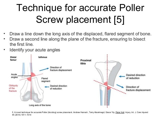
- Poller Screw Placement (Guided by Fluoroscopy):
- Fluoroscopy (real-time X-ray imaging) is crucial. It allows the surgeon to visualize the nail's position within the bone and precisely guide the placement of the Poller screws.
- Entry Points: Small stab incisions (percutaneous) are made in the skin over the intended screw sites.
- Drilling: A small drill bit is used to create pilot holes through the cortical bone, adjacent to the IM nail. The trajectory and depth are carefully controlled to avoid damaging the nail or vital structures.
- Screw Insertion: The Poller screws are then carefully inserted into these pilot holes until they engage the opposite cortex, effectively "blocking" the IM nail.
- Configuration: Poller screws are often placed in pairs (e.g., anterior-posterior or medial-lateral) on one or both sides of the fracture to provide multi-planar stability. The exact number and location depend on the fracture pattern and the surgeon's assessment.
- Post-operative Verification:
- Final fluoroscopic images or post-operative X-rays are taken to confirm optimal fracture reduction, nail position, and Poller screw placement.
Patient Outcome Improvements
The strategic use of Poller screws contributes significantly to superior patient outcomes:
- Enhanced Fracture Stability: Provides a more rigid construct, minimizing unwanted motion at the fracture site.
- Reduced Risk of Malunion/Nonunion: By maintaining precise alignment and stability, Poller screws create an optimal biological environment for bone healing, significantly lowering the incidence of healing complications.
- Improved Biomechanical Environment: Controlled stability promotes better load transfer and early callus formation.
- Potentially Faster Weight-Bearing and Rehabilitation: With increased stability, patients may be able to commence partial or full weight-bearing sooner, accelerating their rehabilitation process and return to daily activities.
- Better Functional Outcomes: Accurate alignment and robust healing lead to improved long-term function of the limb.
- Reduced Need for Revision Surgery: By optimizing the primary fixation, the likelihood of needing subsequent surgeries due to fixation failure is diminished.
Risks, Side Effects, or Contraindications
While Poller screws offer substantial benefits, like any surgical intervention, there are potential risks and considerations. It's important for patients to have a clear understanding of these.
General Surgical Risks (Applicable to any orthopedic surgery)
- Infection: Risk of bacterial contamination at the surgical site.
- Bleeding: Intra-operative or post-operative hemorrhage.
- Nerve or Vessel Damage: Injury to surrounding nerves or blood vessels during screw placement.
- Anesthesia Risks: Adverse reactions to anesthetic agents.
- Scarring: Formation of surgical scars.
Specific Risks Related to Poller Screws
- Screw Breakage: While rare due to the strength of materials, a Poller screw could potentially fracture under extreme stress.
- Soft Tissue Irritation/Pain: If a screw head is prominent, it might irritate overlying soft tissues, leading to localized pain or discomfort.
- Implant Migration: Extremely rare with proper placement, but theoretically a screw could loosen or migrate.
- Over-constraining the Nail: If too many Poller screws are used or placed too tightly, they could potentially impede the natural dynamization (controlled settling) of the fracture, which is sometimes desired for healing.
- Increased Surgical Time: The placement of Poller screws adds a small amount of time to the overall surgical procedure.
- Potential Need for Removal: Similar to other orthopedic implants, if a Poller screw causes symptoms (e.g., pain, irritation), it might need to be removed in a subsequent procedure, typically after the fracture has healed.
Contraindications
- Active Infection: Surgery should not proceed in the presence of an active infection near the fracture site.
- Severe Osteoporosis: In cases of extremely poor bone quality, the screws may not gain adequate purchase, rendering them ineffective or prone to loosening.
- Allergy to Implant Materials: Although rare, patients with known allergies to stainless steel or titanium alloys would require alternative materials or fixation methods.
- Fracture Patterns Where Impeded Reduction: In very specific fracture patterns, the presence of Poller screws might actually hinder the ability to achieve proper fracture reduction.
- Insufficient Medullary Canal Space: If the medullary canal is already very narrow, there might not be enough space to safely place Poller screws alongside the IM nail.
Expert Tips from Dr. Mohammed Hutaif
As an orthopedic specialist, I emphasize that the successful application of Poller (blocking) screws is a blend of art and science, requiring meticulous planning, precise execution, and a deep understanding of fracture biomechanics. Here are my key recommendations:
- Meticulous Pre-operative Planning is Non-Negotiable: "The battle is won before it's fought." Thoroughly review all imaging studies (X-rays, CT scans with 3D reconstructions if available). Understand the fracture morphology, the exact location of fragments, the medullary canal dimensions, and the planned trajectory of the intramedullary nail. Templating is essential to anticipate nail size and optimal Poller screw positions.
- Fluoroscopic Guidance is Paramount: Real-time imaging is not just helpful; it's indispensable. Every step, from pilot hole drilling to screw insertion, must be guided by fluoroscopy to ensure precise placement, avoid neurovascular structures, and prevent damage to the primary nail.
- Strategic Placement is Key – "Less is More, but Right Place is Everything": It's not about how many screws you use, but where you place them. The goal is to create just enough constraint to centralize the nail and block unwanted motion, without over-constraining the system or impeding dynamization if it's desired. Often, two screws placed in opposing planes (e.g., anterior and posterior) at each end of the fracture segment are sufficient.
- Understand the Biomechanics of the Specific Fracture: Each fracture pattern presents unique challenges. For metaphyseal fractures, the focus is often on preventing angulation and translation. For comminuted diaphyseal fractures, it’s about preventing rotation and maintaining length. Your Poller screw placement should directly address these specific biomechanical instabilities.
- Balance Stability with Biological Healing: While Poller screws enhance stability, remember that controlled micromotion (relative stability) is often beneficial for secondary bone healing. The goal is to optimize this environment, not create absolute rigidity that might hinder callus formation.
- Patient Education is Crucial: Clearly explain to the patient why Poller screws are being used, their benefits, and potential risks. Managing expectations regarding recovery, potential for hardware removal, and post-operative pain is vital for patient satisfaction and adherence to rehabilitation protocols.
- Consider Screw Orientation: Screws should be placed as close to the nail as possible without touching it, and their orientation should be chosen to maximize their blocking effect against the anticipated direction of nail migration.
By adhering to these principles, Poller screws can significantly elevate the quality of fracture fixation, leading to superior healing outcomes and a faster, more complete recovery for our patients.
Massive FAQ Section
Q1: What exactly is a Poller screw?
A1: A Poller screw, also known as a blocking screw, is a small, fully threaded screw used in orthopedic surgery alongside a larger primary implant, typically an intramedullary (IM) nail. Its purpose is to act as a "bumper" or "guide" within the bone canal, helping to precisely position the IM nail and prevent unwanted movement of the bone fragments, thereby enhancing the overall stability of the fracture fixation.
Q2: Why are they called "blocking" screws?
A2: They are called "blocking" screws because their primary function is to "block" or "prevent" the intramedullary nail from moving excessively within the bone canal. By creating a tighter, more constrained pathway for the nail, they effectively block translational (sideways) and rotational movements that could lead to malalignment of the fracture.
Q3: Are Poller screws used in all fracture surgeries?
A3: No, Poller screws are not used in every fracture surgery. They are specifically indicated for more complex fracture patterns where the primary implant (like an IM nail) might not achieve sufficient stability or optimal alignment on its own. This includes fractures in wide medullary canals, highly comminuted fractures, or metaphyseal fractures near joints. Their use is a surgical decision based on the specific fracture characteristics.
Q4: What materials are Poller screws made from?
A4: Poller screws are made from high-quality, biocompatible medical-grade materials. The most common materials are stainless steel (e.g., 316L) and titanium alloys (e.g., Ti-6Al-4V). Both materials are chosen for their excellent strength, corrosion resistance, and proven safety within the human body.
Q5: Do Poller screws need to be removed?
A5: In most cases, Poller screws, like other internal fixation hardware, are designed to be left in permanently unless they cause symptoms. If a screw becomes prominent and irritates soft tissues, causes pain, or if there's an infection, then removal might be recommended after the fracture has fully healed. Your surgeon will discuss this with you.
Q6: How do Poller screws improve healing?
A6: Poller screws improve healing by enhancing the stability and precision of the fracture fixation. By preventing excessive movement and maintaining anatomical alignment, they create an optimal biomechanical environment for bone cells to bridge the fracture gap. This controlled stability promotes better blood supply and callus formation, which are essential for robust bone healing.
Q7: Is the surgery more complicated with Poller screws?
A7: The addition of Poller screws does make the surgical procedure slightly more involved, as it requires precise placement under fluoroscopic (real-time X-ray) guidance. However, for an experienced orthopedic surgeon, this is a standard technique. The increased complexity is justified by the significant improvement in fracture stability and potential for better patient outcomes.
Q8: What are the risks associated with Poller screws?
A8: Beyond general surgical risks like infection or bleeding, specific risks for Poller screws include potential for soft tissue irritation if a screw head is prominent, very rare screw breakage, or the theoretical risk of over-constraining the fracture. Your surgeon will discuss all potential risks and benefits with you prior to surgery.
Q9: Can I feel the Poller screws under my skin?
A9: It is possible, but not always. Poller screws are generally small and placed deep within the bone. However, depending on your body habitus and the specific location of the screw, you might be able to feel a slight bump or prominence under the skin. If this causes discomfort, discuss it with your surgeon.
Q10: How long does it take to recover after surgery involving Poller screws?
A10: The recovery time is primarily dictated by the nature and severity of the fracture itself, not solely by the presence of Poller screws. However, because Poller screws enhance stability, they can sometimes allow for earlier weight-bearing and accelerated rehabilitation. Recovery typically involves several months of gradual progression, guided by your surgeon and physical therapist.
Q11: Will Poller screws set off metal detectors?
A11: Yes, it is highly likely that Poller screws, being made of metal (stainless steel or titanium), will trigger metal detectors at airports or other security checkpoints. It's advisable to carry a doctor's note or medical implant card if provided, though typically a brief explanation is sufficient.
Q12: Is there an alternative to using Poller screws?
A12: The decision to use Poller screws is made when conventional intramedullary nailing alone is deemed insufficient for stable fixation. Alternatives might include different types of nails (e.g., larger diameter, different locking mechanisms), plate and screw fixation, or external fixation, depending on the fracture. However, for certain complex IM nailing cases, Poller screws are often considered the superior adjunctive technique for optimizing outcomes.