The PLIF Interbody Cage (Posterior Lumbar): An Expert Guide to Spinal Fusion
1. Comprehensive Introduction & Overview
The Posterior Lumbar Interbody Fusion (PLIF) procedure represents a cornerstone in modern spinal surgery for addressing a variety of degenerative and unstable conditions of the lumbar spine. At its heart lies the PLIF Interbody Cage, an innovative orthopedic implant designed to restore disc height, stabilize the spinal segment, and create an optimal environment for bony fusion. This comprehensive guide delves deep into the specifics of the PLIF interbody cage, exploring its design, applications, biomechanics, and impact on patient outcomes.
Spinal fusion surgery aims to permanently join two or more vertebrae, eliminating motion between them. The interbody fusion technique, where a cage is placed directly into the disc space, has gained immense popularity due to its ability to provide immediate structural support and facilitate a robust fusion. The PLIF approach, characterized by accessing the disc space from a posterior incision, allows for direct decompression of neural structures and direct visualization of the disc space for cage insertion.
The evolution of spinal implants, particularly interbody cages, has been driven by advancements in material science, biomechanical understanding, and surgical techniques. Modern PLIF cages are meticulously engineered to maximize fusion rates, minimize complications, and improve patient recovery.
2. Deep-dive into Technical Specifications & Mechanisms
The efficacy of a PLIF interbody cage is intrinsically linked to its sophisticated design, material composition, and the biomechanical principles it leverages within the spinal column.
2.1. Design Principles and Materials
PLIF interbody cages come in a variety of designs, each optimized for specific surgical needs and patient anatomies.
Key Design Features:
- Shape and Profile:
- Banana or Kidney-shaped: Curved to conform to the natural anterior convex curvature of the vertebral endplates, often inserted obliquely.
- Parallel: Straight-sided cages designed for direct impaction, often used in pairs.
- Lordotic: Tapered designs that help restore or maintain the natural lordotic curvature of the lumbar spine, crucial for sagittal balance.
- Fenestrations: Openings or windows within the cage structure are critical. These allow for packing with bone graft material (autograft, allograft, or synthetic bone substitutes) which then grows through and around the cage, bridging the vertebral bodies to achieve solid fusion.
- Surface Features:
- Teeth or Ridges: Micro- or macro-features on the superior and inferior surfaces of the cage provide immediate primary stability and resist expulsion or migration post-implantation.
- Porous Surfaces: Advanced designs incorporate porous structures or coatings to encourage bone ingrowth and ongrowth, enhancing secondary stability and fusion.
- Sizing: Cages are available in a wide range of heights, widths, and lengths to accommodate varying disc space dimensions and restore optimal anatomical parameters. Trial implants are used intraoperatively to determine the correct size.
Common Materials Used:
| Material Type | Characteristics | Advantages | Disadvantages |
|---|---|---|---|
| PEEK | Polyetheretherketone. Radiotranslucent polymer. Modulus of elasticity similar to bone. | Excellent biocompatibility, radiolucent (allows post-op fusion assessment), good load-sharing. | Hydrophobic surface may limit bone ingrowth, lack of osteoinductivity. |
| Titanium (Ti) | Biocompatible metal. Strong, durable. | High strength, good osteoconductivity, visible on X-ray. | Higher stiffness than bone (stress shielding risk), significant artifact on MRI/CT. |
| Hybrid Designs | PEEK cages with titanium endplates or porous titanium coatings. | Combines benefits: bone-friendly interface of Ti with load-sharing of PEEK, reduced imaging artifact. | More complex manufacturing, potentially higher cost. |
| Porous Titanium | 3D-printed titanium with interconnected pores mimicking trabecular bone structure. | Enhanced bone ingrowth, improved osteointegration, reduced stress shielding. | Manufacturing complexity, potentially higher cost. |
| HA Coatings | Hydroxyapatite (HA) coating on PEEK or titanium surfaces. | Promotes osteoconductivity, encourages bone apposition. | Coating delamination is a rare concern. |
| Tantalum | Highly biocompatible, radiopaque, porous structure. | Excellent bone ingrowth, good imaging characteristics, high strength. | Higher cost, less common. |
2.2. Biomechanical Function
The PLIF interbody cage plays a critical biomechanical role in achieving successful spinal fusion and restoring spinal function.
- Disc Height Restoration: Degenerative disc disease often leads to disc space collapse. The cage restores physiological disc height, decompressing nerve roots in the neuroforamina and indirectly decompressing the central canal.
- Lordosis Maintenance/Restoration: Lumbar lordosis is essential for spinal balance. Lordotic cages or strategic placement help restore the natural curvature, improving spinal alignment and reducing stress on adjacent segments.
- Load Sharing: By bearing a significant portion of the axial compressive load, the cage reduces stress on the posterior instrumentation (pedicle screws and rods), minimizing the risk of hardware failure and promoting fusion through Wolff's Law (bone remodels in response to mechanical stress).
- Immediate Stability: The cage, with its surface features, provides immediate segmental stability, preventing translation and rotation, which is crucial for reducing pain and protecting neural structures post-surgery.
- Facilitating Fusion: The fenestrations of the cage contain bone graft material, creating a contained environment for bone growth across the intervertebral space. This osteoconductive scaffold, combined with osteoinductive signals from the graft, promotes robust arthrodesis.
- Minimizing Subsidence: Appropriate cage sizing and endplate preparation are vital to prevent cage subsidence (sinking into the vertebral body), which can lead to loss of disc height and potential non-union. Modern cages are designed to distribute load evenly across the endplates.
3. Extensive Clinical Indications & Usage
The PLIF interbody cage is indicated for a range of conditions causing lumbar spinal instability, pain, and neurological deficits that have not responded to conservative management.
3.1. Primary Clinical Indications
- Degenerative Disc Disease (DDD): Chronic low back pain originating from a degenerated intervertebral disc, often accompanied by disc height loss and instability.
- Spondylolisthesis:
- Degenerative Spondylolisthesis: Forward slippage of one vertebra over another due to degenerative changes in the facet joints and disc.
- Isthmic Spondylolisthesis: Spondylolisthesis caused by a defect (lysis) in the pars interarticularis.
- Spinal Stenosis with Instability: Narrowing of the spinal canal or neuroforamina, often requiring decompression, where concomitant instability necessitates fusion.
- Post-Laminectomy Instability: When extensive decompression (laminectomy) leads to iatrogenic instability, requiring stabilization.
- Recurrent Disc Herniation with Instability: When a disc herniation recurs at the same level, especially if there are signs of segmental instability.
- Pseudarthrosis (Failed Fusion): In cases where a previous fusion attempt has failed, a revision PLIF may be performed.
3.2. Surgical Procedure: Posterior Lumbar Interbody Fusion (PLIF)
The PLIF procedure is a complex spinal surgery requiring meticulous technique.
General Steps:
- Patient Positioning & Access: The patient is positioned prone on a specialized surgical frame to maintain lumbar lordosis and minimize abdominal pressure. A midline incision is made in the lower back.
- Exposure & Decompression: The paraspinal muscles are retracted to expose the posterior elements of the lumbar spine (lamina, facet joints). A laminectomy, partial facetectomy, and/or foraminotomy are performed to decompress the neural elements (spinal cord and nerve roots).
- Discectomy & Endplate Preparation: The degenerated disc material is completely removed from the intervertebral space. The cartilaginous endplates are meticulously prepared using curettes and rasps to expose the vascular bone, which is crucial for optimal bone graft incorporation and fusion.
- Trial Sizing: Various trial implants are inserted into the disc space to determine the appropriate cage height, width, and lordotic angle that restores disc height and lordosis without overdistraction.
- Bone Graft Packing: The PLIF cage's fenestrations are packed with bone graft material. This can be autograft (patient's own bone, often harvested from the iliac crest or local decompression), allograft (donor bone), or synthetic bone substitutes (e.g., calcium phosphate, BMPs). Additional bone graft is often placed directly into the prepared disc space anterior to the cage.
- Cage Insertion: One or two PLIF cages are carefully impacted into the prepared disc space, typically from a posterior-lateral approach, ensuring proper alignment and engagement with the vertebral endplates.
- Posterior Instrumentation: To provide immediate rigid stabilization, pedicle screws are inserted into the vertebral bodies above and below the fused segment, and rods are connected to the screws. This construct helps compress the interbody cage and resists movement during the fusion process.
- Wound Closure: The surgical site is irrigated, and the incision is closed layer by layer.
3.3. Fitting & Usage Instructions (General Principles)
- Pre-operative Planning: Detailed imaging (X-rays, MRI, CT scans) is critical to assess spinal alignment, disc height, and degree of degeneration. Surgical templates or 3D planning may be used to select appropriate cage sizes and plan trajectories.
- Intraoperative Sizing: The use of trial cages is paramount. Surgeons carefully assess the fit, stability, and restoration of disc height and lordosis. Over-distraction or under-distraction can lead to complications.
- Endplate Preparation: This is a critical step for successful fusion. Removal of cartilage and exposure of bleeding bone ensures direct contact between the bone graft and host bone, promoting osteointegration.
- Bone Graft Selection & Packing: The choice of bone graft and thorough packing of the cage and disc space are essential for promoting fusion.
- Cage Impaction & Placement: Cages must be fully seated within the annulus and aligned correctly to maximize endplate contact and minimize migration risk. Intraoperative fluoroscopy or navigation systems are often used for precise placement.
4. Maintenance & Sterilization Protocols
The PLIF interbody cage itself is a sterile implant and requires no maintenance by the end-user. However, the instruments used for its implantation require rigorous sterilization protocols.
4.1. Implant Sterilization
- Manufacturer Pre-Sterilization: PLIF interbody cages are supplied in sterile packaging by the manufacturer. Common sterilization methods include:
- Gamma Irradiation: Uses gamma rays to kill microorganisms.
- Ethylene Oxide (ETO) Sterilization: A chemical gas sterilization method, often used for heat-sensitive materials.
- Aseptic Handling: In the operating room, implants must be handled using strict aseptic techniques to prevent contamination. The integrity of the sterile packaging must be checked before opening.
4.2. Instrument Sterilization and Maintenance
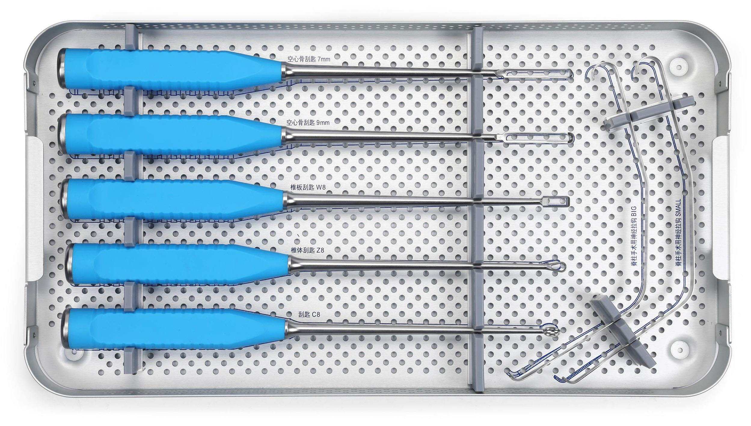
The specialized instruments used to implant PLIF cages (e.g., distractors, curettes, rasps, cage holders, impactors, trial sizers) are reusable and require meticulous care.
- Cleaning:
- Point-of-Use Cleaning: Immediately after surgery, gross soil (blood, tissue) should be removed.
- Manual Cleaning: Instruments are manually scrubbed with enzymatic detergents to remove all organic matter.
- Ultrasonic Cleaning: Instruments are placed in an ultrasonic cleaner to remove microscopic debris from hard-to-reach areas.
- Disinfection: High-level disinfection may precede sterilization for certain instruments, though sterilization is the ultimate goal for surgical implants.
- Sterilization Methods:
- Steam Autoclave (Saturated Steam Sterilization): The most common and effective method for heat-resistant instruments. Parameters typically include specific temperature, pressure, and exposure time.
- Low-Temperature Sterilization (e.g., STERRAD, Steris System): Used for heat-sensitive instruments that cannot withstand steam autoclaving. These systems use hydrogen peroxide plasma or peracetic acid.
- Inspection: After cleaning and before sterilization, instruments are thoroughly inspected for damage, corrosion, or wear. Damaged instruments must be removed from service.
- Maintenance: Lubrication of hinged instruments and ensuring all components are functional are part of routine maintenance.
- Storage: Sterilized instruments must be stored in a clean, dry, and protected environment to maintain sterility until use.
5. Risks, Side Effects, or Contraindications
While PLIF surgery with interbody cages is highly effective, like any surgical procedure, it carries potential risks and contraindications.
5.1. General Surgical Risks
- Infection: Superficial or deep surgical site infection.
- Bleeding: Intraoperative or postoperative hemorrhage.
- Nerve Damage: Injury to spinal nerves or the spinal cord, potentially leading to weakness, numbness, or paralysis.
- Dural Tear: A tear in the dura mater (the membrane surrounding the spinal cord), which can lead to cerebrospinal fluid (CSF) leakage.
- Anesthesia Risks: Allergic reactions, respiratory or cardiac complications.
- Deep Vein Thrombosis (DVT) / Pulmonary Embolism (PE): Blood clots forming in the legs that can travel to the lungs.
5.2. Cage-Specific Risks & Side Effects
- Non-union (Pseudoarthrosis): Failure of the bones to fuse together, leading to persistent pain and instability. This is the most common long-term complication.
- Cage Migration/Expulsion: The cage can shift from its intended position, potentially causing nerve compression or failure of fusion.
- Cage Subsidence: The cage sinking into the vertebral endplates, leading to loss of disc height and potential nerve impingement.
- Hardware Failure: Breakage of screws, rods, or the cage itself, though rare with modern implants.
- Adjacent Segment Disease (ASD): Increased stress on the spinal segments above or below the fused level, potentially leading to accelerated degeneration and the need for further surgery.
- Neurological Deficit Post-op: Can occur due to direct trauma during cage insertion, nerve root irritation, or hematoma formation.
- Chronic Pain: Despite successful fusion, some patients may experience persistent or new pain.
5.3. Contraindications
- Active Systemic or Local Infection: Fusion surgery should be delayed until the infection is resolved.
- Severe Osteoporosis: Compromised bone quality significantly increases the risk of non-union, screw loosening, and cage subsidence.
- Uncontrolled Systemic Disease: Conditions like uncontrolled diabetes, severe cardiovascular disease, or active autoimmune disorders can increase surgical risks and impair healing.
- Allergy to Implant Materials: Although rare, patients with known allergies to PEEK or titanium should not receive implants made from those materials.
- Poor Bone Quality: Any condition that severely compromises the ability of bone to fuse.
- Certain Psychiatric Conditions: Patients with severe psychiatric disorders or unrealistic expectations may not be suitable candidates.
- Morbid Obesity: While not an absolute contraindication, it can increase surgical complexity and complication rates.
- Pregnancy: Elective spinal surgery is generally contraindicated during pregnancy.
6. Patient Outcome Improvements
The primary goal of PLIF with interbody cage implantation is to significantly improve patient quality of life by alleviating pain, restoring function, and achieving long-term spinal stability.
- Pain Relief: The most significant improvement for many patients is the reduction or elimination of chronic axial back pain and radicular (leg) pain caused by nerve compression or instability.
- Neurological Symptom Improvement: Decompression of nerve roots and stabilization of the spinal segment can lead to resolution of neurological deficits such as numbness, tingling, weakness, or foot drop.
- Restoration of Spinal Alignment: The cage helps restore physiological disc height and lumbar lordosis, which is crucial for overall spinal balance, posture, and reducing stress on other spinal segments.
- Improved Functional Mobility and Quality of Life: Patients typically experience increased ability to perform daily activities, return to work, and engage in recreational pursuits, leading to a substantial improvement in their overall quality of life.
- High Fusion Rates: With modern surgical techniques, appropriate bone graft materials, and advanced cage designs, PLIF procedures generally achieve high rates of solid bony fusion, ensuring long-term stability.
- Long-Term Stability: A successful fusion provides a permanent solution to segmental instability, preventing further degeneration at the treated level.
7. Frequently Asked Questions (FAQ)
Q1: What is a PLIF Interbody Cage?
A PLIF (Posterior Lumbar Interbody Fusion) Interbody Cage is a small, hollow device made of materials like PEEK or titanium, designed to be surgically inserted into the disc space between two vertebrae in the lower back. Its purpose is to restore disc height, stabilize the spine, and create a space for bone graft to grow through, ultimately fusing the vertebrae together.
Q2: How is a PLIF cage different from a TLIF cage?
Both PLIF and TLIF (Transforaminal Lumbar Interbody Fusion) cages are used for interbody fusion. The primary difference lies in the surgical approach. In PLIF, the disc space is accessed from a posterior approach, often requiring retraction of nerve roots, and two cages are typically inserted. In TLIF, the approach is posterolateral, usually through a single facet joint, allowing for a single, larger, banana-shaped cage to be inserted obliquely, minimizing nerve root retraction.
Q3: What materials are PLIF cages made from?
Common materials include PEEK (Polyetheretherketone), which is a radiolucent polymer with bone-like elasticity, and Titanium, known for its strength and osteoconductivity. Many modern cages are hybrid designs, combining PEEK with titanium coatings or endplates, or are entirely made from porous titanium to enhance bone ingrowth.
Q4: How long does it take for fusion to occur after a PLIF?
Bony fusion is a biological process that typically takes 3 to 12 months to achieve a solid state. While initial stability is provided by the cage and posterior instrumentation (screws and rods), true fusion requires bone growth across the disc space. Regular follow-up X-rays or CT scans are used to monitor the fusion progress.
Q5: What are the benefits of using a PLIF cage?
Benefits include restoration of disc height (decompressing nerve roots), maintenance or restoration of lumbar lordosis, immediate spinal stability, creation of a favorable environment for bone graft to fuse, and load-sharing to promote bone growth and prevent hardware failure. These ultimately lead to pain relief and improved function.
Q6: Can the PLIF cage be removed?
Generally, PLIF cages are designed to be permanent implants and are not typically removed unless there's a significant complication like migration, infection, or persistent pain directly attributed to the cage itself. Removing a fused cage is a complex procedure.
Q7: What are the potential complications of PLIF surgery with a cage?
Potential complications include non-union (failure to fuse), cage migration or subsidence (sinking into the bone), infection, bleeding, nerve damage, dural tear (CSF leak), hardware failure, and adjacent segment disease (degeneration at levels next to the fusion).
Q8: How long is the recovery after PLIF surgery?
Initial recovery involves a hospital stay of a few days. Patients typically begin walking shortly after surgery. Full recovery and return to normal activities can take several months to a year, depending on the individual's health, commitment to physical therapy, and the extent of the fusion. Light activities are gradually introduced, with strenuous activities restricted for several months.
Q9: Will I set off metal detectors with a PLIF cage?
If your PLIF cage contains titanium or other metallic components (like pedicle screws and rods, which are always used with PLIF), it is possible to set off sensitive metal detectors, such as those at airports. It is advisable to carry a doctor's note or an implant card if available.
Q10: Is PLIF surgery right for everyone with back pain?
No. PLIF surgery is typically reserved for patients with specific degenerative or unstable spinal conditions that have not responded to extensive conservative treatments (e.g., physical therapy, medications, injections). A thorough evaluation by a spine specialist is crucial to determine if PLIF is the appropriate treatment option.
Q11: How do I know if my fusion was successful?
Successful fusion is often indicated by significant reduction in pain and improvement in function. Radiographically, success is determined by the presence of a solid bridge of bone across the fused segment, visible on X-rays or CT scans, and the absence of motion between the vertebrae. This assessment is usually made by your surgeon during follow-up appointments.
Q12: What is the typical lifespan of a PLIF interbody cage?
The PLIF interbody cage itself is designed to be a permanent implant, as its primary role is to facilitate a solid bony fusion between vertebrae. Once fusion is achieved, the biological fusion construct (the new bone) effectively takes over the load-bearing function, and the cage becomes integrated within this fused segment. The material of the cage (PEEK or titanium) is highly durable and biocompatible, intended to last for the patient's lifetime within the fused spine.