The Orthopedic Fracture Table: Precision and Stability in Modern Fracture Surgery
Welcome to this comprehensive guide on the Orthopedic Fracture Table, a cornerstone instrument in advanced orthopedic trauma surgery. At Dr. Mohammed Hutaif's practice, we believe in empowering our patients with knowledge about the tools and techniques that ensure the best possible outcomes for their care. This information is for patient education only and does not constitute medical advice. Always consult with a qualified healthcare professional for any medical concerns.
1. Comprehensive Introduction & Overview
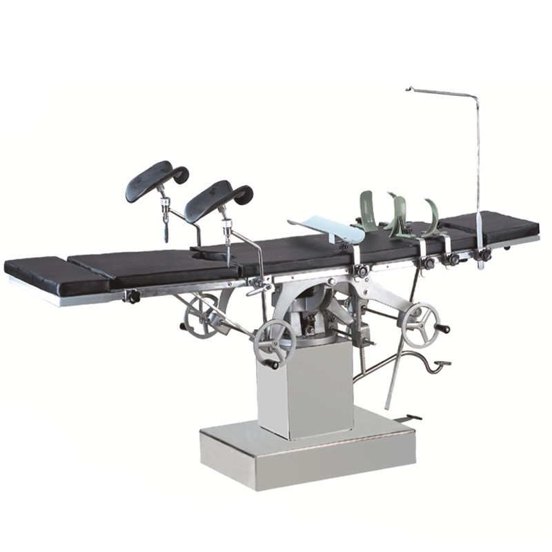
The orthopedic fracture table is a highly specialized operating room table designed specifically to aid surgeons in the accurate reduction and fixation of complex fractures, particularly those of the lower limbs and pelvis. Unlike standard operating tables, a fracture table incorporates unique features such as traction systems, radiolucent (X-ray transparent) components, and modular attachments that allow for precise patient positioning and manipulation during surgery.
Its primary purpose is to provide stable support, controlled traction, and unrestricted access for fluoroscopy (real-time X-ray imaging) throughout the surgical procedure. This combination of capabilities is crucial for achieving optimal anatomical alignment of fractured bones, minimizing surgical invasiveness, and ultimately improving patient outcomes. The evolution of the fracture table has revolutionized orthopedic trauma care, enabling surgeons to tackle highly challenging cases with greater precision and safety.
2. Deep-Dive into Technical Specifications & Mechanisms
Modern orthopedic fracture tables are marvels of engineering, designed to meet the rigorous demands of complex orthopedic surgery.
Design and Materials
- Modular Construction: Fracture tables are inherently modular, allowing for various configurations to suit different fracture types and surgical approaches. Key components include:
- Main Frame: Provides the core stability, often made of high-strength stainless steel or aluminum alloys.
- Radiolucent Top: The patient support surface is typically made from advanced carbon fiber composites. This material is crucial because it allows X-rays to pass through unimpeded, providing clear fluoroscopic images without needing to reposition the patient.
- Perineal Post: A padded support that fits between the patient's legs, providing counter-traction against the applied limb traction. It is often adjustable and designed to minimize pressure points.
- Leg Spars/Outriggers: Adjustable extensions that attach to the main frame, allowing for the attachment of traction boots or ankle clamps. These are also often radiolucent.
- Traction Boots/Ankle Clamps: Specialized padded devices that secure the patient's foot/ankle, allowing for controlled longitudinal traction.
- Arm Boards/Supports: For positioning the upper extremities, often articulating and padded.
- Hydraulic and Electric Systems: Most modern tables utilize advanced hydraulic or electric systems for smooth, precise adjustments of height, Trendelenburg/reverse Trendelenburg, lateral tilt, and often, rotation. These movements are typically controlled via a remote control, allowing the surgical team to make adjustments without contaminating the sterile field.
Detailed Surgical Mechanisms
- Traction System: This is the defining feature of a fracture table. It applies a controlled, sustained pulling force along the axis of the fractured limb.
- Purpose: Traction helps to reduce (realign) the fractured bone fragments by overcoming muscle spasm and restoring the bone's original length and alignment. It also creates a "ligamentotaxis" effect, where surrounding ligaments and soft tissues help guide the fragments back into place, particularly useful in periarticular fractures.
- Mechanism: The traction force is applied via the traction boots/ankle clamps, with the perineal post providing the opposing counter-traction. The amount of traction can be finely adjusted using a crank or electronic controls, often with integrated force indicators to ensure safety and precision.
- Fluoroscopy Access: The radiolucent table top and modular design ensure that the C-arm (a mobile X-ray unit) can be positioned around the patient from virtually any angle. This provides real-time, intraoperative imaging, allowing the surgeon to visualize the fracture reduction and implant placement without interrupting the procedure.
- Biomechanics in Fracture Reduction: The table’s design leverages biomechanical principles to achieve optimal fracture reduction.
- Distraction: Traction not only realigns but can also distract (pull apart) joint surfaces, allowing for better visualization and manipulation of articular fragments.
- Rotation and Angulation Control: The modular leg spars and traction devices often allow for precise control over the rotation and angulation of the limb, which is critical for restoring anatomical alignment, especially in long bone fractures.
- Stability: The rigid frame and secure patient positioning minimize inadvertent movement, providing a stable platform for delicate surgical maneuvers.
Maintenance and Sterilization Protocols
Maintaining the integrity and sterility of a fracture table is paramount for patient safety.
* Cleaning: After each use, the table surfaces are thoroughly cleaned with hospital-grade disinfectants. Non-porous materials facilitate this process.
* Sterilization: Components that come into direct contact with the sterile field (e.g., traction boots, perineal post pads) are often covered with single-use sterile drapes. Some specific components might be designed for autoclaving or chemical sterilization, though sterile draping is the most common method for the main patient contact areas.
* Preventive Maintenance: Regular inspections by biomedical engineers ensure all mechanical, hydraulic, and electrical systems are functioning correctly. This includes checking for wear and tear, lubricating moving parts, and calibrating traction systems.
3. Extensive Clinical Indications & Usage
The orthopedic fracture table is indispensable for a wide range of complex orthopedic trauma surgeries.
Common Clinical Indications
- Femoral Shaft Fractures: Ideal for closed intramedullary nailing, as it allows for precise reduction and maintenance of length and rotation during implant insertion.
- Proximal Femur Fractures: Including intertrochanteric and subtrochanteric fractures, where controlled traction aids in reduction for nailing or plating.
- Tibial Plateau Fractures: Traction can help distract the joint, allowing for better visualization and reduction of articular fragments, often combined with external fixation or ORIF.
- Pilon Fractures (Distal Tibia): Similar to tibial plateau fractures, traction assists in restoring length and reducing articular fragments.
- Acetabular Fractures: Crucial for providing stable positioning and allowing surgeons to approach the pelvis from various angles, facilitating complex internal fixation.
- Pelvic Ring Fractures: Used to stabilize the pelvis, often in conjunction with external fixators or internal fixation for anterior and posterior pelvic injuries.
- Distal Radius Fractures: While less common for the full fracture table, some systems can provide traction for complex distal radius fractures, especially for arthroscopic-assisted reduction.
Fitting/Usage Instructions (General Principles for the Surgical Team)
- Pre-operative Planning: The surgical team, including the surgeon, anesthesiologist, and nurses, meticulously plans the patient's positioning based on the fracture type, surgical approach, and patient comorbidities.
- Patient Transfer: The patient is carefully transferred from the stretcher to the fracture table, ensuring spinal precautions if necessary. Proper padding is applied to all pressure points to prevent skin breakdown.
- Perineal Post Placement: The perineal post is positioned securely and comfortably to provide effective counter-traction, avoiding excessive pressure on neurovascular structures.
- Limb Positioning and Traction Application:
- The fractured limb is carefully placed into the traction boot or ankle clamp.
- Traction is applied gradually and controlled, often with the guidance of fluoroscopy, to achieve anatomical reduction.
- The amount of traction is carefully monitored to ensure adequate reduction without over-distraction or causing neurovascular compromise.
- The uninjured leg is typically placed in a position that provides stability and access, often flexed at the hip and knee, or abducted.
- C-arm Integration: The table's radiolucency and design allow for optimal positioning of the C-arm, ensuring clear, real-time imaging throughout the procedure. The surgical team works collaboratively to manipulate the limb and C-arm to obtain necessary views.
- Sterile Draping: The entire surgical field and relevant parts of the table are meticulously draped to maintain sterility.
- Continuous Monitoring: Throughout the surgery, the patient's vital signs, nerve function, and limb perfusion are continuously monitored, especially when traction is applied.
4. Risks, Side Effects, or Contraindications
While the fracture table significantly enhances surgical precision, its use is associated with potential risks and contraindications that the surgical team carefully manages.
Potential Risks and Side Effects
- Nerve Palsy: Excessive or prolonged traction can stretch or compress nerves, particularly the peroneal nerve (causing foot drop), femoral nerve, or sciatic nerve. Careful padding, monitoring traction force, and limiting operative time are crucial preventative measures.
- Skin Breakdown/Pressure Sores: Prolonged pressure from the perineal post, traction boots, or other support areas can lead to skin irritation or pressure ulcers, especially in elderly or debilitated patients. Meticulous padding and regular inspection are vital.
- Vascular Injury: While rare, improper positioning or excessive traction could theoretically compromise blood flow to the limb.
- Compartment Syndrome: Although not directly caused by the table, excessive traction in certain situations could exacerbate or contribute to the development of compartment syndrome if not managed appropriately.
- Table Malfunction: Mechanical or electrical failure of the table is rare but can occur, necessitating immediate corrective action or transfer to a standard table.
- Infection: As with any surgical procedure, there's a risk of infection, especially if skin integrity is compromised by pressure points.
Contraindications (Relative)
- Patient Instability: Severely unstable patients (e.g., hemodynamically unstable trauma patients) may not tolerate the time required for positioning on a fracture table.
- Extreme Patient Size/Weight: Patients exceeding the table's weight limit or those whose body habitus prevents safe and effective positioning may be better managed on a standard table with alternative traction methods.
- Specific Fracture Patterns: In some highly comminuted articular fractures where ligamentotaxis is not beneficial or where immediate open reduction without traction is preferred, a standard table might be chosen.
- Pre-existing Neurological Deficits: Patients with pre-existing nerve damage may be at higher risk for further injury, requiring extra precautions.
5. Expert Tips from Dr. Mohammed Hutaif
"The orthopedic fracture table is an indispensable tool in my practice, allowing for unparalleled precision in treating complex fractures. However, its effective use goes far beyond simply turning it on. Here are some key principles I always emphasize:
- Prioritize Pre-operative Planning: Every case is unique. A thorough pre-operative plan, including reviewing imaging, anticipating challenges, and briefing the entire surgical team, is crucial. This ensures we select the optimal table configuration and approach.
- Gentle and Controlled Traction: Traction is powerful. It must be applied gradually and precisely, always guided by fluoroscopy and clinical assessment. Over-traction is as detrimental as under-traction.
- Vigilant Neurovascular Monitoring: The safety of the patient's nerves and blood vessels is paramount. Regular checks of sensation, motor function, and peripheral pulses are non-negotiable throughout the procedure.
- Meticulous Padding: Preventing pressure injuries is a team effort. Ensure all potential pressure points, especially the perineum, heels, and malleoli, are adequately padded and inspected.
- Mastering C-arm Integration: The true power of the fracture table lies in its synergy with fluoroscopy. Efficient C-arm positioning and interpretation of real-time images are critical for accurate reduction and implant placement.
- Team Communication: A well-coordinated surgical team is essential. Clear communication regarding table adjustments, traction changes, and patient status ensures a smooth, safe, and efficient operation."
6. Massive FAQ Section
Q1: What is an orthopedic fracture table used for?
A1: An orthopedic fracture table is a specialized operating table used to position patients and apply controlled traction during surgery for complex bone fractures, particularly in the lower limbs and pelvis. It helps surgeons accurately realign fractured bones and maintain stability while implants are inserted.
Q2: How does a fracture table help during surgery?
A2: It provides several key benefits:
* Precise Reduction: Applies controlled traction to realign bone fragments.
* Stability: Holds the limb securely in the desired position.
* Fluoroscopy Access: Its radiolucent design allows for clear, real-time X-ray imaging (C-arm) throughout the surgery, guiding the surgeon.
* Minimally Invasive Surgery: Facilitates closed reduction techniques, reducing the need for large incisions.
Q3: Are all fracture tables the same?
A3: No, while they share core functionalities, fracture tables vary in design, features, and modularity. Some are designed for specific types of fractures, while others are more versatile. Modern tables often have advanced hydraulic/electric controls and carbon fiber components.
Q4: What types of fractures are typically treated on a fracture table?
A4: Common indications include fractures of the femur (thigh bone), tibia (shin bone), pelvis, and acetabulum (hip socket). These are often complex fractures requiring precise length, rotation, and alignment restoration.
Q5: Is the fracture table safe for the patient?
A5: Yes, when used by experienced surgical teams, fracture tables are very safe. The risks, such as nerve injury or skin breakdown, are carefully mitigated through meticulous patient positioning, padding, continuous monitoring, and controlled traction.
Q6: How long does a patient stay on a fracture table during surgery?
A6: The duration varies depending on the complexity of the fracture and the specific surgical procedure. It can range from one to several hours. The surgical team continuously monitors the patient throughout this time.
Q7: What materials are fracture tables made from?
A7: The main frame is typically made from high-strength metals like stainless steel or aluminum alloys for durability. The patient support surface and key attachments are often made from advanced radiolucent materials like carbon fiber to allow X-ray penetration.
Q8: Can a fracture table be used for children?
A8: Yes, fracture tables can be adapted for pediatric patients, often with specialized smaller attachments and careful adjustment of traction forces appropriate for a child's size and bone structure.
Q9: What is "traction" and why is it used?
A9: Traction is the application of a pulling force to a limb. In fracture surgery, it's used to:
* Overcome muscle spasms that pull bone fragments out of alignment.
* Restore the natural length of the bone.
* Realign the fractured fragments, often guided by surrounding soft tissues (ligamentotaxis).
Q10: How is the fracture table cleaned and sterilized?
A10: After each use, all surfaces are thoroughly cleaned with hospital-grade disinfectants. Components directly contacting the sterile field are typically covered with single-use sterile drapes. The table also undergoes regular preventive maintenance and inspection to ensure proper function.
Q11: Does using a fracture table improve surgical outcomes?
A11: Absolutely. By enabling precise fracture reduction, stable positioning, and real-time imaging, fracture tables significantly contribute to:
* More accurate anatomical alignment.
* Reduced surgical time and blood loss.
* Facilitation of minimally invasive techniques.
* Lower complication rates.
* Ultimately, faster recovery and better long-term function for the patient.
Q12: What are the advantages of a modern fracture table compared to older methods?
A12: Modern fracture tables offer superior advantages:
* Enhanced Precision: Fine-tuned traction and positioning controls.
* Improved Imaging: Full radiolucency for comprehensive fluoroscopic views without patient repositioning.
* Versatility: Modular design adapts to various fracture types and patient anatomies.
* Ergonomics: Better access for the surgical team, reducing fatigue.
* Safety: Integrated safety features and robust construction.
This guide aims to provide a comprehensive understanding of the orthopedic fracture table and its critical role in modern fracture care. At Dr. Mohammed Hutaif's practice, we leverage such advanced instruments to ensure our patients receive the highest standard of orthopedic treatment.