The Operating Microscope: A Revolution in Orthopedic and Hand Surgery
1. Comprehensive Introduction & Overview
In the intricate world of orthopedic and hand surgery, precision is not just a preference; it is an absolute necessity. The human hand, a marvel of biological engineering, comprises a complex network of tiny bones, delicate nerves, minuscule blood vessels, and fine tendons. Repairing or reconstructing these structures demands unparalleled visual acuity and steady hands. This is where the operating microscope steps onto the surgical stage, transforming possibilities and redefining patient outcomes.
An operating microscope is a specialized optical instrument designed to provide surgeons with a magnified, three-dimensional view of the surgical field, coupled with intense, coaxial illumination. Far beyond what the naked eye or even standard surgical loupes can offer, these advanced microscopes enable surgeons to visualize structures as small as a fraction of a millimeter, facilitating intricate procedures that would otherwise be impossible.
For patients undergoing hand or specific orthopedic procedures, the use of an operating microscope signifies a commitment to the highest standard of care. It allows for:
* Enhanced Precision: Repairing nerves and blood vessels with sutures finer than a human hair.
* Minimized Trauma: Making smaller, more accurate incisions, reducing damage to surrounding healthy tissue.
* Improved Outcomes: Leading to better functional recovery, reduced scarring, and faster rehabilitation.
This comprehensive guide will delve into the fascinating world of the operating microscope, exploring its sophisticated design, diverse clinical applications, meticulous maintenance, and profound impact on patient care in the specialized fields of orthopedic and hand surgery.
2. Deep-dive into Technical Specifications / Mechanisms
The operating microscope is a marvel of optical and mechanical engineering, meticulously designed for the demanding environment of the operating room.
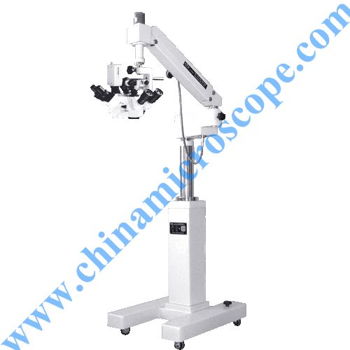
2.1. Design and Materials
Modern operating microscopes are engineered for stability, flexibility, and optical clarity.
- Optical System:
- Lenses: High-quality apochromatic lenses are standard, correcting for chromatic and spherical aberrations to produce a sharp, color-accurate image across the entire field of view.
- Magnification Changer: Surgeons can dynamically adjust magnification, typically from 2x to 40x, using either a step-magnification changer (discrete increments) or a zoom system (continuous adjustment), often controlled by a foot pedal.
- Eyepieces: Ergonomically designed, adjustable eyepieces accommodate different interpupillary distances and diopter settings for individual surgeon comfort.
- Working Distance: The distance between the objective lens and the surgical field is crucial, allowing ample space for surgical instruments. This is often adjustable via different objective lenses.
- Illumination System:
- Light Source: High-intensity LED or Xenon light sources provide bright, white light. LED offers longevity and less heat, while Xenon provides a very natural color rendition.
- Coaxial Illumination: The light path travels along the same axis as the surgeon's line of sight, eliminating shadows and providing uniform illumination deep within cavities.
- Fiber Optics: Light is often transmitted via fiber optic cables to the microscope head, minimizing heat transfer to the surgical site.
- Filters: Integrated filters (e.g., UV, green for vascular visualization, yellow for retinal protection) can be quickly engaged.
- Mounting System:
- Stability: Essential for vibration-free imaging. Common mounts include:
- Floor Stands: Mobile, heavy bases providing excellent stability.
- Ceiling Mounts: Fixed to the ceiling, offering maximum floor space and precise positioning.
- Table Mounts: Less common for major orthopedic surgery, but useful for specific benchtop procedures.
- Articulation: Articulated arms with electromagnetic brakes allow for effortless, precise positioning and locking of the microscope head in any orientation.
- Stability: Essential for vibration-free imaging. Common mounts include:
- Integrated Features:
- Assistant Scopes: Allow a second surgeon or assistant to view the same magnified field.
- Video Cameras: High-definition cameras integrate for recording procedures, teaching, and real-time display on external monitors for the surgical team.
- Image Capture: Capability to capture still images.
- Foot Pedal Controls: Allow hands-free adjustment of magnification, focus, and sometimes XY-positioning.
- Materials: Constructed from high-grade, durable materials such as aluminum alloys, titanium, and medical-grade plastics, ensuring longevity and ease of sterilization. Outer casings are often smooth for easy cleaning and draping.
2.2. Mechanisms of Operation
The core functionality revolves around providing a clear, magnified, and illuminated 3D view.
- Magnification: Achieved through a series of lenses that optically enlarge the object. The surgeon selects the appropriate magnification based on the tissue being worked on – lower power for orientation, higher power for intricate repairs.
- Focusing: Motorized focusing mechanisms, controlled by a foot pedal, allow for precise adjustment of the focal plane, ensuring continuous sharp images even as the surgeon's hands move.
- Stereopsis (3D Vision): The binocular design of the microscope presents a slightly different image to each eye, which the brain interprets as a single, three-dimensional image, crucial for depth perception during delicate maneuvers.
- Illumination: Coaxial light ensures that the deepest parts of the surgical field are brightly and uniformly lit, minimizing shadows that could obscure critical structures.
2.3. Biomechanics and Ergonomics
The design of the operating microscope significantly impacts surgeon comfort and performance, which in turn benefits the patient.
- Surgeon Ergonomics: Adjustable eyepieces, tiltable objective lenses, and flexible mounting arms allow the surgeon to maintain a comfortable, neutral posture, reducing strain on the neck, back, and shoulders during long, complex microsurgical procedures.
- Reduced Fatigue: By minimizing physical discomfort, the microscope helps surgeons maintain peak concentration and precision for extended periods, directly contributing to better surgical outcomes.
- Enhanced Control: Foot pedal controls free the surgeon's hands to manipulate instruments, providing uninterrupted focus on the surgical task.
3. Extensive Clinical Indications & Usage
The operating microscope is indispensable in a wide array of orthopedic and hand surgical procedures, particularly those involving microsurgery.
3.1. Hand Surgery Indications
The hand's delicate anatomy makes it a primary beneficiary of microscope-assisted surgery.
- Nerve Repair (Neurorrhaphy):
- Median, Ulnar, Radial Nerve Lacerations: Precise re-approximation of nerve fascicles (bundles of nerve fibers) and epineurium (outer nerve sheath) using sutures finer than human hair (e.g., 9-0 to 11-0 nylon). This significantly improves the chances of functional recovery and reduces neuroma formation.
- Brachial Plexus Injuries: Complex reconstruction of nerve roots and trunks in the shoulder and arm.
- Nerve Grafting: Harvesting and coaptation of nerve grafts to bridge gaps in damaged nerves.
- Vascular Repair (Microvascular Anastomosis):
- Replantation of Severed Digits/Limbs: Reconnecting arteries and veins (typically 0.5-3mm in diameter) to restore blood flow to a completely detached part, often within a critical time window.
- Free Flap Transfers: Moving tissue (skin, fat, muscle, bone) from one part of the body to another, requiring meticulous connection of its blood supply (artery and vein) to recipient vessels at the new site. Common in reconstructive surgery after trauma or tumor removal.
- Bypass Procedures: Creating new blood flow pathways using small vessels.
- Tendon Repair and Reconstruction:
- Complex Tendon Lacerations: Especially in zones where multiple tendons are in close proximity, requiring precise repair to prevent adhesions and ensure smooth gliding.
- Tenolysis: Releasing adhesions around tendons that restrict movement, often after previous injury or surgery.
- Small Joint Reconstruction and Arthroplasty:
- Digital Joint Arthroplasty/Fusion: Meticulous work on the small joints of the fingers and thumb, requiring precise bone cuts and implant placement.
- Tumor Excision:
- Benign and Malignant Tumors: Precise removal of very small tumors or cysts in the hand, minimizing damage to surrounding vital structures like nerves and vessels, preserving function.
- Congenital Hand Anomalies:
- Syndactyly (Webbed Fingers), Polydactyly (Extra Digits): Delicate reconstruction of digits, often involving separation of fused structures, nerve, and vessel preservation.
3.2. General Orthopedic Indications (where precision is paramount)
While primarily associated with hand surgery, the microscope also plays a role in other orthopedic subspecialties for highly delicate procedures.
- Peripheral Nerve Surgery: Beyond the hand, for nerve entrapments (e.g., severe carpal tunnel release, cubital tunnel release) or nerve tumor excisions in other parts of the limbs.
- Minimally Invasive Spine Surgery: In some specialized cases, for intricate nerve root decompression or microdiscectomy, although endoscopes are often preferred here.
- Pediatric Orthopedics: For delicate corrections in the small bones and joints of children.
3.3. Usage Instructions (Patient's Perspective: What to Expect)
From a patient's viewpoint, understanding the microscope's role means appreciating the meticulous nature of the surgery.
- Pre-operative Setup: Once under anesthesia, the surgical team will carefully position the patient and the affected limb. The microscope will be draped with sterile covers and positioned precisely over the surgical site.
- Surgeon's Focused Work: The surgeon will spend significant time looking through the microscope, performing extremely fine manipulations with specialized micro-instruments. This concentrated focus is critical for the success of the procedure.
- Procedure Duration: Microsurgical procedures typically take longer than conventional surgeries due to the extreme precision required. Patients should be prepared for potentially extended operating times.
- Team Involvement: The entire surgical team, including assistants and nurses, is trained to support the surgeon during microscope-assisted procedures, ensuring a sterile and efficient environment.
- External Monitors: Often, the magnified view from the microscope is displayed on external monitors, allowing the entire surgical team to follow the progress and assist effectively.
4. Risks, Side Effects, or Contraindications
While the operating microscope significantly reduces many surgical risks by enhancing precision, its use introduces certain considerations.
4.1. Patient-Related Considerations
- Prolonged Anesthesia Exposure: Microsurgical procedures can be lengthy, increasing the duration of general anesthesia, which carries its own inherent risks (though these are managed by the anesthesiologist).
- Increased Infection Risk (Minimal): Any prolonged surgical procedure theoretically has a slightly higher exposure time, but strict sterile protocols mitigate this risk significantly.
- Limited Field of View: While providing extreme magnification, the field of view is narrow. The surgeon must be highly skilled at orienting within this magnified view and systematically exploring the surgical area.
- Surgeon Fatigue (Minimized by Ergonomics): Despite ergonomic designs, very long, complex microsurgical cases can still be physically and mentally demanding for the surgeon, emphasizing the need for experienced, well-rested specialists.
4.2. Technical/Equipment-Related Considerations
- Equipment Malfunction: Though rare with modern, well-maintained equipment, any technical issue with the microscope (e.g., light failure, focus malfunction) could potentially delay or complicate the procedure. Redundant systems and regular maintenance protocols minimize this risk.
- Sterilization Breaches: Improper draping or sterilization of the microscope components could introduce infection risk, but operating room protocols are extremely stringent to prevent this.
- Light Intensity: High-intensity illumination, if not properly managed, could theoretically cause tissue desiccation or thermal injury, though modern microscopes have safeguards and surgeons are trained to manage light intensity.
4.3. Contraindications
There are generally no direct contraindications for the use of the operating microscope itself. Rather, contraindications would relate to the underlying surgical procedure, such as:
* Patient Instability: Patients who cannot tolerate prolonged anesthesia or surgery due to severe comorbidities.
* Unrealistic Expectations: Patients with unrealistic expectations about functional recovery following extremely severe injuries.
* The decision to use an operating microscope is typically based on the nature of the injury or condition and the surgeon's assessment of its necessity for achieving the best possible outcome.
5. Expert Tips from Dr. Mohammed Hutaif
Dr. Mohammed Hutaif, a leading expert in orthopedic and hand surgery, shares his insights on the crucial role of the operating microscope:
- "The microscope is an extension of my eyes, not a replacement for my hands. It augments my skill, allowing me to perform with a precision that was unimaginable decades ago."
- "For delicate structures like nerves and tiny vessels, there is simply no substitute for the magnification and illumination an operating microscope provides. It's the difference between guessing and knowing."
- "Microsurgery demands specialized training. It's not just about looking through the eyepiece; it's about developing the fine motor skills and mental discipline to operate in a magnified, three-dimensional world."
- "Patient selection is key. While the microscope vastly improves outcomes for complex injuries, a thorough assessment ensures that the patient is a suitable candidate for a procedure that can be lengthy and demanding."
- "Post-operative care is as critical as the surgery itself. Meticulous wound care, appropriate immobilization, and dedicated rehabilitation are essential to maximize the functional recovery achieved under the microscope."
- "We are not just repairing tissue; we are restoring function and quality of life. The operating microscope is a powerful tool in achieving that goal, allowing for smaller incisions, reduced tissue trauma, and ultimately, better long-term results for our patients."
6. Massive FAQ Section
Q1: What exactly is an operating microscope?
A1: An operating microscope is a specialized surgical instrument that provides surgeons with a highly magnified, three-dimensional view of the surgical field, along with powerful, shadow-free illumination. It's essentially a high-powered binocular microscope mounted on an articulated arm, specifically designed for use in sterile operating environments.
Q2: Why is the operating microscope particularly important in hand surgery?
A2: The human hand contains extremely delicate and small structures like nerves, blood vessels (often less than 1mm in diameter), and fine tendons. Repairing these requires extraordinary precision. The operating microscope magnifies these structures, making them clearly visible and allowing the surgeon to perform repairs with sutures finer than a human hair, significantly improving the chances of restoring function.
Q3: Does using an operating microscope make my surgery safer?
A3: Yes, in many complex procedures, the operating microscope significantly enhances safety. By providing superior visualization, it allows the surgeon to precisely identify and differentiate vital structures (like nerves from tendons) and minimize damage to surrounding healthy tissue. This leads to more accurate repairs, fewer complications, and generally safer outcomes for intricate surgeries.
Q4: Will my surgery take longer if an operating microscope is used?
A4: Typically, yes. Microsurgical procedures performed with an operating microscope often take longer than conventional surgeries. This is due to the extreme precision and meticulous detail required for each step, such as reconnecting tiny blood vessels or nerve fibers. However, this extended time is a necessary investment for achieving the best possible functional results.
Q5: What types of conditions or injuries benefit most from microscope-assisted surgery?
A5: Conditions benefiting most include:
* Nerve injuries: Lacerations or compressions requiring precise repair or grafting.
* Vascular injuries: Replantation of severed fingers/limbs, or free flap transfers for reconstruction.
* Complex tendon repairs: Especially in areas with multiple tendons or significant scarring.
* Removal of very small tumors or cysts: Where preserving surrounding vital structures is critical.
* Congenital hand anomalies: Requiring delicate reconstruction in children.
Q6: Is specialized training required for surgeons to use an operating microscope?
A6: Absolutely. Surgeons who routinely perform microscope-assisted procedures, particularly microsurgery, undergo extensive specialized training, often through dedicated fellowships. This training focuses on developing the fine motor skills, depth perception, and intricate techniques required to operate effectively under high magnification.
Q7: Are there any risks specifically associated with the use of the operating microscope?
A7: The microscope itself carries minimal direct risks. The primary considerations are:
* Longer surgery times: Which can slightly increase general anesthesia exposure.
* Equipment malfunction: Extremely rare with modern, well-maintained equipment.
* Surgeon fatigue: Mitigated by ergonomic designs, but long cases can still be demanding.
The benefits of enhanced precision and reduced tissue trauma far outweigh these minimal considerations.
Q8: How does the operating microscope improve patient outcomes?
A8: The microscope improves outcomes by:
* Enabling successful repairs: Of structures too small to be seen clearly otherwise.
* Reducing complications: Such as nerve damage or poor blood flow.
* Promoting better functional recovery: By facilitating precise anatomical reconstruction.
* Minimizing scarring: Through smaller, more accurate incisions.
* Potentially faster rehabilitation: Due to less overall tissue trauma.
Q9: Can the patient or family members see what the surgeon sees through the microscope?
A9: While you cannot look directly through the microscope during surgery, many modern operating microscopes are equipped with integrated high-definition video cameras. The live, magnified view from the microscope can be displayed on external monitors in the operating room, allowing the surgical team to see, and sometimes recorded for teaching or patient review (with proper consent).
Q10: Is the operating microscope used for all orthopedic surgeries?
A10: No, it is not used for all orthopedic surgeries. Its use is reserved for procedures where extreme magnification and precision are critical, primarily in microsurgery involving small nerves, vessels, and delicate tissues. This includes most hand surgeries, peripheral nerve repairs, and select complex reconstructive or pediatric orthopedic procedures, but not general procedures like knee replacements or fracture fixations of large bones.
Q11: How is the operating microscope kept sterile during surgery?
A11: Sterility is paramount. Before each procedure, the microscope head and any parts that might come into contact with the sterile field are thoroughly draped with sterile, disposable plastic covers. These covers create a barrier between the non-sterile microscope and the sterile surgical environment, ensuring patient safety.
Q12: What's the difference between a standard surgical loupe and an operating microscope?
A12:
| Feature | Surgical Loupe | Operating Microscope |
| :------------------ | :------------------------------------------- | :------------------------------------------------------ |
| Magnification | Low (typically 2x-6x) | High (typically 2x-40x) |
| Field of View | Wide | Narrow (but highly detailed) |
| Illumination | Often external headlamp, or integrated low-power LED | High-intensity, coaxial, shadow-free illumination |
| Stereopsis (3D) | Yes, but less pronounced | Excellent, crucial for depth perception in microsurgery |
| Ergonomics | Worn on surgeon's head, lightweight | Mounted on stand/ceiling, adjustable for posture |
| Application | General surgery, vascular, basic orthopedics | Microsurgery (nerves, vessels), complex hand/orthopedics |
The operating microscope represents the pinnacle of surgical visualization technology, enabling procedures that are truly transformative for patients with complex orthopedic and hand conditions.