Comprehensive Introduction & Overview of the Occipital Plate
The human spine is a marvel of biomechanical engineering, providing both support and flexibility. The craniocervical junction (CCJ), where the skull meets the cervical spine, is a particularly complex and vital region. Instability in this area, whether due to trauma, degenerative disease, congenital anomalies, or tumor resection, can lead to severe neurological deficits and profound disability. In such critical scenarios, robust stabilization is paramount. This is where the occipital plate, a sophisticated orthopedic instrument, plays an indispensable role.
An occipital plate is a specialized implant designed to provide rigid fixation between the occipital bone (the back of the skull) and the upper cervical spine (C1, C2, and often C3 or lower). Its primary purpose is to restore stability, decompress neural structures, and facilitate bony fusion across the craniocervical junction. This guide delves deep into the occipital plate, exploring its intricate design, diverse clinical applications, underlying biomechanics, surgical considerations, potential risks, and the rigorous protocols for its maintenance and sterilization. As expert medical SEO copywriters and orthopedic specialists, our aim is to provide an exhaustive and authoritative resource for clinicians, patients, and medical device professionals alike.
Deep-dive into Technical Specifications / Mechanisms
The efficacy of an occipital plate lies in its meticulous design and the advanced materials used in its construction. Understanding these technical aspects is crucial to appreciating its role in spinal stabilization.
2.1 Design and Materials
The evolution of occipital plates reflects decades of research into biocompatibility, mechanical strength, and surgical ergonomics.
-
Materials:
- Titanium Alloy (Ti-6Al-4V ELI): This is the gold standard for occipital plates. Its exceptional biocompatibility minimizes adverse tissue reactions, while its high strength-to-weight ratio allows for robust fixation with a low profile. Titanium is also largely artifact-free in MRI scans, which is critical for post-operative imaging.
- Cobalt-Chrome Alloys: Less common but sometimes used for specific applications, offering high strength.
- Stainless Steel: Historically used, but largely superseded by titanium due to its lower biocompatibility, higher artifact in MRI, and potential for corrosion over time.
-
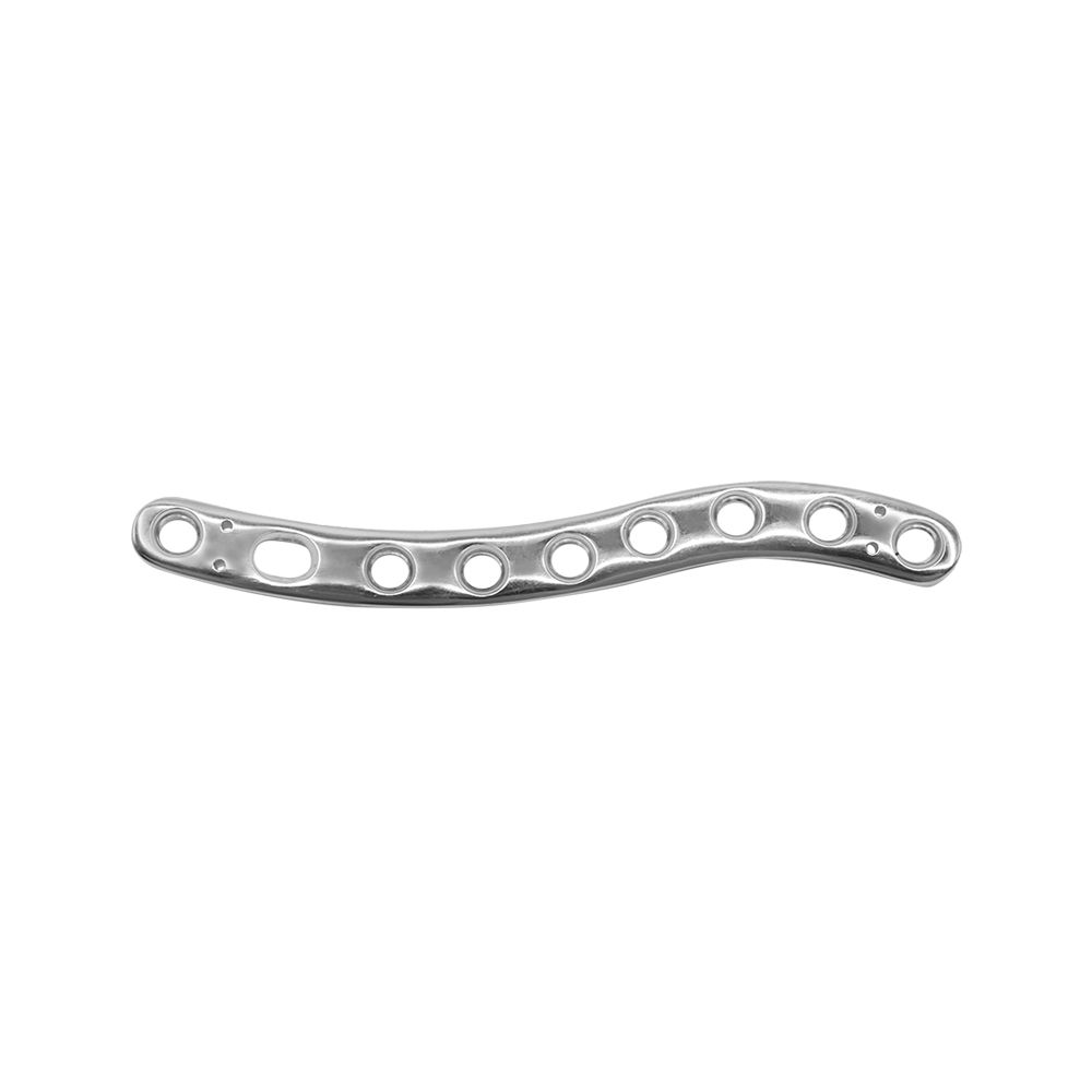
Plate Geometry:
- Contoured Design: Occipital plates are anatomically pre-contoured or designed to be easily contoured intraoperatively to match the complex curvature of the occipital bone. This ensures a broad contact area, distributing stress evenly and minimizing localized pressure points that could lead to bone erosion or skin breakdown.
- Multiple Screw Holes: The plate features numerous strategically placed screw holes, allowing surgeons flexibility in screw placement to achieve optimal purchase in varying bone qualities and anatomical variations, while avoiding critical structures like venous sinuses.
- Low Profile: Modern designs prioritize a low-profile construction to reduce soft tissue irritation and minimize the palpable presence of the implant beneath the skin, enhancing patient comfort.
- Connection Mechanisms: Integrated or modular connectors allow for seamless attachment to cervical rods. These often involve set screws, compression nuts, or proprietary locking mechanisms that securely link the occipital plate to the cervical instrumentation system.
-
Screw Types:
- Self-Tapping/Self-Drilling Screws: Designed to penetrate cortical bone without pre-drilling, simplifying the surgical procedure and reducing operative time.
- Polyaxial (Multiaxial) Screws: These screws allow for angular adjustment (typically 30-45 degrees in any direction) of the screw head relative to the plate, providing significant flexibility in achieving optimal screw trajectory and rod alignment.
- Monoaxial (Fixed Angle) Screws: Offer rigid, fixed-angle fixation, often used when specific, unyielding biomechanical stability is required.
- Locking Screws: Engage with specific features on the plate to create a rigid, fixed-angle construct, preventing screw back-out and enhancing construct stability.
- Variable Lengths and Diameters: Available in a range of sizes to accommodate different patient anatomies and bone densities, ensuring adequate purchase and stability.
-
Modularity: Contemporary systems offer a high degree of modularity, with various plate sizes, shapes (e.g., T-shaped, H-shaped, rectangular), and connection options to address a wide spectrum of craniocervical pathologies and patient anatomies.
2.2 Biomechanics of Occipital Plating
The primary biomechanical objective of an occipital plate is to create a rigid, stable construct that facilitates bony fusion and protects neural elements.
- Rigid Fixation: By securely anchoring to the occiput and extending to the cervical spine, the occipital plate effectively immobilizes the unstable segments. This rigid fixation minimizes micromotion at the fusion site, which is critical for promoting osteointegration and successful arthrodesis.
- Load Sharing and Stress Shielding: The plate bears a significant portion of the mechanical load, thereby reducing stress on the healing bone graft (load sharing). While some stress shielding is inevitable, modern designs aim to optimize this balance to prevent excessive bone resorption while providing sufficient stability.
- Resistance to Multi-Directional Forces: The occipital plate construct is designed to resist the complex forces acting on the craniocervical junction:
- Flexion/Extension: Prevents excessive forward or backward bending.
- Lateral Bending: Resists side-to-side movements.
- Axial Rotation: Limits twisting motions.
- Shear Forces: Counteracts forces that attempt to slide vertebrae past each other.
- Fusion Promotion: By creating a stable environment, the plate minimizes motion that could disrupt the healing process, thereby maximizing the chances of successful bony fusion. This stable environment allows osteoblasts to lay down new bone, bridging the unstable segments.
Extensive Clinical Indications & Usage
Occipital plating is a complex procedure reserved for specific, often severe, conditions affecting the craniocervical junction and upper cervical spine.
3.1 Detailed Surgical Applications
The indications for occipital plate fixation are broad and often involve significant instability or deformity.
-
Craniocervical Junction Instability: This is the most common indication.
- Trauma:
- Occipital Condyle Fractures: Especially unstable types that compromise the ligamentous integrity of the CCJ.
- Atlanto-occipital Dislocation (AOD): A catastrophic injury requiring immediate and robust stabilization.
- C1 (Atlas) Fractures: Burst fractures (Jefferson fractures) with associated transverse ligament disruption or C1-C2 instability.
- C2 (Axis) Fractures: Odontoid fractures (Type II and III) that are unstable or non-union after conservative management, particularly when associated with C1 instability or requiring extension to the occiput for greater stability.
- Rheumatoid Arthritis (RA): Chronic inflammation can lead to pannus formation around the odontoid process, eroding bone and ligaments, causing atlantoaxial subluxation and basilar invagination, necessitating occipitocervical fusion.
- Congenital Anomalies:
- Chiari Malformation: When associated with craniocervical instability or basilar invagination.
- Klippel-Feil Syndrome: Can present with congenital fusion anomalies and compensatory hypermobility at adjacent segments, sometimes requiring occipitocervical fusion.
- Os Odontoideum: A separate ossicle instead of a true odontoid, leading to instability.
- Tumors: Resection of primary or metastatic tumors involving the occiput, C1, or C2 often creates significant instability, requiring reconstructive fusion.
- Infections: Osteomyelitis or other infections that compromise the structural integrity of the craniocervical region.
- Degenerative Conditions: While less common, severe multi-level degenerative disease extending to the occiput, especially with kyphotic deformity, may necessitate occipitocervical fusion.
- Trauma:
-
Complex Cervical Spine Reconstruction:
- Multi-level Cervical Fusion: When extensive anterior or posterior decompression and fusion are required over multiple cervical segments (e.g., C3-C7), and the pathology extends to or requires anchoring from the occiput for enhanced stability.
- Revision Surgery: For failed previous cervical instrumentation, pseudoarthrosis, or recurrent instability, where a more robust construct anchored to the occiput is necessary.
- Deformity Correction: In cases of severe cervical kyphosis or scoliosis that involve the craniocervical junction, an occipital plate can be instrumental in restoring sagittal and coronal balance.
3.2 Fitting and Usage Instructions (General Principles)
The successful implantation of an occipital plate requires meticulous planning and precise surgical technique.
-
Pre-operative Planning:
- Advanced Imaging: High-resolution CT scans with 3D reconstructions are essential to understand the patient's unique occipital anatomy, identify critical venous sinuses, and plan optimal screw trajectories. MRI scans help assess neural compression and soft tissue pathology.
- Patient Positioning: The patient is typically placed in a prone position with the head secured in a Mayfield clamp or similar head holder, allowing for precise control of head position and alignment. Careful attention is paid to protecting pressure points and ensuring proper airway management.
- Surgical Approach: A posterior midline incision is made from the inion (external occipital protuberance) down to the appropriate cervical level. Subperiosteal dissection exposes the occipital bone and posterior elements of the cervical vertebrae.
-
Plate Contouring:
- In-situ Bending: Many plates are designed to be contoured intraoperatively using specialized bending irons to precisely match the individual patient's occipital curvature. This minimizes gaps between the plate and bone, which can lead to stress risers or poor fixation.
- Pre-contoured Plates: Some systems offer a range of pre-contoured plates that can reduce operative time, though fine-tuning may still be necessary. The goal is a snug, anatomical fit.
-
Screw Placement:
- Occipital Screws: Placed into the thickest part of the occipital bone, typically superior to the inion, to achieve bicortical purchase while carefully avoiding the transverse and sagittal venous sinuses and the dura. Trajectory is crucial, often directed slightly inferiorly and laterally.
- Cervical Screws: Depending on the level, these may be C1 lateral mass screws, C2 pedicle or pars screws, or C3-C7 lateral mass screws. Each has specific anatomical landmarks and trajectory considerations to maximize purchase and avoid neurovascular structures.
- Pilot Holes: Often created with a drill, followed by tapping (if not self-tapping screws) to create threads for the screw. Screw depth is measured meticulously.
-
Rod Connection:
- Once occipital and cervical screws are placed, appropriately sized rods are contoured to connect the screw heads. The rods are then secured to the plate and screws using locking caps, set screws, or compression nuts.
- Compression/Distraction: The surgeon may apply gentle compression or distraction along the rod to optimize spinal alignment and stability before final tightening.
-
Intraoperative Imaging:
- Fluoroscopy: Real-time X-ray imaging is routinely used to confirm screw placement, rod alignment, and overall construct integrity throughout the procedure.
- Navigation Systems: Advanced surgical navigation systems (e.g., O-arm, StealthStation) can provide highly accurate 3D guidance for screw placement, significantly improving precision and reducing radiation exposure.
-
Post-operative Management:
- Bracing: Patients often require a rigid cervical collar or halo brace post-operatively for a period to further protect the fusion site during initial healing.
- Rehabilitation: A structured rehabilitation program is initiated to gradually restore function, improve strength, and manage pain.
Risks, Side Effects, or Contraindications
While highly effective, occipital plate surgery is a major undertaking with potential risks and complications.
4.1 Surgical Risks
- Hemorrhage: The occipital region is highly vascular, with significant venous sinuses (transverse, sagittal, torcula) that can lead to massive bleeding if injured.
- Neurological Injury: Damage to the spinal cord, brainstem, or cranial nerves (e.g., hypoglossal, accessory nerves) can occur during dissection, screw placement, or excessive manipulation. This can lead to paralysis, weakness, or sensory deficits.
- Dural Tear/CSF Leak: Accidental puncture of the dura mater can lead to cerebrospinal fluid (CSF) leakage, increasing the risk of infection (meningitis) and persistent headache.
- Infection: Superficial wound infection or deep hardware infection, which can necessitate implant removal.
- Hardware Malposition/Failure:
- Screw Pullout/Loosening: Especially in osteoporotic bone.
- Plate Fracture: Rare, but can occur with excessive stress or material fatigue.
- Rod Dislodgement: If not properly secured.
- Pseudoarthrosis (Non-union): Failure of the bone graft to fuse, which may require revision surgery.
- Dysphagia/Airway Compromise: Swelling in the pharyngeal region post-surgery can temporarily affect swallowing or breathing.
4.2 Post-operative Complications
- Pain: Persistent pain at the surgical site, headache, or neck stiffness.
- Restricted Range of Motion: Fusion inherently limits movement at the fused segments.
- Skin Breakdown: Especially over prominent hardware in thin patients.
- Implant Palpability: The plate may be felt under the skin.
- Adjacent Segment Disease: Increased stress on the unfused segments above or below the fusion, potentially leading to future degeneration.
4.3 Contraindications
- Active Systemic Infection: Increases the risk of hardware infection and poor wound healing.
- Insufficient Bone Quality: Severe osteoporosis or pathological bone destruction may preclude adequate screw purchase, making rigid fixation impossible.
- Severe Allergic Reaction: Documented allergy to implant materials (though rare with titanium).
- Unrealistic Patient Expectations/Non-compliance: Patients must understand the limitations and commitment required for successful recovery.
- Local Skin/Soft Tissue Conditions: Poor skin integrity or active skin infection at the surgical site.
Massive FAQ Section
Here are some frequently asked questions about occipital plates:
1. What is the primary purpose of an occipital plate?
The primary purpose of an occipital plate is to provide rigid stabilization and facilitate bony fusion between the occipital bone (skull) and the upper cervical spine. This is crucial for treating instability, deformity, or severe trauma at the craniocervical junction, protecting the spinal cord and brainstem.
2. What materials are occipital plates typically made from?
Occipital plates are predominantly made from titanium alloy (Ti-6Al-4V ELI). This material is highly biocompatible, strong, lightweight, and largely compatible with MRI scans, making it ideal for long-term implantation.
3. How long does an occipital fusion surgery take?
The duration of occipital fusion surgery can vary significantly depending on the complexity of the case, the number of spinal levels involved, and the surgeon's experience. Typically, these procedures can range from 3 to 6 hours or even longer for very complex reconstructions.
4. What is the recovery time after occipital plate surgery?
Initial recovery often involves a hospital stay of 3-7 days. The bone fusion process can take 6-12 months, during which time physical activity is restricted. Full recovery, including rehabilitation and return to most normal activities, can extend up to a year or more. A cervical collar or brace is often worn for several months.
5. Will I be able to move my head normally after the surgery?
Occipital fusion surgery aims to stabilize segments that were previously unstable. This stabilization, by definition, involves fusing bones together, which will permanently restrict motion at the fused segments. While some compensatory movement may occur at unfused segments, overall head and neck range of motion will be reduced compared to pre-surgery.
6. Are there alternatives to occipital plate fixation?
For significant craniocervical instability, an occipital plate with a rod-screw system is often the most robust and preferred method of fixation. In very specific, less severe cases, non-operative management with external bracing might be considered, but it's rarely sufficient for true instability. Surgical alternatives are limited and often involve variations of posterior fusion techniques.
7. Can an occipital plate be removed? If so, when?
While occipital plates are designed for permanent implantation, removal is possible. It is typically considered only if complications arise, such as deep infection, persistent pain directly attributable to the hardware, or hardware failure after successful fusion. Removal is usually performed after bony fusion is confirmed, which can take 12-24 months.
8. Is an occipital plate MRI compatible?
Yes, modern occipital plates made from titanium alloy are generally considered MRI compatible. Patients should always inform their healthcare providers about their implant before undergoing an MRI, but typically, titanium implants pose no significant safety risk and only minimal image artifact.
9. What are the long-term outcomes for patients with occipital plates?
Long-term outcomes are generally favorable, with good rates of fusion and significant improvement in neurological function and pain for appropriately selected patients. However, patients will experience permanent restriction of head movement. Regular follow-up with the surgeon is essential to monitor for potential long-term complications like adjacent segment disease or hardware issues.
10. How is an occipital plate different from other spinal plates?
An occipital plate is specifically designed to conform to the unique curvature and bone thickness of the occipital bone and to connect to the upper cervical spine. Unlike thoracic or lumbar plates, which primarily stabilize vertebral bodies, the occipital plate must bridge the skull and cervical spine, requiring specific anatomical contours and robust connection mechanisms for rods extending into the neck.
11. What kind of pain management can I expect post-surgery?
Post-operative pain management typically involves a multi-modal approach, including intravenous pain medications initially, transitioning to oral narcotics, muscle relaxants, and non-steroidal anti-inflammatory drugs (NSAIDs) as tolerated. The goal is to control pain effectively to allow for early mobilization and participation in rehabilitation.
12. How do surgeons ensure proper placement of the occipital plate?
Surgeons employ several techniques to ensure proper placement, including meticulous pre-operative planning with advanced imaging (CT, MRI), using anatomical landmarks, intraoperative fluoroscopy (real-time X-ray), and increasingly, advanced surgical navigation systems (e.g., O-arm, StealthStation) that provide 3D imaging and guidance for precise screw trajectory and plate positioning.
Maintenance/Sterilization Protocols for Occipital Plate Systems
The integrity and sterility of orthopedic implants and instruments are paramount to patient safety and surgical success. Strict adherence to maintenance and sterilization protocols is non-negotiable.
6.1 Pre-use Inspection
Before each surgical procedure, all components of the occipital plate system (plates, screws, rods, and associated instruments) must undergo a thorough visual inspection.
* Damage Assessment: Check for any signs of damage such as scratches, nicks, bends, corrosion, or deformation. Damaged implants must be discarded.
* Cleanliness: Ensure all items are visibly clean and free from any residual debris or organic material from previous uses.
* Functionality: Verify that all instruments (e.g., screwdrivers, rod benders, torque wrenches) are in perfect working order.
6.2 Cleaning Protocols
Effective cleaning is the critical first step in preventing surgical site infections.
* Point-of-Use Cleaning: Immediately after use, instruments should be wiped down to remove gross soil and kept moist (e.g., with enzymatic spray or damp cloth) to prevent blood and tissue from drying on surfaces.
* Manual Cleaning:
* Disassemble multi-part instruments.
* Use enzymatic detergents specifically designed for medical instruments to break down organic matter.
* Scrub all surfaces thoroughly with soft brushes, paying close attention to crevices, screw threads, and articulation points.
* Rinse extensively under running water to remove all detergent and debris.
* Automated Cleaning (Washer-Disinfectors/Ultrasonic Cleaners):
* Follow manufacturer's instructions for loading and processing.
* Ultrasonic cleaners use high-frequency sound waves to create cavitation, dislodging microscopic debris from intricate surfaces.
* Washer-disinfectors automate washing, rinsing, and thermal disinfection cycles, providing consistent cleaning.
6.3 Rinsing and Drying
- Thorough Rinsing: All instruments and implants must be rinsed thoroughly with distilled or deionized water to remove all traces of cleaning agents, which can cause corrosion or interfere with sterilization.
- Complete Drying: Instruments must be completely dry before packaging for sterilization. Residual moisture can impede sterilization processes and promote corrosion or microbial growth. Use medical-grade lint-free towels or forced-air drying.
6.4 Inspection Post-Cleaning
A meticulous inspection after cleaning is crucial to ensure that all bioburden has been removed.
* Visual Inspection: Use magnification and adequate lighting to check every surface for residual soil, stains, or damage.
* Functional Check: Reassemble and test instruments to ensure they function correctly. Any item not meeting cleanliness or functional standards must be reprocessed.
6.5 Packaging
Correct packaging is essential to maintain sterility after processing and until the point of use.
* Sterilization Wrap: Use medical-grade sterilization wrap that is permeable to the sterilant but impervious to microorganisms.
* Sterilization Containers: Rigid sterilization containers with filters provide robust protection and are often preferred for sets of implants and instruments.
* Labeling: Each package must be clearly labeled with the contents, date of sterilization, and expiration date.
6.6 Sterilization Methods
The chosen sterilization method must be validated for the specific materials and design of the occipital plate system.
* Steam Sterilization (Autoclaving): This is the most common and preferred method for heat-stable items like titanium occipital plates and most surgical instruments.
* Parameters: Typically involves exposure to saturated steam at specific temperatures (e.g., 121°C for 20-30 minutes or 132-135°C for 4-10 minutes) under pressure.
* Validation: Cycle parameters must be validated to ensure sterility.
* Ethylene Oxide (EtO) Sterilization: Used for heat- and moisture-sensitive items. Requires aeration time to dissipate toxic EtO gas. Less common for titanium implants themselves, but may be used for associated packaging or components.
* Hydrogen Peroxide Plasma Sterilization: A low-temperature method suitable for heat-sensitive instruments. Utilizes hydrogen peroxide vapor to sterilize.
* Sterilization Monitoring: Chemical indicators (internal and external) and biological indicators (spore tests) are used to confirm that sterilization parameters have been met.
6.7 Storage
- Sterile Storage: Sterilized items must be stored in a clean, dry, temperature-controlled environment away from potential contaminants, traffic, and moisture.
- Shelf Life: Adhere to the shelf life specified by the packaging manufacturer or facility policy.
6.8 Traceability
- Lot Numbers and Expiration Dates: Maintain meticulous records of implant lot numbers and expiration dates for every implanted device. This is crucial for tracking in case of recalls or adverse events.
- Documentation: All cleaning, sterilization, and inspection processes must be thoroughly documented to ensure accountability and compliance with regulatory standards.
By adhering to these rigorous protocols, healthcare facilities can ensure that occipital plates and their associated instruments are safe, sterile, and ready to provide critical stabilization for patients in need.