The O-Arm Intraoperative 3D Imaging System: A Revolution in Orthopedic Precision
Welcome to an in-depth exploration of the O-Arm Intraoperative 3D Imaging System, a groundbreaking technology that is transforming the landscape of orthopedic and spinal surgery. At our practice, led by Dr. Mohammed Hutaif, we are committed to utilizing the most advanced tools to ensure the highest levels of precision, safety, and optimal outcomes for our patients. The O-Arm system stands as a testament to this commitment, offering surgeons unparalleled real-time, high-resolution imaging capabilities during complex procedures.
1. Comprehensive Introduction & Overview
The O-Arm system is a state-of-the-art mobile X-ray system designed to provide 2D fluoroscopic and 3D volumetric images of a patient's anatomy during surgery. Unlike traditional C-arms, which primarily offer 2D views, the O-Arm performs a complete 360-degree rotation around the patient, capturing a comprehensive dataset that is then reconstructed into a detailed 3D image. This capability empowers surgeons with an "inside view" of the surgical field, allowing for immediate verification of implant placement, fracture reduction, and anatomical alignment.
The advent of intraoperative 3D imaging has marked a significant paradigm shift in surgical methodology. Historically, surgeons relied on pre-operative imaging and their extensive anatomical knowledge, often confirming results post-operatively. The O-Arm eliminates much of this uncertainty, enabling critical adjustments to be made during the procedure, thereby enhancing patient safety, reducing the need for revision surgeries, and ultimately improving long-term outcomes. For patients, this translates to greater confidence in their surgical care, potentially shorter operating times, and a smoother recovery journey.
2. Deep-dive into Technical Specifications & Mechanisms
The O-Arm system is a sophisticated piece of medical engineering, meticulously designed to integrate seamlessly into the operating room environment.
Design and Materials
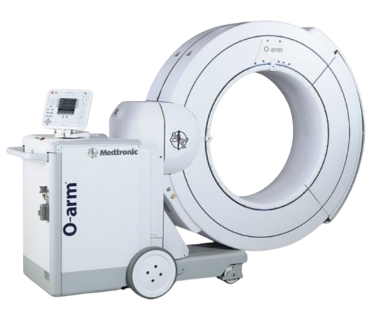
The O-Arm unit consists of several key components:
* Gantry: A large, O-shaped ring that encircles the patient. This gantry houses the X-ray source on one side and the flat-panel detector on the opposite side. It's constructed from robust, medical-grade materials designed for durability and ease of cleaning.
* Flat-Panel Detector: A high-resolution digital detector responsible for capturing the X-ray images. Modern detectors offer superior image quality with reduced radiation dose compared to older image intensifier technologies.
* X-ray Source: Emits controlled X-ray beams through the patient's anatomy to the detector.
* Mobile Workstation: Equipped with powerful computing capabilities and intuitive software for image reconstruction, visualization, and navigation integration. This workstation is mobile, allowing for flexible positioning in the OR.
* Sterile Draping: Critical for maintaining a sterile field, the O-Arm is designed to be easily draped with sterile covers, ensuring patient safety during surgery.
Mechanism of Action
The core mechanism of the O-Arm involves its unique ability to perform a full 360-degree rotation around the patient.
1. Patient Positioning: The patient is positioned on the operating table, and the O-Arm gantry is moved into place to encompass the target area.
2. Image Acquisition: Once activated, the gantry performs a controlled, isocentric rotation. During this rotation, it captures a series of 2D X-ray projections from multiple angles. This process is remarkably fast, often taking less than 30 seconds for a full 3D scan.
3. 3D Reconstruction: The workstation's sophisticated software processes these 2D projections. Using advanced algorithms, it reconstructs a detailed, high-resolution 3D volumetric image of the scanned anatomy. This image can be viewed in axial, sagittal, and coronal planes, providing a comprehensive understanding of the bone structure, implants, and surrounding tissues.
4. Navigation System Integration: A critical advantage of the O-Arm is its seamless integration with surgical navigation systems. Once a 3D scan is acquired, the O-Arm data is automatically transferred to the navigation platform. This allows the surgeon to track instruments in real-time on the 3D model, guiding precise placement with sub-millimeter accuracy.
Key Technical Specifications
- Field of View: Varies by model, but typically large enough to capture entire vertebral segments or complex joint areas.
- Scan Time: Rapid acquisition, typically 13-30 seconds for a full 3D scan.
- Dose Management: Incorporates advanced features like pulsed fluoroscopy, iterative reconstruction, and adjustable dose settings to minimize radiation exposure while maintaining diagnostic image quality, adhering strictly to the ALARA (As Low As Reasonably Achievable) principle.
- Image Resolution: High spatial resolution for clear visualization of fine anatomical details and implant components.
3. Extensive Clinical Indications & Usage
The O-Arm system has revolutionized surgical approaches across various orthopedic and neurosurgical subspecialties, particularly where precision is paramount.
Spinal Surgery
The O-Arm's impact on spinal surgery is perhaps its most celebrated application.
* Pedicle Screw Placement: One of the most common and critical uses. The O-Arm provides real-time 3D verification of pedicle screw trajectory and depth, significantly reducing the risk of neurological or vascular injury. This is crucial in complex cases like severe scoliosis or revision surgeries where anatomy may be distorted.
* Spinal Deformity Correction: For conditions like scoliosis and kyphosis, accurate placement of instrumentation is vital for achieving optimal correction and fusion. The O-Arm ensures screws are placed correctly to maximize corrective forces and minimize complications.
* Spinal Fusion Procedures: Whether for degenerative disc disease, spondylolisthesis, or trauma, confirming the correct placement of interbody cages, rods, and screws is essential for successful fusion.
* Spinal Trauma: In cases of vertebral fractures, the O-Arm assists in precise reduction and stabilization, ensuring optimal alignment and fixation.
* Minimally Invasive Spine Surgery (MISS): The O-Arm is indispensable for MISS, where direct visualization is limited. It allows surgeons to navigate through small incisions with high accuracy, leading to smaller scars, less muscle disruption, reduced blood loss, and faster recovery.
Orthopedic Trauma
For complex fractures, especially those involving joints or the pelvis, the O-Arm offers invaluable guidance.
* Complex Fracture Fixation: Aids in precise reduction and fixation of fractures of the pelvis, acetabulum, and long bones (e.g., tibia, femur).
* Intra-articular Fracture Reduction: Ensures articular surfaces are perfectly realigned, which is critical for preventing post-traumatic arthritis.
* Screw/Plate Placement Verification: Confirms that screws are of appropriate length and trajectory and that plates are positioned optimally for stability.
Neurosurgery
While primarily orthopedic, the O-Arm can also be utilized in certain neurosurgical applications, particularly for cranial navigation and tumor resection, although less commonly than in spine.
Biomechanics and Patient Outcome Improvements
The precise placement of implants, guided by the O-Arm, has profound biomechanical implications:
* Enhanced Stability: Correctly placed screws and plates provide superior biomechanical stability, crucial for successful fusion and fracture healing.
* Reduced Stress Shielding: Optimal implant positioning helps distribute loads more naturally, reducing stress shielding which can lead to bone weakening around implants.
* Optimized Load Bearing: Ensures that weight-bearing forces are transmitted effectively through the reconstructed anatomy, promoting quicker rehabilitation and long-term functional integrity.
* Prevention of Hardware Failure: Minimizes the risk of screw pull-out, breakage, or loosening, which can necessitate revision surgery.
* Improved Fusion Rates: Accurate implant placement and anatomical reduction contribute directly to higher rates of successful spinal fusion.
Fitting/Usage Instructions (Patient Perspective)
From a patient's perspective, the use of an O-Arm during surgery is largely unobtrusive.
* Pre-operative Planning: The surgeon will use pre-operative imaging (MRI, CT) to plan the surgery. The O-Arm data then augments this planning in real-time.
* Intraoperative Setup: Once under anesthesia, the patient is positioned on the operating table. The O-Arm unit is brought into the sterile field and draped.
* Image Acquisition: The O-Arm gantry will momentarily move around the surgical site to acquire 2D and 3D images. This process is quick and silent, lasting only a few seconds.
* Real-time Feedback: The surgeon and surgical team will review the images on the workstation, using them to guide instrument placement and confirm surgical progress. This happens discreetly in the background, allowing the surgeon to make informed decisions without delay.
Maintenance/Sterilization Protocols
While not directly involving the patient, stringent maintenance and sterilization protocols are critical for patient safety and optimal system performance.
* Sterile Draping: Before each use, the O-Arm unit is meticulously covered with sterile drapes to maintain a sterile operating field, preventing infection.
* Regular Maintenance: The system undergoes routine checks and calibration by certified technicians to ensure all components function correctly and image quality is consistent.
* Software Updates: Regular software updates ensure the system benefits from the latest advancements in imaging algorithms and navigation capabilities.
* Cleaning and Disinfection: The non-sterile surfaces of the O-Arm are thoroughly cleaned and disinfected according to hospital protocols after each procedure.
4. Risks, Side Effects, or Contraindications
While the O-Arm significantly enhances surgical safety and precision, it's important to understand the general risks associated with any surgical procedure and those specific to imaging.
- Radiation Exposure: The O-Arm utilizes X-rays, meaning there is some radiation exposure. However, the system is designed with advanced dose reduction technologies (e.g., pulsed fluoroscopy, iterative reconstruction) to keep exposure "As Low As Reasonably Achievable" (ALARA). The benefits of precise surgery and reduced risk of complications generally outweigh the minimal radiation risk.
- General Surgical Risks: The O-Arm itself does not eliminate the inherent risks of surgery, such as:
- Anesthesia complications
- Infection at the surgical site
- Bleeding
- Nerve damage (though O-Arm significantly reduces this risk)
- Blood clots
- Adverse reaction to medications
- Contraindications: There are no specific contraindications for the O-Arm system itself. The decision to use O-Arm-guided surgery is based on the patient's overall health, the specific surgical indication, and the surgeon's clinical judgment. Pregnancy is a relative contraindication due to radiation, but necessary procedures would involve careful risk-benefit analysis.
5. Expert Tips from Dr. Mohammed Hutaif
"As an orthopedic specialist, my primary goal is to provide the safest, most effective care possible. The O-Arm Intraoperative 3D Imaging System is a cornerstone of achieving this for many of my complex spinal and orthopedic cases.
- Unrivaled Precision: The ability to obtain high-resolution 3D images during surgery allows for an unparalleled level of precision. This is particularly critical in delicate areas like the spine, where even a millimeter can make a significant difference in patient outcomes and safety. We can verify screw placement and anatomical alignment in real-time, drastically reducing the chances of misplacement.
- Enhanced Patient Safety: By offering immediate feedback on instrument and implant positioning, the O-Arm helps us avoid potential complications such as nerve damage or vascular injury. This proactive approach to safety is invaluable.
- Optimized Outcomes: For patients, this translates to better long-term results. We see improved fusion rates in spinal surgeries, more accurate fracture reductions, and a reduced likelihood of needing follow-up revision surgeries. This often means less post-operative pain, faster recovery times, and a quicker return to daily activities.
- Minimally Invasive Advantage: For minimally invasive procedures, the O-Arm is indispensable. It allows us to perform complex maneuvers through small incisions with confidence, harnessing the benefits of reduced tissue trauma for our patients.
- Commitment to Advanced Technology: Embracing technologies like the O-Arm reflects our dedication to staying at the forefront of orthopedic care. It's an investment in patient well-being, ensuring that every procedure benefits from the highest available standards of accuracy and safety."
6. Massive FAQ Section
Q1: What exactly is the O-Arm system?
A1: The O-Arm is an advanced mobile X-ray system used during surgery to provide high-resolution 2D and 3D images of a patient's anatomy. It rotates around the patient to capture detailed views, helping surgeons perform procedures with greater precision and safety.
Q2: How does the O-Arm differ from traditional X-rays or C-arms?
A2: Traditional X-rays provide static 2D images. While C-arms offer real-time 2D fluoroscopy, the O-Arm goes a step further by performing a full 360-degree rotation to generate comprehensive 3D volumetric images. This 3D capability, often integrated with navigation systems, provides a much more detailed and accurate view of the surgical field.
Q3: Is the O-Arm safe for me? What about radiation exposure?
A3: Yes, the O-Arm is considered safe. While it uses X-rays, it incorporates advanced dose reduction technologies to minimize radiation exposure to "As Low As Reasonably Achievable" (ALARA). The benefits of increased surgical precision and reduced complications generally outweigh the minimal radiation risks. Your surgical team will always prioritize your safety.
Q4: What types of surgeries benefit most from O-Arm imaging?
A4: The O-Arm is particularly beneficial for complex orthopedic and spinal surgeries where precise implant placement and anatomical alignment are crucial. This includes spinal fusions, pedicle screw placement, scoliosis correction, and complex fracture fixations (e.g., pelvis, acetabulum).
Q5: Does O-Arm guided surgery take longer?
A5: While there's a brief period for image acquisition (typically 13-30 seconds per scan), the overall surgical time can sometimes be reduced. This is because surgeons can verify implant placement immediately, avoiding the need for repeated repositioning or post-operative imaging to check accuracy, which might lead to revision surgery. The efficiency gained from precision often balances or reduces the total time.
Q6: Will I feel anything during the O-Arm scan during surgery?
A6: No, you will be under general anesthesia during the surgery, so you will not feel anything when the O-Arm performs its scan. The system moves around the operating table without touching you directly.
Q7: How does O-Arm improve surgical accuracy?
A7: By providing real-time 3D images, the O-Arm allows surgeons to visualize the exact anatomy and the precise position of surgical instruments and implants. This significantly reduces the margin for error, ensuring implants are placed correctly and fractures are perfectly reduced, often with sub-millimeter accuracy when integrated with navigation.
Q8: What are the benefits of O-Arm for patient recovery?
A8: Improved surgical accuracy leads to better patient outcomes. This can include:
* Reduced risk of complications (e.g., nerve damage, implant malposition)
* Potentially shorter hospital stays
* Less post-operative pain
* Faster recovery and rehabilitation
* Reduced need for revision surgeries
* Better long-term functional results
Q9: Is O-Arm technology available everywhere?
A9: The O-Arm is a sophisticated and significant investment for medical facilities. It is typically found in leading hospitals and specialized orthopedic or neurosurgical centers that prioritize advanced technology for complex procedures. Our practice is proud to offer this cutting-edge technology.
Q10: Can O-Arm be used for children?
A10: Yes, the O-Arm can be used for pediatric orthopedic and spinal surgeries, especially in cases of complex deformities like severe scoliosis. The radiation dose is carefully managed, and the benefits of precision in growing anatomies are often considered to outweigh the minimal risks.
Q11: Does O-Arm affect the cost of surgery?
A11: The use of advanced technology like the O-Arm may contribute to the overall cost of the surgical procedure. However, the potential for reduced complications, fewer revision surgeries, and improved long-term outcomes can lead to cost savings over time by avoiding future medical expenses.
Q12: What specific orthopedic conditions does Dr. Hutaif treat with the O-Arm?
A12: Dr. Mohammed Hutaif utilizes the O-Arm system for a range of complex conditions, primarily focusing on spinal pathologies such as severe scoliosis, kyphosis, degenerative disc disease requiring fusion, spondylolisthesis, and complex spinal trauma. He also employs it for intricate orthopedic trauma cases involving the pelvis, acetabulum, and intra-articular fractures where precise reduction and fixation are critical.
Disclaimer: This content is for patient information only and is not medical advice. Always consult with a qualified healthcare professional for diagnosis and treatment of any medical condition.