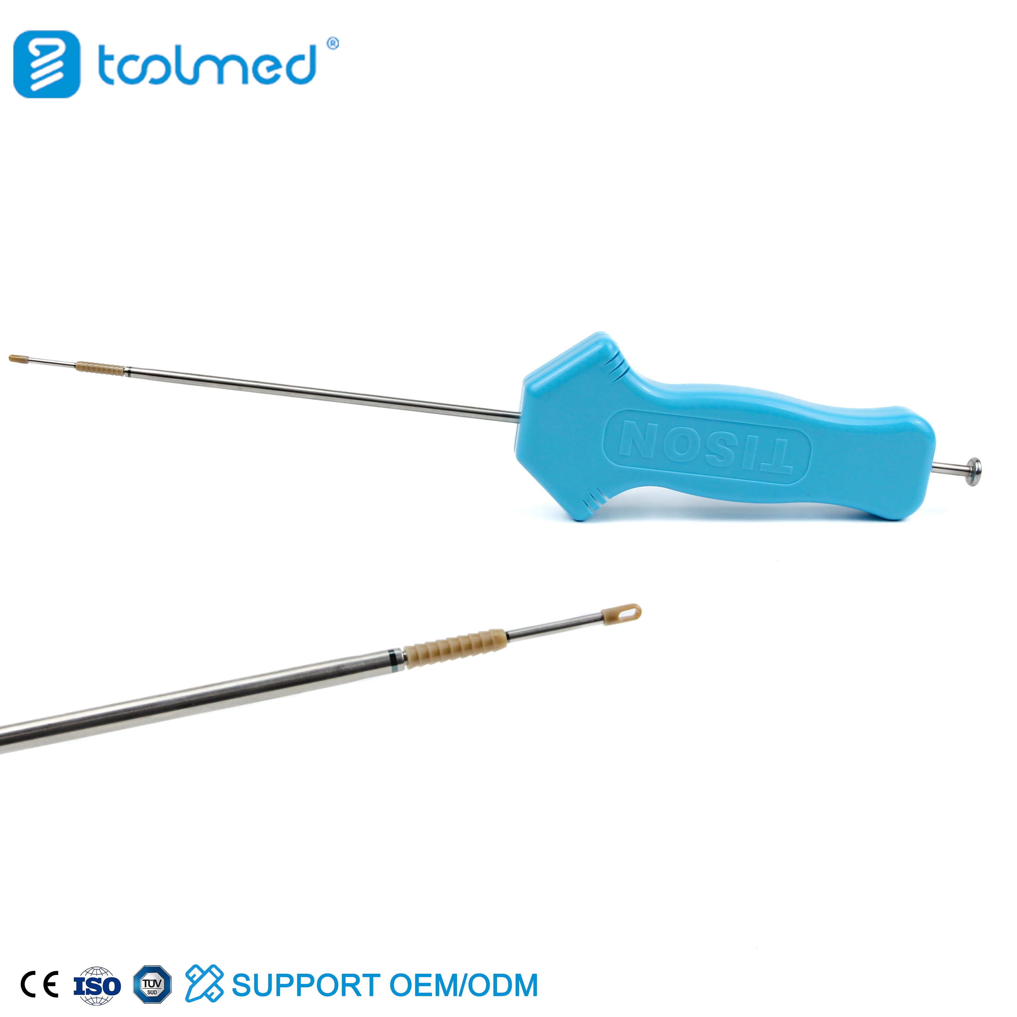
The Innovation of Knotless Suture Anchors: PushLock and SwiveLock Explained
In the evolving landscape of orthopedic surgery, advancements in implant technology continuously seek to improve surgical outcomes, reduce operative time, and enhance patient recovery. Among these innovations, knotless suture anchors represent a significant leap forward, particularly in arthroscopic procedures. This comprehensive guide delves into the world of knotless suture anchors, with a specific focus on the widely utilized PushLock and SwiveLock systems, exploring their design, applications, biomechanics, maintenance, and the profound impact they have on patient care.
1. Comprehensive Introduction & Overview
Knotless suture anchors are specialized orthopedic implants designed to reattach soft tissues (such as tendons, ligaments, and capsules) to bone without the need for traditional knot tying. This eliminates the bulk and potential irritation associated with knots, streamlines the surgical process, and often results in a more consistent and biomechanically sound repair.
Traditional knotted anchors, while effective, can be time-consuming to deploy arthroscopically and the knots themselves can sometimes lead to impingement or soft tissue irritation. Knotless technology addresses these challenges by incorporating an internal locking mechanism that secures the suture within the anchor body once it is seated in the bone.
The PushLock and SwiveLock systems, pioneered by Arthrex, are prominent examples of knotless suture anchor technology. Both offer distinct advantages and mechanisms for tissue fixation, making them versatile tools in the orthopedic surgeon's armamentarium.
Key Advantages of Knotless Suture Anchors:
* Reduced Knot Bulk: Minimizes soft tissue irritation and potential impingement.
* Faster Procedure Time: Eliminates the need for intricate knot tying, especially in confined arthroscopic spaces.
* Consistent Tensioning: Allows for precise and reproducible tensioning of repaired tissues.
* Improved Biomechanics: Often provides a more stable and stronger construct at the repair site.
* Enhanced Arthroscopic Efficiency: Simplifies complex arthroscopic repairs.
This guide will provide a deep dive into the technical aspects, clinical utility, and patient benefits associated with these advanced orthopedic instruments.
2. Deep-Dive into Technical Specifications / Mechanisms
The efficacy of knotless suture anchors lies in their ingenious design and the materials used in their construction. Understanding these elements is crucial for appreciating their clinical value.
### Design and Materials
Knotless suture anchors are typically composed of a body designed for secure bone fixation and an internal mechanism to lock the suture.
Anchor Body Materials:
* PEEK (Polyetheretherketone): A high-performance thermoplastic known for its excellent mechanical strength, biocompatibility, and radiolucency (meaning it's not visible on X-rays, which can be advantageous for post-operative imaging without artifact). PEEK anchors are non-resorbable.
* BioComposite (PLLA/TCP - Poly-L-Lactide/Tricalcium Phosphate): These anchors are designed to be bioabsorbable, gradually breaking down and being replaced by host bone over time. The PLLA provides initial mechanical strength, while TCP encourages bone ingrowth and integration. They are radiolucent initially but may become partially radiopaque as they resorb and are replaced by bone.
* Titanium: A traditional material for orthopedic implants, offering high strength and excellent biocompatibility. Titanium anchors are radiopaque.
Suture Material:
* High-Strength Polyethylene: Often braided, these sutures (e.g., FiberWire, FiberLoop) are designed for superior strength, abrasion resistance, and excellent handling characteristics, crucial for securing tissues.
Mechanism of Action:
#### PushLock Anchor System
The PushLock anchor is an interference fit anchor, meaning it locks into the bone via compression.
* Design: Features a pre-loaded suture (often a FiberLoop or similar) passed through an eyelet at the anchor's tip. The anchor body is typically tapered.
* Insertion: A pilot hole is drilled into the bone. The anchor, with the tissue-passing suture loaded, is inserted into the pilot hole.
* Locking Mechanism: The surgeon tensions the free ends of the suture to achieve the desired tissue reduction and compression. Once tensioned, a driver is used to 'push' the anchor fully into the pilot hole. This action causes the suture to be compressed and locked securely against the anchor's internal cannulation and the bone, creating an interference fit. The excess suture is then trimmed.
#### SwiveLock Anchor System
The SwiveLock anchor is a screw-in anchor that provides fixation through thread engagement with bone.
* Design: Consists of a threaded anchor body and an internal "swivel" mechanism. It also comes pre-loaded with suture, often a FiberTape or FiberWire.
* Insertion: A pilot hole is drilled. The anchor is then screwed into the bone, engaging the threads.
* Locking Mechanism: As the anchor is screwed in, the surgeon tensions the suture to achieve desired tissue compression. Once the anchor is fully seated and the tissue is appropriately reduced, a final turn or engagement of the driver "swivels" and locks the suture within the anchor body, often by trapping it against the anchor's internal wall or a specialized cap. This creates a secure, knotless fixation. The excess suture is then trimmed.
Sizes: Both systems are available in various diameters (e.g., 2.4mm, 2.9mm, 3.5mm, 4.75mm, 5.5mm) to accommodate different bone qualities and surgical applications.
### Biomechanics
The biomechanical performance of knotless suture anchors is a critical factor in their clinical success.
* Load to Failure: Studies consistently show that knotless anchors, particularly those with a screw-in design like SwiveLock, can achieve load-to-failure strengths comparable to or even exceeding traditional knotted anchors, especially in good bone quality. The direct bone-suture interface without a bulky knot can distribute stress more efficiently.
* Cyclic Loading: Knotless anchors demonstrate excellent resistance to cyclic loading, which simulates the repetitive stresses experienced by repairs during rehabilitation and daily activities. The secure locking mechanism prevents suture slippage, maintaining consistent tension.
* Bone-Anchor Interface: For bio-composite anchors, the osteoconductive properties of TCP promote bone ingrowth and eventual osseointegration, leading to a biological fixation that further strengthens the repair over time. PEEK and Titanium anchors rely on mechanical fixation, but their robust design ensures long-term stability.
* Reduced Soft Tissue Irritation: The absence of knots eliminates a potential source of friction and impingement against surrounding soft tissues, which can contribute to post-operative pain and stiffness.
* Improved Footprint Compression: Knotless anchors, especially when used in conjunction with tape-like sutures (e.g., FiberTape), can provide a broad, even compression of the tendon or ligament onto the bone, optimizing the healing environment.
3. Extensive Clinical Indications & Usage
Knotless suture anchors have revolutionized the repair of soft tissue injuries across various joints, offering versatility and improved outcomes.
### Detailed Surgical Applications
#### Shoulder Surgery
- Rotator Cuff Repair:
- Arthroscopic Single-Row Repair: Anchors are used to secure the torn rotator cuff tendon directly back to the humeral head. Knotless anchors facilitate precise tensioning and footprint coverage.
- Double-Row Repair: Involves medial row fixation (often with knotted anchors or knotless anchors) and a lateral row of knotless anchors (e.g., PushLock or SwiveLock) to create a "compression" or "transosseous equivalent" repair, maximizing contact pressure and blood supply to the healing tendon.
- Labral Repair (SLAP, Bankart Lesions): Used to reattach the glenoid labrum to the glenoid rim. Their small profile and precise locking mechanism are ideal for these delicate repairs.
- Biceps Tenodesis: Securing the long head of the biceps tendon into the humerus.
- Capsular Shift/Plication: For shoulder instability, to tighten the joint capsule.
#### Knee Surgery
- ACL/PCL Reconstruction: While primary fixation is often with screws or suspensory devices, knotless anchors can be used for supplemental fixation of hamstring grafts or for meniscal root repairs.
- Meniscal Repair: Especially for meniscal root tears where the meniscus detaches from its bony insertion, knotless anchors provide strong, reproducible fixation.
- Patellar Tendon Repair: Reattaching the patellar tendon to the patella or tibia following rupture.
- MCL/LCL Repair: For collateral ligament repairs or reconstructions.
#### Hip Surgery
- Labral Repair: Similar to the shoulder, knotless anchors are excellent for reattaching the hip labrum to the acetabular rim.
- Capsular Repair: Following hip arthroscopy, for capsular closure or plication.
- Gluteus Medius/Minimus Repair: For tears of the hip abductor tendons, reattaching them to the greater trochanter.
#### Foot & Ankle Surgery
- Achilles Tendon Repair: Reattaching the Achilles tendon to the calcaneus.
- Lateral Ankle Ligament Reconstruction (e.g., Brostrom procedure): Securing the ATFL and CFL back to the fibula.
- Midfoot/Forefoot Ligament Repair: For Lisfranc injuries or other ligamentous disruptions.
### Fitting/Usage Instructions (General Principles)
While specific surgical techniques vary by procedure and surgeon preference, the general steps for using knotless suture anchors are as follows:
- Preparation:
- Identify the anchor placement site.
- Use appropriate drill guides to ensure accurate pilot hole placement and angle.
- Drill a pilot hole of the specified diameter and depth for the chosen anchor. Bone quality dictates drill technique (e.g., slower speed in dense bone).
- Suture Management:
- Pass the pre-loaded suture(s) through the soft tissue to be repaired using appropriate suture passers.
- Ensure the tissue is adequately mobilized and positioned for reduction to the bone.
- Anchor Insertion (PushLock):
- Load the PushLock anchor onto its specific inserter.
- Guide the anchor through the cannula (if arthroscopic) into the prepared pilot hole.
- Tension the free suture limb(s) to reduce the tissue to the bone, ensuring proper compression.
- While maintaining tension, firmly push the inserter handle to fully seat and lock the anchor within the bone. A tactile "click" or firm resistance indicates locking.
- Verify secure fixation and trim excess suture flush with the anchor.
- Anchor Insertion (SwiveLock):
- Load the SwiveLock anchor onto its specific inserter.
- Guide the anchor through the cannula into the prepared pilot hole.
- Begin screwing the anchor into the bone, using the inserter.
- As the anchor seats, tension the free suture limb(s) to reduce the tissue to the bone.
- Once the anchor is fully seated and the desired tissue compression is achieved, perform the final "swivel" rotation or engagement of the inserter to lock the suture within the anchor body.
- Verify secure fixation and trim excess suture.
- Post-Insertion:
- Confirm stability of the repair by gently challenging the tissue.
- Proceed with wound closure.
Crucial Considerations:
* Bone Quality: Assess bone density pre-operatively. In osteoporotic bone, larger anchors or alternative fixation strategies may be necessary to ensure adequate purchase.
* Tissue Tensioning: Proper tensioning is paramount. Too little tension may lead to gaps and poor healing; too much can strangulate the tissue or pull out the anchor.
* Driver Compatibility: Always use the manufacturer-specified driver/inserter for each anchor type and size to ensure proper deployment and locking.
### Patient Outcome Improvements
The adoption of knotless suture anchors has demonstrably contributed to better patient outcomes:
* Reduced Operative Time: Shorter surgery means less anesthesia exposure and potentially faster recovery.
* Minimally Invasive Surgery: Facilitates arthroscopic procedures, leading to smaller incisions, less post-operative pain, and reduced scarring.
* Decreased Post-Operative Pain: Elimination of knot bulk reduces potential irritation and impingement, contributing to a more comfortable recovery.
* Enhanced Repair Strength and Stability: Consistent and reproducible tensioning, combined with robust fixation, can lead to stronger initial repairs and potentially lower re-rupture rates.
* Accelerated Rehabilitation: A stable repair allows for earlier, more aggressive rehabilitation protocols, leading to faster return to function and activities.
* Improved Patient Satisfaction: A combination of less pain, faster recovery, and improved functional outcomes translates directly into higher patient satisfaction.
4. Risks, Side Effects, or Contraindications
While knotless suture anchors offer significant advantages, it is important to be aware of potential risks and contraindications.
### Risks and Side Effects
- Anchor Pull-Out/Loosening: Can occur if bone quality is poor, if the anchor is not properly seated, or due to excessive post-operative loading.
- Suture Breakage: Although high-strength sutures are used, breakage can occur due to improper tensioning, sharp edges during passage, or excessive force.
- Bone Fracture During Insertion: Especially a risk in osteopenic or osteoporotic bone if the pilot hole is too small or if excessive force is used during insertion.
- Infection: As with any surgical procedure involving implants, there is a risk of surgical site infection.
- Neurovascular Injury: Risk during drilling or anchor insertion, particularly in anatomically complex regions.
- Allergic Reaction: Rare, but patients may have sensitivities to implant materials (e.g., PEEK, PLLA).
- Pain/Stiffness: General post-operative surgical risks.
- Re-rupture/Failure of Repair: Despite optimal fixation, biological healing processes are variable, and re-injury or repair failure can occur.
- Implant Migration: Extremely rare, but possible if initial fixation is compromised.
### Contraindications
- Active Infection: Any active infection at or near the surgical site is an absolute contraindication, as it significantly increases the risk of implant-related infection.
- Insufficient Bone Quality/Density: While not an absolute contraindication, extremely poor bone quality (severe osteoporosis) may preclude secure fixation, requiring alternative surgical strategies or careful selection of larger anchors.
- Known Allergy to Implant Materials: Patients with documented allergies to PEEK, PLLA, TCP, or titanium should not receive implants made from those materials.
- Unwillingness/Inability to Follow Post-Operative Protocols: Successful healing heavily relies on patient compliance with immobilization, weight-bearing restrictions, and rehabilitation. Non-compliant patients may be at higher risk of repair failure.
- Extreme Bone Loss: Situations where there is insufficient bone stock to achieve stable anchor purchase.
5. Maintenance/Sterilization Protocols
Knotless suture anchors themselves are typically supplied as sterile, single-use implants. However, the instruments used for their insertion (e.g., drivers, drill guides, cannulas) are often reusable and require rigorous maintenance and sterilization protocols.
### For Reusable Instruments (Drivers, Drill Guides, etc.)
- Point-of-Use Cleaning: Immediately after surgery, remove gross contaminants (blood, tissue) with a damp cloth. This prevents drying and makes subsequent cleaning easier.
- Disassembly: If instruments are designed for disassembly, take them apart to expose all surfaces for cleaning.
- Manual Cleaning:
- Immerse instruments in an enzymatic detergent solution.
- Brush all surfaces, lumens, and crevices thoroughly using soft brushes. Pay close attention to threads, hinges, and mating surfaces.
- Rinse thoroughly with purified water.
- Ultrasonic Cleaning (Optional but Recommended):
- Place cleaned instruments in an ultrasonic cleaner with an appropriate enzymatic solution.
- Follow manufacturer's recommended cycle time and temperature.
- Rinse thoroughly with purified water.
- Inspection:
- Visually inspect all instruments for cleanliness, damage (e.g., cracks, bends, corrosion, dullness of cutting edges), and proper function.
- Ensure all moving parts operate smoothly. Remove any damaged instruments from service.
- Packaging:
- Package instruments in appropriate sterilization wraps, pouches, or containers as per facility protocols and manufacturer instructions.
- Ensure proper labeling with date, contents, and sterilizer used.
- Sterilization:
- Autoclave (Steam Sterilization): This is the most common method. Follow validated parameters for temperature, pressure, and exposure time (e.g., 132°C (270°F) for 4 minutes for wrapped instruments, or as per manufacturer's IFU).
- Ensure sterilizer is properly maintained and monitored.
- Storage:
- Store sterilized instruments in a clean, dry, temperature-controlled environment, away from direct sunlight and potential contaminants.
- Respect the shelf-life of the sterile package.
### For Disposable Knotless Suture Anchors
- Single-Use Only: Knotless suture anchors are designed and packaged for single use. They must never be re-sterilized or reused.
- Sterile Packaging: Always verify the integrity of the sterile packaging before opening. Do not use if the package is compromised, torn, or expired.
- Proper Disposal: After use, dispose of the anchor and any associated single-use components (e.g., drill bits) as biohazardous waste according to institutional guidelines.
Adherence to these stringent protocols is essential to prevent surgical site infections and ensure the longevity and proper function of reusable surgical instrumentation.
6. Massive FAQ Section
### Frequently Asked Questions About Knotless Suture Anchors
Q1: What is a knotless suture anchor?
A knotless suture anchor is a surgical implant designed to reattach soft tissues (like tendons or ligaments) to bone without the need to tie traditional knots. It uses an internal locking mechanism to secure the suture, streamlining procedures and reducing knot bulk.
Q2: How do PushLock and SwiveLock anchors differ in their locking mechanism?
The PushLock anchor uses an interference fit: the suture is tensioned, and the anchor is then "pushed" into a pilot hole, compressing and locking the suture against the anchor body and bone. The SwiveLock anchor is a screw-in device: it's screwed into the bone, and then a final "swivel" or turn of the driver locks the suture securely within the anchor body.
Q3: What materials are knotless anchors typically made from?
Common materials include PEEK (Polyetheretherketone) for non-resorbable anchors, BioComposite (e.g., PLLA/TCP) for bioabsorbable anchors that resorb and are replaced by bone, and less commonly, Titanium.
Q4: Are knotless anchors stronger than traditional knotted anchors?
Biomechanically, knotless anchors can achieve comparable or often superior load-to-failure strengths and resistance to cyclic loading compared to traditional knotted anchors. The absence of knot slippage and consistent tensioning contribute to their robust fixation.
Q5: What are the main advantages of using knotless anchors in surgery?
Key advantages include reduced operative time, elimination of knot bulk (minimizing irritation), consistent and reproducible tissue tensioning, improved biomechanical strength, and facilitation of complex arthroscopic repairs, all leading to potentially better patient outcomes.
Q6: Which surgical procedures commonly use knotless suture anchors?
Knotless anchors are widely used in arthroscopic shoulder surgery (e.g., rotator cuff repair, labral repair, biceps tenodesis), knee surgery (e.g., meniscal root repair, patellar tendon repair), hip surgery (e.g., labral repair, gluteus medius repair), and foot & ankle surgery (e.g., Achilles tendon repair, lateral ankle ligament repair).
Q7: Can knotless anchors be removed?
Generally, knotless anchors are designed to be permanent or bioabsorbable. PEEK and Titanium anchors are typically left in place indefinitely unless complications arise. BioComposite anchors gradually resorb and are replaced by bone over time. Removal is usually not necessary unless there's an issue like infection or persistent irritation, which is rare.
Q8: What are the potential complications associated with knotless anchors?
Potential complications include anchor pull-out, suture breakage, bone fracture during insertion, infection, neurovascular injury, allergic reaction to materials (rare), and re-rupture of the repaired tissue. These risks are generally low but are inherent to any surgical procedure.
Q9: How does bone quality affect the use of these anchors?
Bone quality is a critical factor. In osteoporotic or poor-quality bone, there is an increased risk of anchor pull-out or bone fracture during insertion. Surgeons must carefully assess bone density pre-operatively and may opt for larger anchors, different anchor types, or alternative fixation methods in compromised bone.
Q10: What is the typical recovery time after surgery involving knotless anchors?
Recovery time varies significantly depending on the specific surgical procedure, the extent of the injury, patient age, overall health, and adherence to rehabilitation protocols. For example, rotator cuff repair might involve 4-6 weeks of immobilization followed by several months of physical therapy. It's crucial to follow your surgeon's specific post-operative instructions.
Q11: Are knotless anchors MRI-safe?
Yes, the vast majority of knotless suture anchors made from PEEK, BioComposite, or Titanium are considered MRI-safe. PEEK and BioComposite are non-metallic and produce no artifact. Titanium is also non-ferromagnetic and safe in an MRI environment, though it might cause a small amount of artifact directly at the implant site. Always confirm with your surgeon or the implant manufacturer if you have concerns.
Q12: How do bio-composite anchors differ functionally from PEEK anchors?
Bio-composite anchors (e.g., PLLA/TCP) are designed to be bioabsorbable. Over time, they gradually break down and are replaced by new, healthy bone, promoting a more biological repair. PEEK anchors, on the other hand, are non-resorbable and remain permanently in the bone, providing stable mechanical fixation throughout the patient's life. Both offer excellent initial mechanical strength.