Inside-Out Meniscal Repair Needles: A Comprehensive Orthopedic Guide
Meniscal injuries are among the most common orthopedic pathologies, particularly in active individuals. The meniscus plays a crucial role in knee joint health, acting as a shock absorber, load distributor, and stabilizer. Preserving meniscal tissue through repair, rather than removal (meniscectomy), is paramount for long-term knee function and preventing early onset osteoarthritis. The "Inside-Out" meniscal repair technique stands as a gold standard in orthopedic surgery, offering robust fixation and excellent healing potential. Central to the success of this technique are the specialized instruments known as Inside-Out Meniscal Repair Needles.
This exhaustive guide delves into every facet of Inside-Out Meniscal Repair Needles, from their intricate design and biomechanical principles to their detailed surgical application, maintenance protocols, and the profound impact they have on patient outcomes. As expert medical SEO copywriters and orthopedic specialists, we aim to provide an authoritative resource for surgeons, medical professionals, and informed patients alike.
Comprehensive Introduction & Overview
The evolution of meniscal repair has seen significant advancements, moving from open procedures to minimally invasive arthroscopic techniques. The Inside-Out technique, first popularized in the 1980s, revolutionized meniscal repair by allowing surgeons to place sutures precisely from within the joint capsule (the "inside") and retrieve them externally through a small incision (the "outside"). This approach provides unparalleled control over suture placement and tension, crucial for creating a stable repair environment conducive to healing.
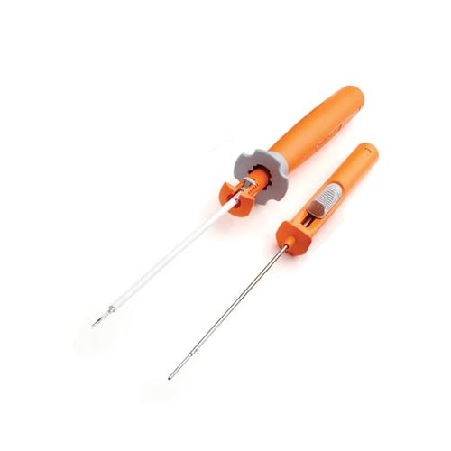
Inside-Out Meniscal Repair Needles are specifically engineered instruments designed to facilitate this precise suture passage. They are characterized by their slender profile, controlled curvature, and mechanisms for securely delivering suture material through the torn meniscal tissue and out of the joint. Their design directly addresses the challenges of working within the confined space of the knee joint while ensuring the structural integrity of the repair.
Key Advantages of the Inside-Out Technique:
- Superior Biomechanical Strength: Allows for strong, reproducible suture configurations.
- Versatility: Applicable to a wide range of meniscal tear types and locations.
- Direct Visualization: Enables precise suture placement under arthroscopic guidance.
- Proven Efficacy: Extensive long-term studies support its high success rates in appropriate cases.
- Meniscal Preservation: Promotes healing of the native meniscus, mitigating the risk of future degenerative changes.
Deep-Dive into Technical Specifications & Mechanisms
The efficacy of Inside-Out Meniscal Repair Needles is rooted in their meticulous design and the materials used in their construction.
Design & Materials
Inside-Out needles are precision instruments, often comprising several key features:
- Needle Characteristics:
- Length: Typically ranges from 15 cm to 25 cm, allowing reach to various meniscal locations.
- Gauge: Fine gauges (e.g., 18G, 20G) minimize tissue trauma while maintaining rigidity.
- Curvature: Precisely engineered curves (e.g., 20°, 45°, 90°) facilitate access to posterior horn tears, body tears, and even anterior horn tears, navigating around articular cartilage and bony structures.
- Tip Design:
- Trocar/Cutting Tip: Sharp, beveled tips allow for easier penetration through dense meniscal tissue.
- Blunt Tip: Less common for the needle itself, but blunt cannulas are used to protect soft tissues.
- Suture Passage Mechanism:
- Eyelet/Hole: A small aperture near the tip through which the suture is threaded.
- Slotted Design: A channel or slot along the needle shaft allows the suture to be "loaded" and then pushed through the tissue.
- Pre-loaded Needles: Some systems come with sutures pre-threaded, streamlining the process.
- Material Composition:
- Medical-Grade Stainless Steel (e.g., 304, 316L): Chosen for its exceptional strength, rigidity, corrosion resistance, and biocompatibility. It can withstand repeated sterilization cycles without degradation.
- Titanium Alloys: In some specialized instruments, titanium offers superior strength-to-weight ratio and enhanced biocompatibility, particularly for components that might remain in contact with tissue for longer periods, though less common for the needles themselves.
- Handle Design:
- Ergonomic: Designed for comfortable grip and precise control during manipulation.
- Single-Use vs. Reusable: Many needles are single-use to ensure optimal sharpness and sterility. Reusable handles may be paired with disposable needle tips.
- Attachment Points: Securely connect to cannulas, suture passers, and sometimes knot-tying devices.
- Associated Components:
- Cannulas: Hollow tubes inserted through arthroscopic portals, providing a protected pathway for the needles into the joint and preventing damage to articular cartilage.
- Suture Retrievers/Graspers: Instruments used externally to grasp and retrieve the suture ends once they exit the skin.
- Knot Pushers: Used arthroscopically to advance and secure knots inside the joint.
Biomechanics of Inside-Out Repair
The biomechanical superiority of the Inside-Out technique lies in its ability to create stable, load-sharing repairs.
- Suture Configurations:
- Vertical Mattress Suture: The most common and biomechanically strongest configuration. It compresses the tear edges, creating a broad contact area and providing excellent resistance to pull-out forces.
- Horizontal Mattress Suture: Offers good compression along the tear line, particularly useful for more stable, horizontal tears.
- Figure-of-Eight: A variation that can provide excellent stability for certain tear patterns.
- Load Distribution: By placing multiple sutures across the tear, the technique ensures that stress is distributed evenly, minimizing localized strain on the healing tissue.
- Pull-Out Strength: Studies consistently show that Inside-Out repairs, especially with vertical mattress sutures, exhibit superior pull-out strength compared to other techniques, critical for resisting the forces encountered during knee movement and rehabilitation.
- Healing Environment: The compression provided by the sutures brings the torn edges into apposition, facilitating the migration of healing cells and growth factors into the repair site, especially in the vascularized "red-red" and "red-white" zones of the meniscus.
- Comparison to Other Techniques:
- All-Inside Repair: Utilizes implants (suture anchors, all-suture devices) fully contained within the joint. While less invasive externally, it may offer less versatility in suture configurations and potentially higher material costs. Biomechanically, it can be comparable, but the surgeon has less direct control over the "outside" component of the repair.
- Outside-In Repair: Sutures are passed from the outside of the knee into the joint, then retrieved arthroscopically. It is useful for anterior and middle segment tears but can be challenging for posterior horn tears and offers less direct control over the external knot tying.
Extensive Clinical Indications & Usage
Selecting the appropriate meniscal repair technique hinges on a thorough understanding of the tear characteristics and patient factors.
Clinical Indications
Inside-Out meniscal repair is indicated for a variety of tear types, primarily those with good healing potential:
- Tear Morphology:
- Longitudinal Tears: Vertical tears running along the circumference of the meniscus, especially those greater than 1 cm.
- Bucket-Handle Tears: A type of longitudinal tear where a portion of the meniscus is displaced into the intercondylar notch, often requiring reduction before repair.
- Radial Tears: Tears extending from the free edge towards the periphery, if they extend into the vascularized zone.
- Root Tears: Tears at the meniscal attachments to the tibial plateau, though specialized root repair techniques are often combined.
- Tear Location:
- Peripheral Tears (Red-Red Zone): Tears within the vascularized outer third of the meniscus, which have the highest healing potential.
- Red-White Zone Tears: Tears extending into the junction of the vascularized and avascular zones, also suitable for repair.
- Patient Factors:
- Age: Younger, biologically active patients generally have better healing potential.
- Activity Level: Active individuals who wish to return to sports benefit most from meniscal preservation.
- Concomitant Injuries: Often performed in conjunction with anterior cruciate ligament (ACL) reconstruction, as the presence of ACL reconstruction can enhance meniscal healing due to increased vascularity and stability.
- Meniscal Stability: Tears that are unstable and displace with probing.
Detailed Surgical Applications (Fitting/Usage Instructions)
The Inside-Out technique requires meticulous surgical planning and execution.
- Patient Positioning & Arthroscopic Portal Placement:
- Patient typically supine with the knee flexed, often in a leg holder.
- Standard anteromedial and anterolateral arthroscopic portals are established for visualization and instrument access.
- Accessory portals (e.g., posteromedial) may be used for specific tear locations.
- Meniscal Visualization & Debridement:
- A diagnostic arthroscopy is performed to assess the tear pattern, size, and location.
- Unstable tear edges are débrided to create fresh, bleeding surfaces, which enhances healing.
- Synovial rasping or trephination of the meniscal rim may be performed to promote vascular access to the repair site.
- Needle Insertion through Cannula:
- A specialized cannula is inserted through an arthroscopic portal (e.g., anteromedial for medial meniscus, anterolateral for lateral meniscus).
- The Inside-Out repair needle, pre-loaded with suture, is then passed through the cannula into the joint.
- Crucial Step: Under direct arthroscopic visualization, the surgeon carefully guides the needle to penetrate the superior and inferior surfaces of the meniscal tear, typically 2-3 mm from the tear edge and 3-5 mm apart for each stitch.
- Precise Suture Placement:
- The needle is advanced through the meniscal tissue, across the tear, and out through the meniscal rim.
- For the "outside" component, a small accessory incision (typically 1-2 cm) is made on the skin corresponding to the exit point of the needle.
- Neurovascular Protection: A critical step involves using a blunt retractor or specialized skin protector (e.g., a small cannula or finger) to protect underlying neurovascular structures (e.g., saphenous nerve and vein for medial repairs, peroneal nerve for lateral repairs) as the needle exits the joint capsule and skin.
- The suture is then advanced, and the needle tip exits the skin incision.
- Retrieval of Sutures on the "Outside":
- Once the needle has exited, the suture ends are grasped with a hemostat or suture retriever on the "outside" of the knee.
- The needle is then carefully withdrawn, leaving the suture traversing the meniscal tear.
- Multiple sutures are placed in a similar fashion, typically 3-5 sutures depending on the tear length, often in a vertical mattress configuration.
- Knot Tying Techniques:
- The suture ends are then tied securely over the joint capsule on the "outside" of the knee. This can be done either openly with standard surgical knots or using specialized knot-tying instruments.
- Arthroscopic knot pushers are used inside the joint to advance the knot and ensure adequate tension.
- The knots are typically tied firmly but without excessive tension that could strangulate the meniscal tissue.
- Post-operative Assessment:
- The knee is cycled through a range of motion to assess the stability of the repair and ensure no impingement or excessive tension.
- The arthroscope confirms accurate reduction and stable fixation of the meniscal tear.
Patient Outcome Improvements
The decision to repair a torn meniscus using Inside-Out needles, rather than resecting it, has profound long-term benefits for the patient:
- Higher Healing Rates: Studies consistently demonstrate high healing rates (70-90%) for appropriately selected tears, especially in conjunction with ACL reconstruction.
- Preservation of Meniscal Function: Maintaining the native meniscus preserves its critical roles in:
- Shock Absorption: Distributes compressive loads across the knee joint, reducing peak stresses on articular cartilage.
- Load Distribution: Increases the contact area between the femur and tibia, preventing focal loading.
- Joint Stability: Contributes to the overall stability of the knee, particularly in conjunction with ligaments.
- Reduced Risk of Osteoarthritis Progression: By preserving the meniscal tissue and its biomechanical functions, the risk of developing early-onset osteoarthritis, a common sequela of meniscectomy, is significantly reduced.
- Improved Long-Term Knee Health & Functional Scores: Patients undergoing successful meniscal repair often report better long-term pain relief, functional outcomes, and satisfaction compared to those who undergo meniscectomy.
- Faster Return to Activity (with appropriate rehab): While rehabilitation for repair is longer and more restrictive than for meniscectomy, it allows for a more complete return to high-level activities in the long run.
Maintenance & Sterilization Protocols
Proper maintenance and sterilization of reusable Inside-Out Meniscal Repair Needles and their associated components are paramount to patient safety and instrument longevity.
Cleaning
- Immediate Post-Operative Cleaning: As soon as possible after use, instruments should be wiped clean of gross debris with a damp cloth.
- Soaking: Instruments should be soaked in an enzymatic detergent solution to prevent blood and tissue from drying onto surfaces.
- Manual Cleaning: Thorough manual cleaning with soft brushes and specialized cleaning solutions is essential to remove all visible organic material, especially from lumens, crevices, and articulation points.
- Rinsing: Rinse thoroughly with demineralized or distilled water to remove all detergent residue.
Inspection
- Visual Inspection: Before and after sterilization, each instrument must be meticulously inspected for:
- Damage: Bends, cracks, burrs, or deformities in the needle or handle.
- Dullness: Needle tips must be sharp for effective tissue penetration. Dull needles can cause excessive tissue trauma.
- Corrosion: Any signs of rust or pitting indicate material degradation and potential compromise of sterility and function.
- Functionality: Ensure all moving parts (if any) operate smoothly.
Sterilization
- Autoclave (Steam Sterilization): This is the most common and effective method for heat-stable instruments like stainless steel needles.
- Parameters: Typically involves exposure to saturated steam at specific temperatures and pressures for a defined duration (e.g., 121°C (250°F) for 15-30 minutes or 132°C (270°F) for 4-10 minutes, followed by a drying cycle).
- Validation: Sterilization cycles must be validated and routinely monitored using biological and chemical indicators.
- Flash Sterilization: Rapid steam sterilization for immediate use, typically for unwrapped instruments. While sometimes necessary, it should be used cautiously and only when standard sterilization is not feasible, as it offers less assurance of sterility.
- Ethylene Oxide (EtO) Sterilization: Used for heat-sensitive instruments, though less common for metal needles themselves. Requires specific aeration times to remove toxic gas residues.
- Packaging: Instruments must be packaged in sterile pouches, wraps, or rigid containers that allow steam penetration but maintain sterility until use.
- Storage: Sterilized instruments should be stored in a clean, dry, and protected environment to prevent recontamination.
Shelf Life & Reusability
- Single-Use Instruments: Many Inside-Out needles are designed for single use to ensure optimal sharpness and minimize infection risk. These should be disposed of immediately after use.
- Reusable Instruments: If a needle or its components are designed for reuse, the manufacturer's instructions for the maximum number of reprocessing cycles must be strictly adhered to. Exceeding these limits can compromise instrument integrity and patient safety.
Risks, Side Effects, or Contraindications
While Inside-Out meniscal repair is highly effective, like any surgical procedure, it carries potential risks and is not suitable for all patients or tear types.
Risks & Side Effects
- Neurovascular Injury: This is the most significant specific risk associated with the Inside-Out technique, due to the suture needles exiting the joint capsule and potentially coming close to nerves (e.g., saphenous nerve, peroneal nerve) and blood vessels (e.g., saphenous vein, popliteal artery/vein). Meticulous surgical technique and neurovascular protection are paramount.
- Infection: As with any invasive procedure, there is a risk of surgical site infection, though rare with proper sterile technique.
- Suture Breakage/Loosening: The sutures may break or loosen, leading to failure of the repair.
- Re-Tear/Failure of Repair: Despite successful initial repair, the meniscus may re-tear due to trauma or failure of biological healing.
- Pain, Swelling, Stiffness: Common post-operative symptoms that typically resolve with time, rehabilitation, and pain management.
- Hardware Irritation: If suture anchors or other implants are used in conjunction, they can sometimes cause irritation, though this is less common with Inside-Out needles themselves.
- Puncture of Articular Cartilage: Improper needle trajectory can damage healthy articular cartilage.
Contraindications
- Degenerative Tears with Extensive Damage: Tears that are chronic, degenerative, and involve significant tissue loss or poor tissue quality have limited healing potential.
- Tears in Avascular Zones: Tears entirely within the inner, avascular "white-white" zone typically have a very low potential for healing and are generally not amenable to repair.
- Severe Osteoarthritis: Patients with advanced knee osteoarthritis may not benefit from meniscal repair, as the underlying joint degeneration is the primary source of symptoms.
- Poor Tissue Quality: Severely macerated, frayed, or calcified meniscal tissue is unlikely to hold sutures effectively.
- Patient Non-Compliance: A strict post-operative rehabilitation protocol is crucial for successful meniscal repair. Patients unwilling or unable to adhere to these restrictions may have a higher risk of failure.
- Certain Systemic Conditions: Conditions that impair wound healing (e.g., severe diabetes, chronic steroid use, significant malnutrition) may be relative contraindications.
Massive FAQ Section
Q1: What is an Inside-Out Meniscal Repair?
A1: An Inside-Out meniscal repair is an arthroscopic surgical technique where sutures are passed from inside the knee joint, through the torn meniscus, and then retrieved outside the joint capsule through a small skin incision. This allows for strong, precise fixation of the torn meniscal edges.
Q2: Why choose Inside-Out over other meniscal repair techniques?
A2: The Inside-Out technique is often preferred for its superior biomechanical strength, versatility in addressing various tear types and locations (especially posterior horn tears), and the surgeon's ability to achieve precise suture placement and tension under direct arthroscopic visualization. It has a long track record of high success rates.
Q3: Are Inside-Out Meniscal Repair Needles reusable or single-use?
A3: Many Inside-Out Meniscal Repair Needles are designed for single use to ensure optimal sharpness, sterility, and to mitigate the risk of cross-contamination. Some systems may feature reusable handles with disposable needle tips. Always refer to the manufacturer's instructions for specific product guidelines.
Q4: What materials are Inside-Out needles typically made from?
A4: They are predominantly made from high-quality medical-grade stainless steel (e.g., 304 or 316L). This material is chosen for its strength, rigidity, corrosion resistance, and biocompatibility, allowing for effective tissue penetration and repeated sterilization if applicable.
Q5: What are the main advantages of using these needles in surgery?
A5: The main advantages include enabling precise and strong suture placement, allowing for various suture configurations (like vertical mattress), facilitating repair of tears in challenging locations, contributing to high healing rates, and ultimately preserving the meniscal tissue to prevent future osteoarthritis.
Q6: What are the potential complications associated with Inside-Out repair using these needles?
A6: The most notable complication is the risk of neurovascular injury (damage to nerves or blood vessels) as the needle exits the joint capsule. Other risks include infection, suture breakage, re-tear, pain, swelling, and stiffness. Meticulous surgical technique is crucial to minimize these risks.
Q7: How long does recovery typically take after an Inside-Out meniscal repair?
A7: Recovery is a gradual process. Patients typically use crutches for 4-6 weeks with limited weight-bearing. Return to light activities usually occurs around 3-4 months, and full return to sports may take 6 months or longer, depending on the individual, the tear, and adherence to a structured rehabilitation program.
Q8: Can all meniscal tears be repaired using the Inside-Out method?
A8: No, not all meniscal tears are suitable for Inside-Out repair. It is most effective for tears located in the vascularized (red-red or red-white) zones of the meniscus, longitudinal tears, bucket-handle tears, and some radial tears. Degenerative tears, tears in the avascular "white-white" zone, or severely damaged tissue often have poor healing potential and may not be candidates for repair.
Q9: What is the role of a cannula in this surgical technique?
A9: A cannula acts as a protective sleeve or conduit. It is inserted through the arthroscopic portal into the knee joint, providing a clear and protected pathway for the Inside-Out needle. This prevents damage to the articular cartilage and surrounding soft tissues as the needle is advanced and withdrawn.
Q10: How do surgeons ensure neurovascular safety during the "outside" component of the repair?
A10: Surgeons employ several critical steps: using blunt retractors or specialized skin protectors to shield neurovascular structures, making a small, controlled external incision, careful anatomical dissection, and having a thorough understanding of the regional anatomy to avoid vital structures. Direct visualization and palpation are key.
Q11: What is the typical success rate of an Inside-Out meniscal repair?
A11: When performed on appropriately selected tears and patients, the success rate for Inside-Out meniscal repair can range from 70% to over 90%, especially when performed concurrently with an ACL reconstruction, which can enhance healing.
Q12: How are these instruments cleaned and sterilized for reuse?
A12: Reusable instruments undergo a multi-step process: immediate post-operative cleaning to remove debris, soaking in enzymatic detergents, thorough manual cleaning, rinsing, meticulous inspection for damage, and finally, steam sterilization (autoclaving) at specific temperature and pressure parameters. Strict adherence to manufacturer guidelines and hospital protocols is essential.
This comprehensive guide underscores the critical role of Inside-Out Meniscal Repair Needles in modern orthopedic surgery, facilitating effective meniscal preservation and significantly improving long-term patient outcomes.