The Humeral Intramedullary Nail: A Comprehensive Orthopedic Guide
1. Introduction & Overview
The humerus, the long bone of the upper arm, is frequently subjected to traumatic injuries, with fractures of the humeral shaft being a common occurrence. Effective management of these fractures is paramount to restoring arm function and preventing long-term disability. While various treatment modalities exist, the Humeral Intramedullary (IM) Nail has emerged as a gold standard for many humeral shaft fractures due to its biomechanical advantages, minimally invasive nature, and excellent patient outcomes.
An intramedullary nail is a load-sharing implant inserted into the medullary canal (the hollow center) of a long bone to stabilize fractures. For the humerus, these nails provide robust internal fixation, promoting healing while allowing for early mobilization. This comprehensive guide delves into the intricate details of humeral intramedullary nails, covering their design, clinical applications, surgical techniques, biomechanics, maintenance, and the significant improvements they bring to patient recovery and quality of life.
2. Deep-Dive into Technical Specifications & Mechanisms
The efficacy of a humeral intramedullary nail is rooted in its sophisticated design and material properties. These implants are engineered to integrate seamlessly with the bone's anatomy and biological healing processes.
2.1 Design Principles and Materials
Humeral IM nails are not simply straight rods; their design incorporates anatomical curvatures and features optimized for humeral fixation.
- Materials: The vast majority of modern humeral nails are manufactured from biocompatible, high-strength alloys, primarily:
- Titanium Alloys (e.g., Ti-6Al-4V): Known for their excellent strength-to-weight ratio, corrosion resistance, and superior biocompatibility, titanium alloys are the preferred choice. Their modulus of elasticity is closer to cortical bone than stainless steel, which can lead to more favorable load sharing and reduced stress shielding.
- Stainless Steel (e.g., 316LVM): While still used, stainless steel is less common for primary humeral nailing due to its higher stiffness and potential for greater stress shielding.
- Nail Geometry:
- Anatomical Curvature: Nails are pre-bent to match the natural anterior bow of the humerus, reducing stress concentrations and facilitating easier insertion.
- Cross-Section: Most nails have a circular or cloverleaf cross-section, which optimizes fit within the medullary canal.
- Cannulation: Many nails are cannulated (hollow) to allow for insertion over a guide wire, which aids in precise placement and minimally invasive techniques. Solid nails offer slightly greater bending stiffness.
- Length and Diameter: Available in a wide range of lengths (typically 180-320 mm) and diameters (6-10 mm) to accommodate varying patient anatomies and fracture patterns.
- Locking Mechanisms: Interlocking screws are critical for preventing shortening, rotation, and angulation of the fracture fragments.
- Proximal Locking: Often features multi-planar screw options (e.g., two oblique screws and one transverse screw) to maximize fixation in the metaphyseal bone and resist rotational forces. Some designs incorporate specific holes for rotator cuff sparing techniques.
- Distal Locking: Typically involves one or two transverse screws to secure the distal fragment. Dynamic locking options may also be available, allowing for controlled axial micromotion to promote secondary healing.
- Screw Types: Self-tapping, self-drilling screws are common, designed for optimal purchase in cortical bone.
2.2 Biomechanics of Humeral Intramedullary Nailing
The biomechanical advantages of IM nailing stem from its central placement and load-sharing capabilities.
- Load Sharing: Unlike plates, which are load-bearing constructs placed on the bone surface, intramedullary nails share axial loads with the bone itself. This reduces stress shielding, a phenomenon where the implant bears too much load, leading to bone resorption and weakening. Load sharing promotes earlier cortical remodeling and stronger callus formation.
- Stability: The nail provides inherent stability against bending and shear forces due to its central position within the medullary canal.
- Rotational Control: Interlocking screws are crucial for providing rotational stability, preventing the fracture fragments from twisting relative to each other. The number and configuration of locking screws directly impact rotational stiffness.
- Working Length: The segment of the nail not supported by bone (between the fracture fragments) is known as the working length. A shorter working length generally correlates with greater stiffness and stability, although some micromotion can be beneficial for secondary bone healing.
- Minimally Invasive: The percutaneous nature of IM nailing preserves the periosteal blood supply, which is vital for bone healing, unlike extensive open plating which can disrupt it. This leads to reduced soft tissue damage and potentially lower infection rates.
3. Extensive Clinical Indications & Usage
The humeral intramedullary nail is a versatile implant suitable for a broad spectrum of humeral shaft fractures and other pathological conditions.
3.1 Indications for Humeral IM Nailing
- Humeral Shaft Fractures:
- Transverse and Short Oblique Fractures (AO/OTA 12-A): Excellent stability and compression can be achieved.
- Spiral and Long Oblique Fractures (AO/OTA 12-B): Locking screws effectively prevent shortening and rotation.
- Comminuted Fractures (AO/OTA 12-C): The nail acts as an internal splint, maintaining length and alignment while preserving surrounding soft tissue.
- Segmental Fractures: Multiple fracture lines within the shaft.
- Pathological Fractures: Due to metastatic disease or primary bone tumors, where the nail provides immediate stability and pain relief.
- Impending Pathological Fractures: Prophylactic nailing in cases of significant cortical destruction.
- Non-unions and Malunions: Revision surgery to achieve union or correct deformity.
- Polytrauma Patients: Allows for early mobilization and weight-bearing (if other injuries permit), crucial for overall patient recovery.
- Open Fractures (Gustilo-Anderson Type I & II): Often preferred over plates due to lower risk of soft tissue complications and infection, especially after debridement.
3.2 Detailed Surgical Technique (Fitting/Usage Instructions)
The successful implantation of a humeral intramedullary nail requires meticulous pre-operative planning and precise surgical execution.
- Pre-operative Planning:
- Imaging: Anteroposterior and lateral X-rays of the entire humerus, including the shoulder and elbow joints, are essential. CT scans may be useful for complex fractures or articular involvement.
- Nail Selection: Determine appropriate nail length and diameter based on canal width (measured from X-rays or templating) and fracture location.
- Patient Positioning:
- Antegrade Nailing (Proximal Entry): Typically beach chair position or lateral decubitus, allowing full access to the shoulder and arm. Fluoroscopy is positioned to obtain AP and lateral views of the humerus.
- Retrograde Nailing (Distal Entry): Lateral decubitus or supine with the arm draped across the chest, allowing access to the olecranon fossa.
- Antegrade Nailing Technique:
- Incision: A small incision (2-5 cm) is made over the greater tuberosity.
- Entry Portal: The deltoid muscle is split, and the rotator cuff (supraspinatus tendon) is either split or a rotator cuff sparing approach (e.g., lateral to the bicipital groove) is used to create an entry portal into the medullary canal.
- Guide Wire Insertion: A guide wire is inserted down the medullary canal, crossing the fracture site under fluoroscopic guidance.
- Reaming (Optional): If a larger nail is desired or the canal is narrow, sequential reaming over the guide wire may be performed to prepare the canal.
- Nail Insertion: The selected nail is mounted on an insertion handle and carefully advanced over the guide wire across the fracture, ensuring proper reduction.
- Proximal Locking: Using a specialized jig attached to the insertion handle, proximal interlocking screws are placed through small stab incisions. Fluoroscopy confirms screw position.
- Distal Locking: Once the nail is fully inserted and the fracture reduced, distal locking screws are placed. This can be done with a jig (less common for distal humerus) or a freehand technique using fluoroscopy.
- Verification: Final AP and lateral fluoroscopic images confirm fracture reduction, nail position, and screw placement.
- Wound Closure: Incisions are closed in layers.
- Retrograde Nailing Technique:
- Indications: Primarily for distal humeral shaft fractures, floating elbow injuries, or when antegrade nailing is contraindicated.
- Entry Portal: A small incision is made in the posterior aspect of the elbow, and the entry portal is created in the olecranon fossa, just proximal to the olecranon.
- Guide Wire & Nail Insertion: Similar to antegrade, the guide wire is advanced proximally, and the nail is inserted across the fracture.
- Locking: Proximal and distal locking screws are placed as described above.
- Post-operative Care:
- Pain Management: Appropriate analgesia is crucial.
- Immobilization: Often minimal, a sling may be used for comfort for a short period.
- Rehabilitation: Early range of motion exercises for the shoulder and elbow are typically initiated within days of surgery, progressing to strengthening as tolerated and with bone healing progression.
- Weight-bearing: Gradual progression of weight-bearing and activity is guided by fracture stability and radiographic evidence of healing.
- Follow-up: Regular clinical and radiographic evaluations are necessary to monitor healing and detect complications.
4. Risks, Side Effects, or Contraindications
While highly effective, humeral intramedullary nailing is not without potential risks and contraindications.
4.1 Potential Risks and Complications
- Shoulder Pain/Impingement: Particularly with antegrade nails, the proximal end of the nail or locking screws can irritate the rotator cuff or subacromial bursa, leading to pain and impingement. Rotator cuff sparing techniques aim to mitigate this.
- Nerve Injury:
- Radial Nerve: Most commonly injured during distal locking, due to its proximity to the distal humerus.
- Axillary Nerve: Risk during proximal approach, especially with excessive retraction.
- Musculocutaneous Nerve: Less common, but possible.
- Vascular Injury: Rare, but potential for damage to the brachial artery.
- Infection: As with any surgical procedure involving implants, superficial or deep infection can occur, potentially requiring hardware removal.
- Non-union or Delayed Union: Despite stable fixation, biological factors can lead to delayed or absent healing. Smoking, poor nutrition, and severe comminution are risk factors.
- Malunion: Incorrect alignment (angulation, rotation, or shortening) of the healed fracture. Rotational malunion is a common concern if not carefully monitored during surgery.
- Hardware Failure: Breakage of the nail or interlocking screws, often due to non-union or excessive early loading.
- Intra-articular Penetration: Proximal or distal migration of the nail or screws into the shoulder or elbow joint, causing pain and cartilage damage.
- Refracture: After hardware removal, the bone may be temporarily weaker, leading to a risk of refracture.
4.2 Contraindications
- Active Infection: Absolute contraindication. Infection must be cleared before implanting hardware.
- Severe Open Fractures (Gustilo-Anderson Type III): While Type I & II can be nailed, severe Type III open fractures with extensive soft tissue damage and contamination may initially be better managed with external fixation.
- Fractures Extending Too Close to Articular Surfaces: If the fracture line extends into the shoulder or elbow joint, or very close to it, IM nailing may not provide adequate fixation, or the nail could compromise joint function.
- Extremely Wide Medullary Canal: If the canal is too wide, the nail may not achieve sufficient cortical contact for stable fixation.
- Patient Medical Instability: Patients unable to tolerate anesthesia or prolonged surgery.
- Pre-existing Shoulder/Elbow Pathology: Severe osteoarthritis or stiffness that would be exacerbated by the surgical approach or implant.
- Proximal or Distal Comminution at Entry Point: If the bone at the planned entry site is severely fractured, it may not provide a stable portal for nail insertion.
5. Maintenance & Sterilization Protocols (for Surgical Instruments)
Humeral intramedullary nails are supplied sterile and are single-use devices. Therefore, maintenance and sterilization protocols apply to the reusable surgical instrumentation used for their implantation. Strict adherence to these protocols is critical to prevent surgical site infections.
5.1 Pre-use Handling of the Implant
- Integrity Check: Before opening, visually inspect the sterile packaging for any tears, punctures, or signs of compromise. Do not use if packaging is damaged.
- Storage: Store implants according to manufacturer's instructions, typically in a cool, dry place away from direct sunlight.
- Aseptic Technique: Once the sterile barrier is broken in the operating room, all handling of the nail and its components must be performed using strict aseptic technique by scrubbed personnel.
5.2 Cleaning and Sterilization of Reusable Instruments
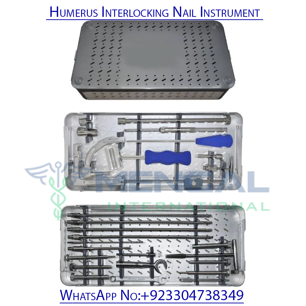
The associated surgical instrument sets (e.g., reamers, guide wire handles, insertion handles, locking jigs, screw drivers) require rigorous processing.
- Point-of-Use Treatment: Immediately after use, instruments should be wiped to remove gross contaminants and kept moist (e.g., with enzymatic spray or damp towel) to prevent blood and tissue from drying onto surfaces.
- Decontamination:
- Manual Cleaning: Instruments are thoroughly cleaned by hand using brushes, enzymatic detergents, and warm water. All lumens and crevices must be meticulously cleaned.
- Automated Cleaning: Washer-disinfectors are often used, following specific cycles for surgical instruments. Ultrasonic cleaners can be used to remove microscopic debris from intricate parts.
- Inspection: After cleaning, each instrument must be visually inspected for cleanliness, damage, corrosion, and proper function. Damaged instruments should be removed from circulation.
- Packaging: Instruments are typically assembled into trays, often with specific layouts, and then wrapped in sterilization wraps or placed into rigid sterilization containers. Packaging must allow sterilant penetration and maintain sterility until use.
- Sterilization:
- Steam Sterilization (Autoclaving): This is the most common and effective method. Parameters vary by device and facility, but typical cycles include:
- Pre-vacuum (Dynamic-Air-Removal) Cycle: 132-135°C (270-275°F) for 4 minutes at 27-30 psi, followed by a drying phase.
- Gravity Displacement Cycle: 121°C (250°F) for 30 minutes.
- Low-Temperature Sterilization: For heat-sensitive instruments, methods like hydrogen peroxide gas plasma or ethylene oxide (EtO) may be used, following specific manufacturer guidelines.
- Steam Sterilization (Autoclaving): This is the most common and effective method. Parameters vary by device and facility, but typical cycles include:
- Storage of Sterile Instruments: Sterilized instrument sets are stored in a designated, clean, dry, and secure area, protected from environmental contaminants.
- Traceability: Each sterilization cycle and instrument set should be documented, allowing for traceability in case of contamination or adverse events.
6. Patient Outcome Improvements
The adoption of humeral intramedullary nailing has significantly advanced the treatment of humeral shaft fractures, leading to tangible improvements in patient outcomes.
- Reduced Hospital Stay: The minimally invasive nature and stable fixation often allow for earlier discharge compared to more extensive open reduction and plating procedures.
- Faster Return to Function: Stable internal fixation facilitates early mobilization of the shoulder and elbow, preventing stiffness and atrophy. This leads to a quicker return to activities of daily living, work, and recreational pursuits.
- Improved Pain Control: A stable fracture is inherently less painful. IM nailing provides immediate stability, significantly reducing post-operative pain and reliance on strong analgesics.
- Higher Union Rates: Studies consistently show high union rates for humeral shaft fractures treated with IM nails, often exceeding those of non-operative management for displaced fractures.
- Lower Complication Rates (in specific contexts): Compared to external fixation or open plating for certain fracture types (e.g., open fractures), IM nailing can have lower rates of infection and soft tissue complications.
- Better Cosmetic Outcomes: Smaller incisions result in less scarring compared to the long incisions required for plate osteosynthesis.
- Enhanced Quality of Life: By restoring function, reducing pain, and facilitating a quicker return to normal activities, humeral IM nailing significantly improves the overall quality of life for patients recovering from these debilitating injuries.
7. Frequently Asked Questions (FAQ)
Q1: What is a humeral intramedullary nail?
A: A humeral intramedullary nail is a surgical implant, typically made of titanium alloy, inserted into the hollow center (medullary canal) of the humerus (upper arm bone) to stabilize fractures. It acts as an internal splint to hold the bone fragments in place while they heal.
Q2: When is a humeral intramedullary nail used?
A: It is primarily used for fractures of the humeral shaft (the middle part of the upper arm bone), including transverse, oblique, spiral, and comminuted fractures. It's also used for pathological fractures, non-unions, and in polytrauma patients.
Q3: What are the advantages of IM nailing for a humeral fracture?
A: Key advantages include its minimally invasive nature (smaller incisions, less soft tissue disruption), load-sharing biomechanics (promoting natural bone healing), high union rates, ability to allow early functional rehabilitation, and often a quicker return to daily activities.
Q4: What are the risks associated with humeral IM nailing?
A: Potential risks include shoulder pain (especially with antegrade nails), nerve injury (e.g., radial nerve), infection, non-union or delayed healing, malunion (incorrect alignment), and hardware failure (e.g., screw breakage).
Q5: How long does recovery take after humeral IM nailing?
A: Recovery time varies depending on the fracture severity, individual healing capacity, and adherence to physical therapy. While early shoulder and elbow motion typically begins within days, full bone healing can take 3-6 months or longer. Return to strenuous activities is usually gradual.
Q6: Will I need to have the nail removed?
A: Not always. If the nail is not causing pain or problems, it can often remain in place indefinitely. However, if it causes discomfort, impingement, or other complications, surgical removal may be recommended, typically after the fracture has fully healed (usually 1-2 years post-op).
Q7: What materials are humeral nails made from?
A: Most modern humeral intramedullary nails are made from titanium alloys (e.g., Ti-6Al-4V) due to their excellent strength, corrosion resistance, and biocompatibility.
Q8: Can I undergo an MRI with a humeral nail?
A: Generally, yes. Titanium alloy nails are non-ferromagnetic and considered safe for MRI scans. However, it's crucial to inform your healthcare provider about the implant before any MRI procedure. Artifacts from the metal may obscure imaging in the immediate vicinity of the nail.
Q9: What is the difference between an antegrade and retrograde nail?
A: An antegrade nail is inserted from the top of the humerus, near the shoulder (proximal entry). A retrograde nail is inserted from the bottom of the humerus, near the elbow (distal entry). The choice depends on the fracture location and surgeon preference.
Q10: How do I care for my arm after surgery?
A: Post-operative care typically involves pain management, keeping the surgical incision clean and dry, and wearing a sling for comfort as advised. Most importantly, early and consistent physical therapy exercises are crucial to regain range of motion and strength.
Q11: What kind of physical therapy is involved?
A: Physical therapy usually starts with gentle passive and active-assisted range of motion exercises for the shoulder and elbow, progressing to active motion, strengthening exercises, and eventually functional training as bone healing progresses and pain subsides.
Q12: How does the nail stabilize the bone?
A: The nail stabilizes the bone by being inserted into the medullary canal, acting as an internal splint. Interlocking screws are then placed through the bone and nail, both proximally and distally to the fracture, preventing shortening, rotation, and angulation of the bone fragments.