The Flexible Osteotome System: Revolutionizing Precision Bone Surgery
1. Comprehensive Introduction & Overview
In the dynamic field of orthopedic surgery, precision and minimal invasiveness are paramount to achieving optimal patient outcomes. The traditional osteotome, a fundamental instrument for cutting or shaping bone, has long been a staple in the operating room. However, its rigid design often presents challenges when navigating complex anatomical structures or performing intricate cuts in confined spaces.
Enter the Flexible Osteotome System – a groundbreaking evolution in orthopedic instrumentation designed to overcome these limitations. This innovative system represents a significant leap forward, offering surgeons unparalleled control, access, and precision in bone manipulation. By integrating flexible shaft technology with specialized cutting tips, the Flexible Osteotome System empowers surgeons to perform highly accurate osteotomies with reduced invasiveness, leading to improved surgical efficiency and superior patient recovery profiles.
This comprehensive guide delves deep into the Flexible Osteotome System, exploring its sophisticated design, diverse clinical applications, meticulous usage protocols, stringent maintenance requirements, underlying biomechanical advantages, and the transformative impact it has on patient care.
2. Deep-dive into Technical Specifications & Mechanisms
The Flexible Osteotome System is a testament to advanced engineering, combining biocompatible materials with innovative design principles to deliver superior surgical performance.
2.1. Design & Materials
The system typically comprises several key components, each engineered for specific functions:
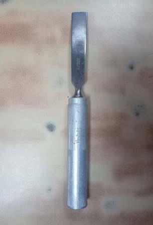
- Ergonomic Handle: Designed for comfortable, secure grip, often made from lightweight, high-strength polymers or anodized aluminum. It provides the surgeon with tactile feedback and reduces hand fatigue during prolonged procedures.
- Flexible Shaft: This is the core innovation. The shaft connects the handle to the cutting tip and is designed to bend and conform to anatomical contours.
- Materials: Often constructed from high-grade, super-elastic alloys such as Nitinol (Nickel-Titanium alloy) or specialized multi-strand stainless steel wires. These materials offer exceptional flexibility, torsional strength, and resistance to kinking, while maintaining biocompatibility.
- Design: May feature a segmented design, a coiled spring-like structure, or a series of interlocking segments to achieve controlled flexibility without compromising cutting force transmission.
- Interchangeable Cutting Tips/Blades: A variety of tips are available to suit different surgical requirements.
- Materials: Typically medical-grade stainless steel (e.g., 420 or 440C for sharpness retention) or titanium alloys (for strength and radiolucency).
- Types:
- Chisel Tips: For precise linear cuts.
- Gouge Tips: For scooping or creating channels.
- Curved Tips: Specifically designed for curvilinear osteotomies.
- Serrated/Oscillating Tips: For more aggressive bone removal when coupled with a power source.
- Ultrasonic Tips: For highly precise, soft tissue-sparing osteotomies when integrated with an ultrasonic generator.
- Connection Mechanism: A robust and secure coupling mechanism ensures a stable connection between the handle, flexible shaft, and cutting tip, often featuring quick-release or screw-lock designs for efficient tip exchange.
- Depth Markers/Gauges: Many systems incorporate laser-etched markings on the shaft or integrated depth stops on the tips to allow for precise control over the depth of bone penetration, crucial for preventing over-penetration and protecting vital structures.
2.2. Mechanism of Action
The primary mechanism of the Flexible Osteotome System revolves around its ability to deliver controlled cutting force to precise anatomical locations that are inaccessible with rigid instruments.
- Controlled Force Transmission: Despite its flexibility, the system is engineered to efficiently transmit the surgeon's applied force (manual or powered) directly to the cutting tip, ensuring effective bone penetration and cutting.
- Navigational Prowess: The flexible shaft allows the cutting tip to be maneuvered around anatomical obstacles, through small incisions, or into curved pathways within bone, significantly expanding surgical access.
- Precision and Minimally Invasive Access: By enabling access through smaller portals and around critical structures, the system facilitates truly minimally invasive approaches. This reduces the need for extensive soft tissue dissection and retraction, thereby minimizing trauma.
- Integration with Power Systems: While some flexible osteotomes are manually operated, many advanced systems are designed to connect to powered handpieces (e.g., oscillating saws, reciprocating saws, ultrasonic generators). The flexible shaft must be capable of transmitting these high-frequency vibrations or reciprocating motions without losing structural integrity or efficiency.
2.3. Biomechanics
The biomechanical advantages of the Flexible Osteotome System are profound and directly contribute to improved surgical outcomes:
- Reduced Stress on Surrounding Tissues: The ability to navigate complex pathways minimizes the need for aggressive retraction of soft tissues, nerves, and vessels, thus reducing iatrogenic injury and post-operative pain.
- Precision in Complex Geometries: The flexible shaft allows the cutting tip to follow intricate bone contours, enabling highly precise osteotomies (e.g., curved cuts, multi-planar cuts) that are difficult or impossible with rigid instruments. This ensures optimal bone-to-bone contact in reconstructive procedures.
- Controlled Bone Removal: The tactile feedback provided by the system, coupled with depth control features, allows for extremely controlled and incremental bone removal, preventing uncontrolled fractures or over-resection.
- Preservation of Periosteum and Vascularity: Minimally invasive access often means less disruption to the periosteum and surrounding vascular supply, which is critical for bone healing and reducing the risk of avascular necrosis.
- Even Stress Distribution: The controlled nature of the cut, especially in curved osteotomies, can lead to a more even distribution of stress across the bone interface post-operatively, promoting stronger fusion or healing.
3. Extensive Clinical Indications & Usage
The versatility and precision of the Flexible Osteotome System make it invaluable across a broad spectrum of orthopedic and related surgical disciplines.
3.1. Orthopedic Surgery
- Deformity Correction Osteotomies:
- High Tibial Osteotomy (HTO): For correcting varus or valgus deformities of the knee, the flexible system allows precise, curved osteotomies to realign the joint axis, particularly useful in closed-wedge or dome osteotomies.
- Femoral Osteotomy: Used in hip preservation surgeries or for correcting rotational/angular deformities of the femur.
- Foot & Ankle Surgery: Calcaneal osteotomies (e.g., for flatfoot correction), bunionectomies (for precise metatarsal head osteotomies), and other forefoot/midfoot reconstructive procedures requiring intricate bone cuts.
- Joint Preservation Surgery:
- Osteochondral Defect Repair: Creating precise recipient sites for grafts in areas like the femoral condyle or talus, especially in curved surfaces.
- Meniscal Root Repair: Preparing bone tunnels for suture anchor placement in meniscal root avulsions.
- Fracture Fixation:
- Precisely shaping bone fragments for optimal reduction and seating of plates or screws, especially in periarticular fractures.
- Removing malunion or non-union tissue for revision surgeries.
- Spinal Surgery:
- Laminectomy/Laminoplasty: Precise bone removal in complex spinal anatomy, minimizing dural or neural tissue damage.
- Osteophytectomy: Removing bone spurs that impinge on nerves or vessels.
- Vertebral Column Resection (VCR): In complex deformity correction, though more specialized tools might be involved, flexible osteotomes can assist in initial bone cuts or precise shaping.
- Interbody Fusion: Preparing precise endplate surfaces for interbody cage placement, particularly in oblique or transforaminal approaches.
- Hand & Wrist Surgery:
- Corrective osteotomies for carpal bones or metacarpals, addressing malunions or deformities in small, confined joints.
3.2. Maxillofacial & Craniofacial Surgery
- Orthognathic Surgery: Performing precise osteotomies (e.g., Le Fort I, bilateral sagittal split osteotomy) for jaw repositioning, especially when navigating around dental roots and nerves.
- Craniofacial Reconstruction: Shaping bone grafts or performing precise cuts in complex cranial vault reconstructions.
3.3. Fitting & Usage Instructions (General Principles)
Proper usage is critical for safety and efficacy. While specific protocols vary by manufacturer and surgical procedure, general guidelines include:
- Pre-operative Planning:
- Thorough review of imaging (X-rays, CT, MRI) to map out osteotomy lines and identify critical anatomical structures.
- Consideration of surgical approach and instrument trajectory.
- System Assembly:
- Carefully unpack sterile components.
- Connect the selected cutting tip to the flexible shaft and then to the handle, ensuring all connections are secure and audible clicks are heard if applicable.
- If powered, connect the flexible shaft to the appropriate powered handpiece, verifying compatibility and secure attachment.
- Surgical Exposure:
- Establish the surgical field and achieve adequate exposure, often through minimally invasive portals or small incisions.
- Bone Cutting Technique:
- Controlled Application: Apply gentle, controlled pressure. Avoid excessive force, which can lead to uncontrolled bone fracture or instrument damage.
- Multi-directional Control: Utilize the flexibility to approach the osteotomy line from optimal angles, following the planned trajectory.
- Irrigation: Continuously irrigate the surgical site with saline to cool the bone and instrument, preventing thermal necrosis and clearing debris, especially with powered systems.
- Depth Control: Constantly monitor the depth of cut using visual markers on the instrument, fluoroscopy, or integrated navigation systems.
- Oscillation/Reciprocation: If using a powered system, activate the power source only when the cutting tip is in contact with bone, and use a controlled, sweeping motion.
- Post-Osteotomy:
- Verify the completeness and accuracy of the osteotomy.
- Carefully remove the instrument from the surgical site.
- Disassembly & Initial Cleaning:
- Immediately after use, disassemble the system.
- Begin preliminary cleaning to prevent blood and tissue from drying on the surfaces.
4. Maintenance & Sterilization Protocols
Maintaining the integrity and sterility of the Flexible Osteotome System is paramount for patient safety and instrument longevity.
4.1. Cleaning
- Point-of-Use Cleaning: Immediately after surgery, wipe down instruments to remove gross contaminants. Flush lumens (if present) with sterile water.
- Manual Cleaning: Immerse instruments in an enzymatic detergent solution. Use soft brushes to thoroughly scrub all surfaces, paying close attention to crevices, connections, and the flexible shaft segments. Rinse thoroughly with purified water.
- Ultrasonic Cleaning: Place disassembled components in an ultrasonic cleaner with appropriate detergent for the recommended cycle time, ensuring all air bubbles are removed from lumens. Rinse thoroughly.
- Automated Washer-Disinfectors: If compatible, instruments can be processed in an automated washer-disinfector following manufacturer guidelines, using validated cycles.
- Final Rinse: Always use critical water (e.g., sterile distilled or reverse osmosis water) for the final rinse to prevent mineral deposits.
- Drying: Thoroughly dry all components with a lint-free cloth or filtered compressed air.
4.2. Inspection
After cleaning and before sterilization, each component must be meticulously inspected:
- Cutting Tips: Check for sharpness, burrs, nicks, bends, or signs of wear. Replace dull or damaged tips.
- Flexible Shaft: Inspect for kinking, cracks, fraying, loss of flexibility, or any signs of material fatigue. Ensure smooth articulation.
- Connections: Verify that all connection points are intact, free of damage, and function securely.
- Overall Integrity: Check for corrosion, discoloration, or any other physical damage. Discard or send for repair any compromised instruments.
4.3. Sterilization
- Packaging: Place instruments in appropriate sterilization wraps, pouches, or rigid containers, ensuring proper spacing for steam penetration.
- Steam Sterilization (Autoclave): This is the most common and preferred method.
- Parameters: Follow manufacturer's validated cycles for specific instruments. Typical parameters include:
- Gravity Displacement: 15-30 minutes at 121°C (250°F) or 4-10 minutes at 132°C (270°F).
- Pre-vacuum: 4 minutes at 132°C (270°F).
- Drying Time: Ensure adequate drying time to prevent condensation and maintain sterility.
- Parameters: Follow manufacturer's validated cycles for specific instruments. Typical parameters include:
- Ethylene Oxide (ETO): May be used for heat-sensitive components if steam sterilization is contraindicated, but less common for metallic osteotomes. Requires aeration time.
- Low-Temperature Plasma (Hydrogen Peroxide): A viable option for certain heat-sensitive items, offering faster cycles and no toxic residuals. Compatibility must be confirmed by the manufacturer.
4.4. Storage
- Store sterilized instruments in a clean, dry, and secure environment, away from extreme temperatures and humidity, until needed for surgery.
- Use dedicated trays or storage systems to protect instruments from damage during handling and transport.
5. Patient Outcome Improvements
The adoption of the Flexible Osteotome System translates directly into tangible benefits for patients, enhancing both short-term recovery and long-term functional results.
- Reduced Post-operative Pain: Minimally invasive approaches lead to less soft tissue dissection and retraction, resulting in significantly less tissue trauma and, consequently, reduced post-operative pain. This often decreases the need for opioid analgesics.
- Faster Recovery and Rehabilitation: Less trauma accelerates the healing process, allowing patients to mobilize earlier and progress faster through rehabilitation protocols, leading to a quicker return to daily activities and work.
- Smaller Incisions and Less Scarring: The ability to access surgical sites through smaller portals results in smaller skin incisions, leading to more cosmetically appealing scars and reduced risk of wound complications.
- Lower Risk of Infection: Smaller incisions and reduced tissue exposure inherently lower the risk of surgical site infections.
- Improved Functional Outcomes: By enabling more precise osteotomies and better anatomical alignment, the system contributes to superior biomechanical reconstruction, leading to improved joint mechanics, range of motion, and overall functional performance.
- Reduced Blood Loss: Minimally invasive techniques typically result in less intraoperative blood loss, reducing the need for blood transfusions and associated risks.
- Potential for Outpatient Procedures: For certain indications, the reduced invasiveness and faster recovery may allow for the procedure to be performed in an outpatient setting, reducing hospital stay costs and patient inconvenience.
- Enhanced Surgical Accuracy: The controlled nature and flexible access improve the accuracy of bone cuts, minimizing the risk of incomplete osteotomies or damage to adjacent neurovascular structures.
6. Risks, Side Effects, or Contraindications
While the Flexible Osteotome System offers significant advantages, it is essential to be aware of potential risks, side effects, and contraindications.
6.1. Risks
- Nerve or Vascular Damage: Despite enhanced precision, there remains a risk of injury to nearby nerves or blood vessels, especially in anatomically complex regions.
- Uncontrolled Fracture: Excessive force or improper technique can lead to unintended bone fractures beyond the planned osteotomy line.
- Infection: As with any surgical procedure, there is a risk of surgical site infection, though MIS techniques can lower this risk.
- Instrument Breakage: Although designed for durability, instruments can break or deform if subjected to extreme forces, improper use, or material fatigue. A broken fragment can necessitate further surgery for retrieval.
- Incomplete Osteotomy: Insufficient bone cutting can lead to an incomplete osteotomy, requiring further intervention.
- Thermal Necrosis: If powered systems are used without adequate irrigation, heat generation can damage bone cells, leading to delayed healing or non-union.
- Delayed Healing/Non-union: While precision can improve healing, individual patient factors and complications can still lead to delayed healing or non-union of the osteotomy site.
6.2. Side Effects
- Post-operative Pain: While generally reduced, some level of pain, swelling, and bruising is expected after any bone surgery.
- Scarring: Even with smaller incisions, a scar will form.
- Temporary Numbness/Paresthesia: Due to nerve manipulation or irritation during surgery.
6.3. Contraindications
- Severe Bone Fragility (Osteoporosis): In cases of severe osteoporosis, bone may be too fragile to withstand the forces of osteotomy, regardless of instrument flexibility, increasing the risk of comminuted fractures.
- Active Infection at Surgical Site: Performing an osteotomy in the presence of active infection can lead to spread of infection and poor healing.
- Inadequate Bone Stock: Insufficient healthy bone stock for the planned osteotomy or fixation.
- Patient Inability to Tolerate Surgery: General contraindications to anesthesia or major surgery apply.
- Uncooperative Patient: Patients unable to comply with post-operative instructions or rehabilitation protocols may have poorer outcomes.
- Known Allergy to Instrument Materials: Though rare with medical-grade materials, material allergies could be a contraindication.
7. Massive FAQ Section
Q1: What is a Flexible Osteotome System?
A1: A Flexible Osteotome System is an advanced orthopedic surgical instrument featuring a flexible shaft that connects a handle to an interchangeable cutting tip. This design allows surgeons to perform precise bone cuts (osteotomies) in anatomically challenging or confined spaces with greater control and minimal invasiveness compared to traditional rigid instruments.
Q2: How does it differ from traditional osteotomes?
A2: The key difference lies in the flexible shaft. Traditional osteotomes are rigid chisels or saws, limiting their access and maneuverability to straight-line cuts or open surgical fields. The flexible system's shaft can bend and conform to anatomical contours, enabling access through smaller incisions and around critical structures, facilitating complex, curved, or multi-planar osteotomies.
Q3: What types of surgeries benefit most from this system?
A3: Surgeries requiring precise bone cuts in complex or confined anatomical areas benefit significantly. This includes deformity correction (e.g., high tibial osteotomy), joint preservation, spinal surgery (e.g., laminectomy, osteophytectomy), foot and ankle reconstruction, and maxillofacial surgery.
Q4: Is the Flexible Osteotome System compatible with all power systems?
A4: Not necessarily. Flexible Osteotome Systems designed for powered use are typically compatible with specific handpieces (oscillating, reciprocating, or ultrasonic) from the same or designated manufacturers. Surgeons must always verify compatibility according to the instrument and power system manufacturers' instructions.
Q5: What materials are typically used in its construction?
A5: High-grade, biocompatible materials are used. The flexible shaft often utilizes super-elastic alloys like Nitinol or specialized stainless steel. Cutting tips are usually medical-grade stainless steel or titanium alloys, while handles may be made of high-strength polymers or anodized aluminum.
Q6: How is the Flexible Osteotome System sterilized?
A6: The primary method is steam sterilization (autoclaving), following manufacturer-validated cycles for temperature and duration. Thorough cleaning (manual, ultrasonic, or automated) and meticulous inspection must precede sterilization to ensure efficacy and instrument longevity.
Q7: Are there different cutting tip types available for the system?
A7: Yes, a wide array of interchangeable cutting tips is available. These include chisel, gouge, curved, serrated, and ultrasonic tips, each designed for specific bone cutting tasks and anatomical requirements.
Q8: What are the main patient benefits of using this system?
A8: Patients typically experience reduced post-operative pain, faster recovery times, smaller incisions leading to less scarring, lower infection risk, and improved functional outcomes due to enhanced surgical precision and reduced tissue trauma.
Q9: Can the Flexible Osteotome System be used in minimally invasive surgery (MIS)?
A9: Absolutely. Its design, particularly the flexible shaft, is ideally suited for MIS techniques. It allows surgeons to perform complex bone work through smaller surgical portals, reducing the need for extensive soft tissue dissection and retraction.
Q10: What are the safety considerations for surgeons when using this instrument?
A10: Surgeons must undergo proper training, adhere strictly to manufacturer's instructions, use appropriate depth control mechanisms, maintain continuous irrigation during powered cutting, and ensure instruments are meticulously cleaned, inspected, and sterilized to prevent risks like instrument breakage, thermal necrosis, or infection.
Q11: How does the flexibility of the osteotome improve surgical precision?
A11: The flexibility allows the surgeon to maneuver the cutting tip around anatomical obstacles and into curved or tight spaces that rigid instruments cannot reach. This enables the creation of highly accurate, contoured osteotomies that precisely match the surgical plan, leading to better bone alignment and reconstruction.
Q12: Is specialized training required to use a Flexible Osteotome System?
A12: Yes, specialized training is highly recommended, and often mandatory. While the core principles of osteotomy apply, mastering the nuances of flexible instrument handling, power system integration, and navigating complex anatomy through smaller fields requires specific training and practice to ensure optimal safety and efficacy.