Femoral Retrograde Nail: A Comprehensive Orthopedic Guide
1. Comprehensive Introduction & Overview
The Femoral Retrograde Nail (FRN) stands as a cornerstone in modern orthopedic trauma surgery, offering a robust and biomechanically sound solution for a specific subset of femoral fractures. Unlike its antegrade counterpart, which is inserted from the greater trochanter, the retrograde nail is inserted through the distal femur, typically through an incision in the knee region. This approach has revolutionized the treatment of distal femoral fractures, periprosthetic fractures around the knee, and certain ipsilateral femur and tibia fractures ("floating knee" injuries).
Initially developed to address the limitations of plating systems and antegrade nailing in the distal femur, the FRN provides intramedullary fixation, harnessing the load-sharing capabilities of the bone. This method distributes stress across the implant and the bone, promoting biological healing and allowing for earlier mobilization and weight-bearing compared to traditional external fixation or plate osteosynthesis in some scenarios. The evolution of FRN technology has led to sophisticated designs that offer enhanced stability, improved anatomical fit, and reduced surgical morbidity, cementing its role as a preferred treatment option for complex distal femoral injuries.
2. Deep-dive into Technical Specifications / Mechanisms
The efficacy of a Femoral Retrograde Nail is rooted in its sophisticated design, choice of materials, and the biomechanical principles it leverages.
2.1 Design and Components
Modern FRNs are highly engineered implants, consisting of several critical components:
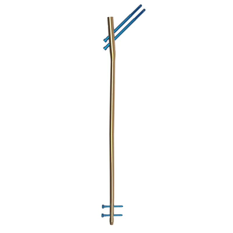
- Nail Body:
- Material: Primarily constructed from biocompatible alloys such as titanium alloy (e.g., Ti-6Al-4V) or medical-grade stainless steel (e.g., 316L). Titanium alloys are favored for their superior strength-to-weight ratio, excellent biocompatibility, and low artifact interference in MRI scans.
- Cannulation: Most FRNs are cannulated, allowing for insertion over a guide wire, which facilitates precise placement and minimizes soft tissue disruption.
- Curvature & Diameter: Designed to match the natural anatomical bow of the distal femur, minimizing cortical impingement. Diameters typically range from 9mm to 14mm, with various lengths to accommodate different fracture patterns and patient anatomies.
- Distal Taper/Flared Design: Often features a flared or tapered distal end to enhance fit and stability within the metaphyseal bone.
- Locking Screws: Crucial for achieving multi-planar stability and preventing rotational instability and axial collapse.
- Proximal Locking: Multiple screw holes (typically 2-4) in various planes (medial-lateral, anterior-posterior) allow for rigid fixation in the diaphyseal segment, controlling rotation and maintaining length.
- Distal Locking: Often features divergent or multi-planar screws that engage the condyles, providing robust fixation in the cancellous bone of the distal femur. These screws are typically smaller in diameter than proximal screws.
- Screw Types: Cortical screws (fully threaded), cancellous screws (partially threaded), and locking screws (threaded into the nail) are used.
- End Cap: An optional component that can be placed at the insertion site of the nail. Its primary functions include preventing tissue ingrowth into the nail's cannulation, providing additional compression across the fracture site (if designed for dynamic compression), and sealing the nail.
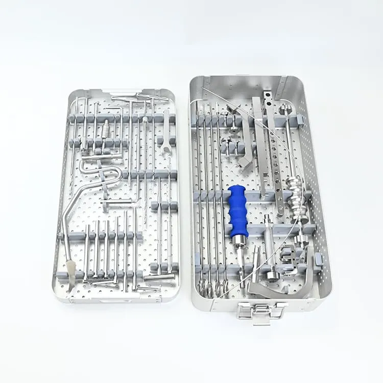
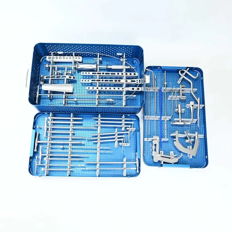
- Instrumentation: A comprehensive set of specialized instruments is required for FRN implantation, including:
- Guide wires (ball-tipped, olive-tipped)
- Reamers (flexible or rigid)
- Introducer and aiming guides/jigs
- Impactor/extractor
- Screw drivers and measuring devices
- C-arm fluoroscopy for real-time imaging
2.2 Materials Science
The selection of materials for FRNs is paramount, directly influencing their mechanical properties, biocompatibility, and longevity within the human body.
- Titanium Alloys (e.g., Ti-6Al-4V):
- Biocompatibility: Excellent, minimizing adverse tissue reactions.
- Strength-to-Weight Ratio: High, allowing for strong yet relatively light implants.
- Corrosion Resistance: Superior resistance to physiological fluids.
- Elastic Modulus: Closer to that of cortical bone compared to stainless steel, potentially reducing stress shielding.
- MRI Compatibility: Generally considered MRI safe with minimal artifact.
- Stainless Steel (e.g., 316L):
- Strength: High mechanical strength.
- Cost-Effectiveness: Generally less expensive than titanium.
- Corrosion Resistance: Good, but generally inferior to titanium in the long term in some applications.
- MRI Compatibility: Can produce significant artifacts and may be contraindicated for certain MRI sequences due to its ferromagnetic properties.
- Surface Treatments: While less common for the entire nail body, specific surface treatments may be applied to locking screws or specific sections to enhance osteointegration or reduce friction. Anodization of titanium implants can improve wear resistance and fatigue strength.
3. Extensive Clinical Indications & Usage
The Femoral Retrograde Nail is a versatile implant, indicated for a range of femoral fractures, particularly those involving the distal segment.
3.1 Primary Indications
- Distal Femur Fractures:
- Supracondylar Fractures: Fractures occurring above the femoral condyles (AO/OTA type 33-A).
- Intercondylar Fractures: Fractures extending into the knee joint between the condyles (AO/OTA type 33-C), often combined with supracondylar extension.
- Condylar Fractures: Isolated condylar fractures (AO/OTA type 33-B) if suitable for nailing, though often managed with plates.
- Periprosthetic Fractures: Fractures occurring around a pre-existing knee arthroplasty (total knee replacement), especially those involving the femoral component where antegrade nailing is difficult or contraindicated due to the prosthesis.
- Ipsilateral Tibia and Femur Fractures ("Floating Knee" Injury): The retrograde approach allows for simultaneous or staged fixation of both the tibia and femur without requiring repositioning or sacrificing access to one fracture for the other.
- Pathological Fractures: Fractures through diseased bone (e.g., metastatic lesions, primary bone tumors) in the distal femur, providing immediate stability and pain relief.
- Non-union/Malunion: For failed previous fixation or malaligned healing of distal femur fractures, requiring revision surgery.
- Polytrauma Patients: Particularly advantageous in patients with associated injuries that make antegrade nailing challenging, such as:
- Pelvic or hip fractures (acetabulum, femoral neck, greater trochanter).
- Severe obesity or pregnancy where supine positioning is preferred or antegrade access is difficult.
- Concomitant vascular injuries requiring repair, where a less invasive approach to the knee is beneficial.
3.2 Surgical Technique (Fitting/Usage Instructions)
The successful implantation of an FRN requires meticulous surgical technique, precise anatomical knowledge, and fluoroscopic guidance.
- Patient Positioning: The patient is typically placed supine on a radiolucent operating table. The knee is flexed approximately 30-90 degrees to facilitate access to the distal femur and allow for fracture reduction.
- Incision: A small infrapatellar incision (medial or lateral parapatellar approach) is made, typically 2-4 cm in length. The patellar tendon is identified and often split longitudinally or retracted to gain access to the intercondylar notch.
- Entry Point: The crucial step is identifying the correct entry point. This is usually located just anterior to the posterior cruciate ligament (PCL) insertion, within the intercondylar notch, and slightly medial or lateral to the midline depending on the nail design and surgeon preference. The entry point must be central to the medullary canal in both AP and lateral fluoroscopic views to avoid iatrogenic fracture or malalignment.
- Guide Wire Insertion: A ball-tipped guide wire is carefully advanced retrograde from the entry point, across the fracture site, and up the femoral shaft, ensuring it remains central in the medullary canal. C-arm fluoroscopy is essential to confirm correct wire placement.
- Reaming: Sequential reaming of the medullary canal is performed over the guide wire to the appropriate diameter, typically 1-2mm larger than the chosen nail diameter. This prepares the canal for nail insertion and removes any cortical bone irregularities.
- Nail Insertion: The selected FRN is then carefully inserted over the guide wire, either manually or with an impactor. It is advanced until the distal tip is flush with or slightly recessed below the articular surface of the femoral condyles, and the proximal end is at the desired depth in the femoral shaft.
- Fracture Reduction: Throughout the guide wire insertion and nailing process, meticulous fracture reduction is maintained using traction, manual manipulation, and specialized reduction clamps or external fixators if needed.
- Distal Locking: Once the nail is in place, distal locking screws are inserted using a targeting device or freehand technique under fluoroscopic guidance. Multiple screws in divergent planes provide rotational and axial stability in the distal fragment.
- Proximal Locking: Proximal locking screws are then inserted, again using a targeting device or freehand, to secure the nail in the proximal femoral shaft, preventing shortening and maintaining rotational control.
- End Cap Placement: An end cap may be placed at the nail's distal end if desired, providing additional stability or compression.
- Wound Closure: The incision is closed in layers after ensuring hemostasis and irrigating the wound.
- Post-operative Care: This involves appropriate pain management, early knee range of motion exercises, and a prescribed weight-bearing protocol that varies based on fracture stability, bone quality, and patient factors.
3.3 Advantages of Retrograde Nailing
- Minimally Invasive: Smaller incision compared to open plating, leading to less soft tissue damage, reduced blood loss, and potentially faster healing.
- Early Mobilization: The stable fixation allows for early weight-bearing and knee range of motion, crucial for preventing stiffness and promoting functional recovery.
- Ideal for Polytrauma: Especially beneficial for patients with ipsilateral hip/pelvic injuries or other conditions that preclude antegrade nailing or prone positioning.
- Load Sharing: Intramedullary implants share stress with the bone, promoting biological healing and reducing the risk of implant fatigue compared to stress-shielding plates.
- High Union Rates: For appropriately selected fractures, FRNs demonstrate excellent union rates.
- Improved Knee Function: Early range of motion contributes to better long-term knee function.
4. Risks, Side Effects, or Contraindications
While highly effective, FRN implantation is not without potential risks and contraindications.
4.1 Potential Risks & Complications
- Intraoperative:
- Iatrogenic Fracture: During guide wire insertion, reaming, or nail insertion.
- Malreduction/Malalignment: Incorrect alignment of fracture fragments.
- Articular Cartilage Damage: Injury to the knee joint cartilage during entry point creation or nail insertion.
- Nerve/Vascular Injury: Rare, but possible damage to popliteal vessels or nerves.
- Infection: Superficial or deep surgical site infection.
- Postoperative:
- Non-union or Delayed Union: Failure of the fracture to heal.
- Malunion: Healing in an unacceptable alignment.
- Hardware Failure: Screw breakage, nail breakage, or loosening of implants.
- Anterior Knee Pain: A common complication, often related to the entry point, patellar tendon irritation, or hardware prominence.
- Knee Stiffness/Reduced Range of Motion: Despite early mobilization protocols.
- Quadriceps Weakness: Due to surgical trauma or prolonged immobilization.
- Infection: Deep or superficial surgical site infection.
- Fat Embolism/DVT/PE: General risks associated with long bone fracture fixation.
- Patellofemoral Pain: Specific to the retrograde approach, due to irritation of the patellofemoral joint.
- Heterotopic Ossification: Formation of bone in soft tissues around the knee.
4.2 Contraindications
- Active Infection: In the knee joint, surrounding soft tissues, or systemic infection.
- Severe Open Fractures: With extensive contamination, where primary nailing may increase infection risk (relative contraindication, may require staged treatment).
- Highly Comminuted Intra-articular Fractures: Where anatomical reduction of the articular surface cannot be adequately achieved with an intramedullary nail, and a plate might offer better direct visualization and fixation.
- Severe Osteopenia/Osteoporosis: Where bone quality is insufficient to provide adequate purchase for locking screws, leading to hardware loosening or pull-out.
- Pre-existing Severe Knee Stiffness or Arthritis: Patients with limited pre-operative knee range of motion may experience exacerbated stiffness.
- Very Small Medullary Canal: If the canal diameter is too small to accommodate even the smallest available nail, reaming may weaken the bone excessively.
- Inability to Achieve Fracture Reduction: If the fracture fragments cannot be reduced adequately, nailing is contraindicated.
- Distal Femoral Metastatic Lesions: If the bone integrity distal to the fracture is severely compromised.
5. Maintenance/Sterilization Protocols
It's crucial to distinguish between the implant itself and the reusable surgical instruments.
5.1 Implant Handling and Sterilization
- Single-Use, Pre-Sterilized: Femoral Retrograde Nails are always supplied as single-use, sterile implants by the manufacturer. They are typically sterilized using methods like gamma irradiation or ethylene oxide (ETO) gas.
- Aseptic Technique: During surgery, the implant must be handled using strict aseptic technique to maintain sterility and prevent contamination.
- Traceability: Each implant comes with unique lot numbers and expiration dates, which must be meticulously recorded for patient safety and inventory management.
- Storage: Implants must be stored in their original, unopened sterile packaging in a clean, dry environment, protected from extreme temperatures and physical damage, until the point of use.
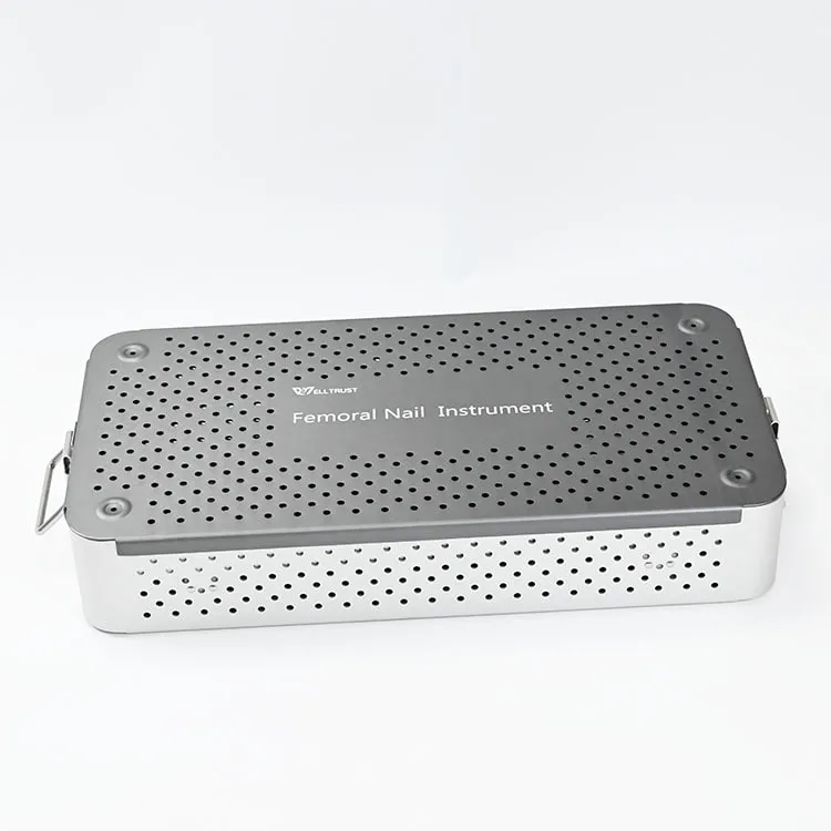
5.2 Reusable Instrument Maintenance and Sterilization
The instruments used to insert the FRN are reusable and require rigorous cleaning and sterilization protocols to prevent surgical site infections.
- Pre-Cleaning/Decontamination:
- Immediately after use, instruments should be wiped clean of gross contamination (blood, tissue).
- Soaked in enzymatic detergent solutions to prevent drying of organic material.
- Cleaning:
- Manual Cleaning: Thorough scrubbing with brushes and appropriate detergents, especially for cannulated or intricate instruments.
- Automated Cleaning: Ultrasonic cleaners or automated washer-disinfectors are commonly used, following manufacturer's instructions for cycle times and detergent concentrations.
- Inspection: After cleaning, instruments are meticulously inspected for cleanliness, functionality, and damage (e.g., bent shafts, dull tips, corrosion). Damaged instruments must be removed from circulation.
- Packaging: Cleaned and inspected instruments are packaged in appropriate sterile barriers (e.g., sterilization wrap, rigid containers) that allow steam penetration while maintaining sterility until use.
- Sterilization:
- Autoclaving (Steam Sterilization): This is the most common and effective method for heat- and moisture-stable instruments.
- Parameters: Specific temperature, pressure, and exposure times are dictated by the instrument manufacturer and national guidelines (e.g., 121°C for 30 minutes, or 132°C for 4 minutes, with appropriate drying cycles).
- Other Methods: Less common for FRN instruments but may include low-temperature sterilization (e.g., hydrogen peroxide plasma) for heat-sensitive items.
- Autoclaving (Steam Sterilization): This is the most common and effective method for heat- and moisture-stable instruments.
- Storage: Sterilized instrument sets are stored in a clean, dry, and secure environment, protected from environmental contaminants, until they are needed for surgery. The integrity of the sterile barrier must be maintained.
6. Biomechanics & Patient Outcome Improvements
The biomechanical principles underlying the FRN contribute significantly to its clinical success and improved patient outcomes.
6.1 Biomechanical Principles
- Load Sharing: The intramedullary position of the nail allows it to share axial and bending loads with the surrounding cortical bone. This physiological load transfer promotes "stress-shielding" in the bone, stimulating callus formation and accelerating fracture healing, unlike plates which bear most of the load.
- Stability:
- Axial Stability: Provided by the tight fit of the nail within the medullary canal and the locking screws, preventing shortening or distraction of the fracture.
- Rotational Stability: Achieved primarily through the multi-planar locking screws, which prevent the fracture fragments from rotating relative to each other. The reamed fit also contributes.
- Bending Stability: The nail's high stiffness in bending, combined with its central placement, resists bending forces across the fracture site.
- Dynamic vs. Static Locking: Some FRNs allow for either static (fixed length) or dynamic (controlled shortening/compression) locking. Dynamic locking can promote axial micromotion at the fracture site, which is beneficial for callus formation, but must be used judiciously to avoid excessive shortening.
- Minimizing Stress Risers: The smooth, contoured design of the nail and the absence of external hardware minimize stress risers on the bone, potentially reducing refracture risk after implant removal.
6.2 Patient Outcome Improvements
The biomechanical advantages of FRNs translate directly into tangible benefits for patients:
- Early Mobilization and Weight-Bearing: The stable fixation achieved with FRNs often allows for early protected weight-bearing and initiation of knee range of motion exercises within days of surgery. This significantly reduces complications associated with prolonged immobilization (e.g., muscle atrophy, joint stiffness, DVT).
- Reduced Pain: Stable fixation minimizes pain from fracture movement, leading to better post-operative pain control.
- Faster Recovery and Return to Function: Patients typically experience a quicker return to activities of daily living, work, and sports compared to less stable fixation methods.
- Lower Incidence of Non-union and Malunion: High primary stability and load-sharing properties contribute to excellent fracture union rates and proper anatomical alignment.
- Reduced Surgical Morbidity: The minimally invasive nature of the retrograde approach results in smaller incisions, less soft tissue dissection, and potentially reduced blood loss and infection rates compared to extensive open approaches.
- Improved Knee Range of Motion: Early, guided rehabilitation, facilitated by stable internal fixation, helps prevent knee stiffness and preserves functional knee articulation.
- Cosmetic Advantage: Smaller, less conspicuous scars.
7. Massive FAQ Section
Q1: What is a Femoral Retrograde Nail?
A1: A Femoral Retrograde Nail (FRN) is an orthopedic implant, typically a metal rod, inserted into the hollow medullary canal of the distal femur (thigh bone) through an incision near the knee. It's used to stabilize fractures of the distal femur and certain other conditions.
Q2: What types of fractures does a Femoral Retrograde Nail treat?
A2: FRNs are primarily used for distal femur fractures, including supracondylar, intercondylar, and certain condylar fractures. They are also effective for periprosthetic fractures around knee replacements, ipsilateral tibia and femur fractures ("floating knee"), and pathological fractures in the distal femur.
Q3: How is the Femoral Retrograde Nail surgery performed?
A3: The surgery involves making a small incision near the knee, typically through the patellar tendon. A guide wire is inserted into the femur's medullary canal, followed by reaming to prepare the canal. The nail is then inserted over the guide wire, and locking screws are placed both proximally and distally to secure the nail and stabilize the fracture. The procedure is guided by real-time X-ray (fluoroscopy).
Q4: What are the advantages of a retrograde nail over a plate for distal femur fractures?
A4: Advantages include a minimally invasive approach, allowing for earlier weight-bearing and knee range of motion, load-sharing properties that promote bone healing, and suitability for patients with associated hip or pelvic injuries that make antegrade nailing difficult.
Q5: What are the risks associated with Femoral Retrograde Nail surgery?
A5: Potential risks include infection, non-union or malunion of the fracture, hardware failure (screw breakage, nail loosening), anterior knee pain (a common side effect), knee stiffness, nerve or vascular injury, and articular cartilage damage.
Q6: How long is the recovery period after FRN surgery?
A6: Recovery varies based on the fracture's severity, bone quality, and patient factors. Generally, patients can start early range of motion exercises and protected weight-bearing within days or weeks. Full recovery, including return to activities, can take 3 to 6 months or longer, with bone healing typically occurring over 6-12 weeks.
Q7: Will the Femoral Retrograde Nail need to be removed?
A7: Removal of the FRN is not always necessary. It may be considered if the patient experiences persistent pain (especially anterior knee pain), infection, or mechanical irritation from the implant after the fracture has healed. Removal is a separate surgical procedure.
Q8: Can I bear weight on my leg immediately after surgery?
A8: In many cases, early protected weight-bearing (e.g., touch-down weight-bearing or partial weight-bearing) is encouraged to promote healing and maintain muscle strength. The specific weight-bearing protocol will be determined by your surgeon based on the stability of the fracture fixation and your individual healing progress.
Q9: What materials are Femoral Retrograde Nails made from?
A9: Femoral Retrograde Nails are typically made from high-strength, biocompatible medical-grade alloys, most commonly titanium alloys (e.g., Ti-6Al-4V) or stainless steel (e.g., 316L). Titanium is often preferred for its excellent strength-to-weight ratio, corrosion resistance, and MRI compatibility.
Q10: Is anterior knee pain common after this surgery?
A10: Yes, anterior knee pain is a relatively common complaint after Femoral Retrograde Nailing, affecting a significant percentage of patients. It can be caused by irritation from the nail's entry point, patellar tendon inflammation, or impingement of the end cap.
Q11: How does a retrograde nail differ from an antegrade nail?
A11: The primary difference is the insertion point. An antegrade femoral nail is inserted from the hip region (greater trochanter) and extends down the femur, typically used for diaphyseal and proximal femoral fractures. A retrograde nail is inserted from the knee region (distal femur) and extends upwards, primarily used for distal femoral fractures.
Q12: What is the role of physical therapy in recovery?
A12: Physical therapy is crucial for optimal recovery. It focuses on restoring knee range of motion, strengthening the quadriceps and hamstring muscles, improving gait, and progressively increasing weight-bearing tolerance. Early and consistent therapy helps prevent stiffness, improve function, and ensures a better long-term outcome.