The Distal Femur Lateral Locking Plate: A Comprehensive Orthopedic Guide
The distal femur, the segment of the thigh bone just above the knee joint, is a critical anatomical region. Fractures in this area, often resulting from high-energy trauma or falls in osteoporotic patients, present significant challenges to orthopedic surgeons. These fractures can severely impact knee function and patient mobility. The Distal Femur Lateral Locking Plate (DFLLP) represents a cornerstone in modern orthopedic trauma management, offering robust and stable fixation for these complex injuries.
This exhaustive guide, crafted by an expert Medical SEO Copywriter and Orthopedic Specialist, delves into every facet of the Distal Femur Lateral Locking Plate, from its innovative design and materials to its intricate surgical applications, biomechanical principles, maintenance protocols, and profound impact on patient outcomes.
1. Comprehensive Introduction & Overview
A Distal Femur Lateral Locking Plate is an advanced orthopedic implant specifically designed for the internal fixation of fractures of the distal femur. Unlike traditional non-locking plates that rely solely on friction between the plate and bone for stability, locking plates feature screws that thread directly into the plate, creating a fixed-angle construct. This angular stability is paramount in managing comminuted fractures, osteoporotic bone, and situations where strong axial and rotational stability is required.
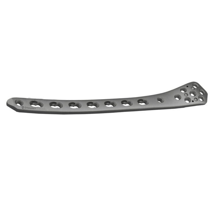
The DFLLP is typically placed on the lateral aspect of the distal femur, conforming to the natural anatomical contour of the bone. Its design often incorporates specific features to support the condyles and provide a stable platform for fracture healing, facilitating early mobilization and improved functional recovery for patients.
Key Advantages of DFLLP:
* Angular Stability: Screws lock into the plate, creating a rigid construct independent of plate-to-bone compression.
* Enhanced Fixation in Poor Bone Quality: Ideal for osteoporotic patients where traditional screws might pull out.
* Indirect Reduction Capabilities: Facilitates Minimally Invasive Plate Osteosynthesis (MIPO) techniques, preserving soft tissue and blood supply.
* Improved Load Sharing: Distributes stress more effectively across the fracture site.
* Anatomical Pre-Contouring: Reduces the need for intraoperative plate bending, saving time and maintaining material integrity.
2. Deep-Dive into Technical Specifications & Mechanisms
The efficacy of the Distal Femur Lateral Locking Plate lies in its sophisticated design, choice of materials, and the underlying biomechanical principles of its locking mechanism.
2.1. Design & Materials
DFLLPs are engineered with precision to address the unique anatomical and biomechanical demands of the distal femur.
Design Features:
* Anatomical Pre-Contouring: Plates are typically pre-contoured for either the left or right femur, matching the complex curves of the lateral distal femoral metaphysis and condyles. This minimizes the need for intraoperative bending, which can weaken the plate.
* Low-Profile Design: Minimizes soft tissue irritation and prominence, reducing the likelihood of requiring hardware removal.
* Combi-Holes: Many DFLLPs feature "combi-holes" that allow for both dynamic compression (using non-locking screws) and angular stability (using locking screws) within the same hole. This provides flexibility during surgery to achieve primary compression where desired, followed by locked stability.
* Multi-Planar Screw Trajectories: The plate head often incorporates multiple locking screw holes angled in various directions to capture as many fragments as possible and provide robust fixation within the distal condyles.
* Threaded Locking Holes: Each locking hole is threaded to accept the corresponding threaded head of a locking screw, ensuring a secure, fixed-angle construct.
* Shaft Holes: The shaft of the plate features multiple holes, typically combi-holes, to secure the plate to the femoral diaphysis.
* Aiming Guides: Specialized guides are often provided to assist in precise screw placement, particularly for the multi-directional screws in the plate head.
Materials:
The choice of material is critical for biocompatibility, strength, and fatigue resistance.
| Material Type | Properties | Advantages | Disadvantages |
| :------------------------- | :------------------------------------------------------------------------------------------------------ | :----------------------------------------------------------------------------------- | :-------------------------------------------------------------------------------- |
| Medical-Grade Stainless Steel (e.g., 316L) | High strength, good corrosion resistance, cost-effective. Contains nickel. | Well-established clinical history, strong, affordable. | Can cause artifacts on MRI, potential for nickel allergy, slightly less biocompatible. |
| Titanium Alloys (e.g., Ti-6Al-4V ELI) | Excellent biocompatibility, high strength-to-weight ratio, superior fatigue resistance, non-ferromagnetic. | Reduced MRI artifacts, superior biocompatibility, lighter, excellent fatigue life. | More expensive, can be more challenging to remove due to bone ongrowth. |
2.2. Biomechanics of Locking Plate Fixation
The biomechanical superiority of locking plates stems from their fundamental principle of angular stability.
- Fixed-Angle Construct: Unlike traditional screws that rely on compression of the plate against the bone for stability, locking screws "lock" into the plate, creating a fixed-angle device. This effectively converts the screw-plate interface into a rigid unit.
- Internal Fixator Concept: The locking plate acts as an "internal fixator," providing stability without requiring direct compression of the plate onto the bone. This allows for indirect reduction techniques, preserving the periosteal blood supply to the bone fragments.
- Load Sharing vs. Load Bearing: Locking plates are designed to share the load with the healing bone, rather than bear the entire load. This promotes secondary bone healing through callus formation.
- Reduced Stress Risers: The secure locking mechanism minimizes micromotion at the screw-plate interface, reducing stress concentrations and the risk of screw loosening or pull-out, especially in compromised bone.
- Working Length: The distance between the innermost screws on either side of the fracture gap is known as the working length. An appropriate working length is crucial; too short can lead to plate stiffness and stress shielding, while too long can result in insufficient stability.
- Screw Density: The number and distribution of screws play a vital role. While adequate screws are necessary, overtightening or placing too many screws in a small segment can create stress risers. The goal is often to achieve sufficient fixation without "over-stuffing" the bone.
3. Extensive Clinical Indications & Usage
The Distal Femur Lateral Locking Plate is a versatile implant indicated for a broad spectrum of distal femur fractures.
3.1. Clinical Indications
DFLLPs are primarily used for fractures classified under the AO/OTA system as 33-A (extra-articular distal femur fractures) and 33-C (intra-articular distal femur fractures).
Specific Indications Include:
* Supracondylar Femur Fractures: Fractures occurring just above the femoral condyles, often extending into the metaphysis.
* Intercondylar Femur Fractures: Fractures that involve the articular surface of the femoral condyles and extend proximally into the metaphysis.
* Periprosthetic Fractures: Fractures occurring around a pre-existing knee prosthesis (e.g., total knee arthroplasty), especially those involving the distal femur. The locking plate's ability to fix in osteoporotic bone and avoid components makes it ideal.
* Fractures in Osteoporotic Bone: The angular stability is particularly beneficial in patients with poor bone quality, where traditional screws might not achieve sufficient purchase.
* Open Fractures of the Distal Femur (Gustilo-Anderson Types I, II, III): After thorough debridement and management of soft tissue, the DFLLP can provide stable fixation.
* Non-unions or Malunions: For failed previous fixation or improperly healed distal femur fractures requiring revision surgery.
* Pathological Fractures: Due to tumors or other bone-weakening conditions.
3.2. Detailed Surgical Applications & Technique
The surgical application of a DFLLP requires meticulous planning and execution.
1. Pre-operative Planning:
* Radiographic Assessment: High-quality X-rays (AP, lateral, oblique views) and CT scans with 3D reconstructions are essential to understand the fracture pattern, comminution, and articular involvement.
* Templating: Digital or physical templating helps select the appropriate plate length, screw types, and positioning.
* Patient Positioning: Typically supine on a radiolucent operating table, often with a bump under the ipsilateral hip to allow for internal rotation and easier lateral access. Traction tables may be used for indirect reduction.
2. Surgical Approach:
* Lateral Subvastus Approach: The most common approach. An incision is made on the lateral aspect of the thigh, and the vastus lateralis muscle is either retracted anteriorly or split longitudinally to expose the distal femur. This approach aims to preserve the blood supply to the bone fragments.
* Minimally Invasive Plate Osteosynthesis (MIPO): Often preferred to minimize soft tissue dissection. Small proximal and distal incisions are made, and the plate is tunneled submuscularly. This technique relies heavily on fluoroscopic guidance.
3. Fracture Reduction:
* Indirect Reduction: The preferred method, using ligamentotaxis, external traction, or temporary K-wire fixation to restore length, alignment, and rotation without directly exposing the fracture site.
* Direct Reduction (Limited): May be necessary for articular fragments, which are typically reduced and fixed with lag screws before plate application.
4. Plate Application & Screw Insertion:
* Plate Placement: The anatomically pre-contoured plate is positioned on the lateral aspect of the distal femur, ensuring proper alignment with the femoral shaft and condyles.
* Provisional Fixation: K-wires or non-locking screws can be used to temporarily hold the plate in position.
* Distal Fixation (Condylar Screws): Locking screws are inserted into the distal fragment(s) first, ensuring bicortical purchase where possible and avoiding articular penetration. Specialized aiming guides facilitate accurate placement.
* Shaft Fixation (Proximal Screws): Locking screws are then inserted into the proximal femoral shaft. Combi-holes allow for initial compression with non-locking screws if desired, followed by locking screws for definitive fixation.
* Fluoroscopic Guidance: Essential throughout the procedure to confirm plate and screw positioning, fracture reduction, and to rule out articular penetration.
5. Post-operative Management:
* Wound Closure: Meticulous closure of muscle layers and skin.
* Pain Management: Multimodal analgesia.
* Weight-Bearing Protocol: Varies based on fracture stability, bone quality, and patient compliance. Non-weight bearing or touch-down weight bearing is often prescribed initially, progressing to partial and then full weight bearing as healing progresses.
* Rehabilitation: Early range of motion exercises for the knee are crucial to prevent stiffness. Physical therapy focuses on strengthening and regaining function.
3.3. Fitting/Usage Instructions
Proper fitting and usage are paramount for successful outcomes.
- Pre-operative Templating: Always template using high-quality radiographs to estimate plate length and screw requirements.
- Intraoperative Fluoroscopy: Use C-arm fluoroscopy extensively to confirm reduction, plate position, and screw lengths. Multiple views (AP, lateral, oblique) are necessary.
- Specialized Instrumentation: Utilize the manufacturer-specific instruments, including:
- Aiming Guides: For precise drilling and screw insertion, especially in the multi-planar condylar holes.
- Drill Sleeves: To protect soft tissues and guide the drill bit accurately.
- Depth Gauges: To measure screw length accurately.
- Torque-Limiting Drivers: To prevent overtightening of locking screws, which can strip the plate threads or break the screw.
- Screw Selection: Choose locking screws for angular stability and non-locking screws for compression where appropriate (e.g., across the fracture line in combi-holes).
- Avoid Articular Penetration: This is a critical error. Use fluoroscopy in multiple planes to confirm screws are not entering the joint space.
- Maintain Working Length: Ensure sufficient distance between proximal and distal screws to allow for controlled micromotion and callus formation.
4. Maintenance/Sterilization Protocols
Maintaining the sterility and integrity of orthopedic implants and instruments is non-negotiable to prevent surgical site infections.
4.1. Manufacturer Guidelines
Always adhere strictly to the manufacturer's instructions for cleaning, sterilization, and storage of both the Distal Femur Lateral Locking Plates and their associated instruments. Deviations can compromise product performance and patient safety.
4.2. Cleaning
- Point-of-Use Cleaning: Immediately after surgery, remove gross debris (blood, tissue) from instruments and implants using sterile water and brushes. This prevents drying and adherence of biological material.
- Manual Cleaning: Thoroughly clean all surfaces of reusable instruments with enzymatic detergents and appropriate brushes to remove all visible soil. Pay special attention to lumens, crevices, and threaded areas.
- Ultrasonic Cleaning: Follow manual cleaning with ultrasonic cleaning to dislodge microscopic debris from hard-to-reach areas. Use approved detergents and proper cycle times.
- Rinsing: Rinse instruments thoroughly with purified water to remove all detergent residues.
4.3. Sterilization
- Method: Steam sterilization (autoclaving) is the most common and validated method for DFLLPs and their instruments.
- Parameters: Specific temperature, pressure, and exposure times are mandated by the manufacturer. Typical cycles include:
- Gravity Displacement: 121°C (250°F) for 30 minutes.
- Pre-vacuum: 132°C (270°F) for 4 minutes.
- Biological indicators and chemical integrators must be used to verify sterility.
- Packaging: Implants and instruments must be placed in appropriate sterile barrier systems (e.g., sterilization wrap, rigid containers) that allow for steam penetration while maintaining sterility until the point of use.
- Single-Use Implants: DFLLPs are typically provided sterile and are single-use items. Do not re-sterilize or reuse implants.
- Traceability: Maintain strict records of lot numbers for all implants used in surgery for traceability in case of a product recall or adverse event.
4.4. Storage
- Store sterile implants and instruments in a clean, dry, and controlled environment, protected from dust, moisture, and extreme temperatures.
- Respect the shelf life indicated on the packaging for sterile implants.
5. Risks, Side Effects, or Contraindications
While the Distal Femur Lateral Locking Plate offers significant benefits, it is not without potential risks and contraindications.
5.1. General Surgical Risks
These are inherent to any orthopedic surgical procedure:
* Infection: Superficial wound infection or deep surgical site infection (osteomyelitis).
* Bleeding/Hematoma: Post-operative bleeding leading to hematoma formation.
* Nerve/Vascular Injury: Damage to surrounding nerves (e.g., peroneal nerve) or blood vessels.
* Anesthesia Risks: Adverse reactions to anesthetic agents.
* Thromboembolic Events: Deep vein thrombosis (DVT) or pulmonary embolism (PE).
5.2. Implant-Specific Risks & Side Effects
- Non-union or Delayed Union: Failure of the fracture to heal within the expected timeframe.
- Malunion: Healing of the fracture in an unacceptable alignment (e.g., varus/valgus deformity, rotational malalignment).
- Implant Failure:
- Plate Breakage: Due to excessive stress or premature weight-bearing before adequate bone healing.
- Screw Loosening/Pull-out: Although less common with locking screws, can occur in severely osteoporotic bone or with improper technique.
- Screw Breakage: Rare, but can occur with high stress concentrations.
- Stiffness/Loss of Range of Motion: Particularly in the knee joint, due to prolonged immobilization, scarring, or articular involvement.
- Hardware Prominence/Irritation: The plate or screw heads may become palpable or cause discomfort, necessitating hardware removal.
- Avascular Necrosis: Rare, but can occur if the blood supply to a bone fragment is compromised during the injury or surgery.
- Periprosthetic Fracture: A new fracture occurring around the implant.
- Allergic Reaction: Extremely rare, but possible reaction to implant materials (e.g., nickel in stainless steel).
5.3. Contraindications
- Absolute Contraindications:
- Active Infection: At the fracture site or adjacent soft tissues.
- Severe Soft Tissue Compromise: Extensive open wounds, devitalized tissue, or severe swelling that precludes safe surgical closure.
- Relative Contraindications:
- Insufficient Bone Stock: Inability to achieve adequate screw purchase.
- Patient Non-compliance: Unwillingness or inability to adhere to post-operative weight-bearing restrictions and rehabilitation protocols.
- Severe Comorbidities: Medical conditions that significantly increase surgical risk and outweigh the benefits of fixation.
- Allergy to Implant Material: If a known allergy exists (e.g., nickel), an alternative material like titanium must be used.
6. Massive FAQ Section
Here are frequently asked questions about the Distal Femur Lateral Locking Plate:
1. What is a Distal Femur Lateral Locking Plate (DFLLP)?
A DFLLP is an orthopedic implant used to stabilize fractures of the distal part of the thigh bone (femur), just above the knee. It's designed with screws that "lock" into the plate, providing strong angular stability.
2. How is a locking plate different from a traditional non-locking plate?
Traditional plates rely on the plate being compressed against the bone for stability. Locking plates, however, have screws that thread into the plate itself, creating a fixed-angle construct. This provides superior stability, especially in complex fractures or poor bone quality, without requiring direct compression of the plate to the bone.
3. What types of fractures does the DFLLP treat?
It's primarily used for supracondylar (above the condyles) and intercondylar (involving the joint surface) fractures of the distal femur. It's also effective for periprosthetic fractures (around knee replacements) and fractures in osteoporotic patients.
4. Is the surgery to implant a DFLLP painful?
As with any major surgery, there will be post-operative pain. However, modern pain management techniques, including nerve blocks and multimodal analgesia, are used to keep discomfort to a minimum.
5. How long does it take to recover after DFLLP surgery?
Recovery varies based on the fracture severity, bone quality, and individual patient factors. Initial healing can take 6-12 weeks, with full weight-bearing often allowed after 3-4 months. Complete functional recovery, including rehabilitation, can take 6-12 months.
6. Will I need to have the DFLLP removed?
Not always. If the plate causes no symptoms and the fracture has healed well, it can remain in place indefinitely. However, if it causes irritation, pain, or infection, or if there's a need for future surgery, it may be removed, typically 12-18 months after initial surgery.
7. What are the potential complications of DFLLP surgery?
Complications can include infection, non-union (failure of the bone to heal), malunion (healing in poor alignment), hardware failure (plate or screw breakage), nerve or blood vessel injury, stiffness of the knee, and hardware prominence.
8. Can I put weight on my leg immediately after DFLLP surgery?
Typically, immediate full weight-bearing is not allowed. Your surgeon will provide a specific weight-bearing protocol, often starting with non-weight bearing or touch-down weight bearing, gradually progressing as the fracture heals.
9. How is the Distal Femur Lateral Locking Plate sterilized before surgery?
DFLLPs are usually supplied pre-sterilized by the manufacturer. If not, or for reusable instruments, they undergo rigorous cleaning and steam sterilization (autoclaving) according to strict protocols to ensure they are completely sterile before implantation.
10. What materials are these plates made from?
DFLLPs are commonly made from medical-grade stainless steel (e.g., 316L) or titanium alloys (e.g., Ti-6Al-4V ELI). Titanium is often preferred due to its excellent biocompatibility and reduced artifacts on MRI scans.
11. Is the DFLLP visible on X-rays?
Yes, both stainless steel and titanium plates are radiopaque and clearly visible on X-rays, allowing surgeons to monitor fracture healing and implant position.
12. How does the DFLLP improve patient outcomes?
By providing stable and anatomically appropriate fixation, the DFLLP facilitates early mobilization, reduces the risk of non-union or malunion, and helps restore knee function. This leads to better long-term functional outcomes, reduced pain, and improved quality of life for patients with complex distal femur fractures.