The Ring Curette with Depth Markings: A Cornerstone of Precision Orthopedic Surgery
Welcome to this comprehensive guide on a crucial instrument in modern orthopedic surgery: the Curette with Depth Markings, often referred to as a "Ring Curette." As experts in orthopedic care, we understand the profound impact that precision and control have on surgical outcomes and patient recovery. This guide aims to demystify this specialized tool, explaining its design, applications, and the significant role it plays in ensuring the highest standards of patient safety and efficacy.
1. Comprehensive Introduction & Overview
In the intricate world of orthopedic surgery, the ability to precisely remove diseased or unwanted tissue while preserving healthy structures is paramount. The curette, a handheld surgical instrument designed for scraping or debriding tissue, has been a staple for centuries. However, the Ring Curette with Depth Markings represents a significant evolution, offering unparalleled control and accuracy.
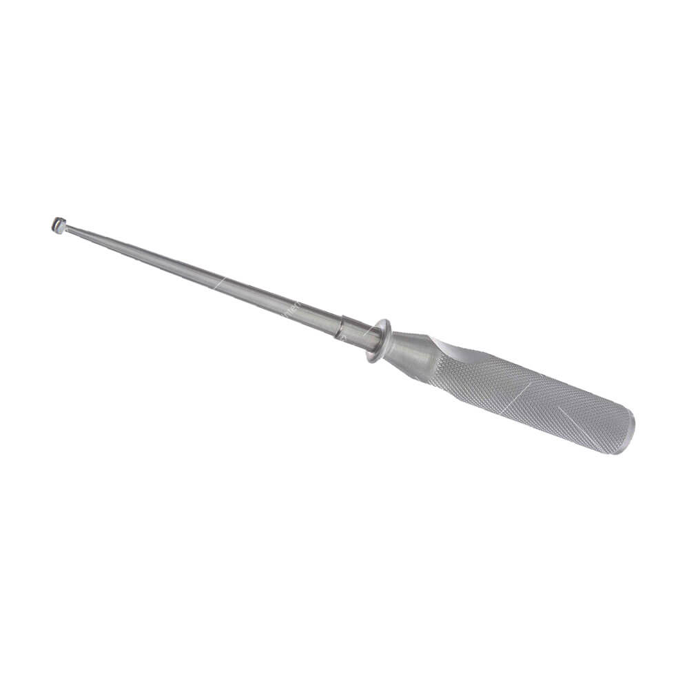
This specialized curette features a distinctive ring-shaped working end, allowing for efficient circumferential scraping and scooping. What truly sets it apart are the integrated depth markings, typically laser-etched in millimeter increments along its shaft. These markings provide the surgeon with real-time, visual feedback on the depth of tissue removal, transforming a skill-dependent maneuver into a more standardized and reproducible procedure.
The primary goal of employing such a precise instrument is to minimize collateral damage to surrounding healthy bone, cartilage, or soft tissues. This precision directly translates into improved patient outcomes, including reduced pain, faster healing, and a lower risk of complications. For patients, understanding the tools their surgeons use can build confidence and highlight the commitment to advanced, patient-centric care.
2. Deep-Dive into Technical Specifications & Mechanisms
The efficacy of the Ring Curette with Depth Markings lies in its meticulously engineered design and the advanced materials used in its construction.
Design Features: The Anatomy of Precision
- Ring-Shaped Working End:
- Configuration: The working end is typically a sharp-edged loop or ring, available in various sizes (e.g., 2mm, 4mm, 6mm diameter) and angles (straight or angled to access difficult areas).
- Function: This design allows for effective scraping, debridement, and scooping of bone, cartilage, or soft tissue from cavity walls or surfaces. The circumferential edge ensures thorough removal with fewer passes compared to a spoon-shaped curette.
- Depth Markings:
- Mechanism: Laser-etched or engraved markings, usually in 1mm or 2mm increments, extend from the working tip along the shaft.
- Importance: These markings serve as a visual caliper, enabling the surgeon to accurately gauge the depth of tissue penetration and removal. This is critical for procedures where precise depth control is essential, such as debriding bone cysts or preparing bone for graft insertion. It prevents both over-resection (weakening bone) and under-resection (leaving diseased tissue behind).
- Shaft:
- Variations: Available in different lengths and degrees of rigidity, often with slight curves or angles to facilitate access to deep or anatomically challenging areas.
- Ergonomics: Designed to provide stable transmission of force from the handle to the working end.
- Handle:
- Material & Grip: Typically made from a knurled or textured stainless steel for a secure, non-slip grip, even when wet. Ergonomic design reduces surgeon fatigue during lengthy procedures.
- Balance: Properly weighted and balanced to enhance tactile feedback and control.
Materials: Durability, Biocompatibility, and Sterilization
The choice of materials is fundamental to the instrument's performance and safety.
- Medical-Grade Stainless Steel:
- Types: Primarily surgical-grade stainless steel such as 420 (for hardness and sharpness) or 304 (for corrosion resistance).
- Properties: Offers excellent durability, high tensile strength, resistance to corrosion from bodily fluids and sterilization processes, and is fully biocompatible. It can withstand repeated sterilization cycles without degradation.
- Titanium (Less Common but Used):
- Properties: Lighter than steel, non-magnetic (MRI-compatible), and even more corrosion-resistant.
- Application: Sometimes used for specialized instruments or for patients with specific metal sensitivities, though less common for standard curettes due to higher cost.
Biomechanics and Mechanism of Action
The biomechanics of the Ring Curette are centered on controlled force application and efficient tissue removal:
- Scraping/Debridement: The sharp, circular edge is drawn across the target tissue (e.g., cyst lining, necrotic bone), effectively detaching and collecting it. The ring shape allows for a continuous, circumferential motion.
- Scooping: Once detached, the tissue can be scooped out of the surgical site.
- Depth Control: The depth markings, combined with the surgeon's tactile feedback, allow for precise, layer-by-layer removal. This minimizes the risk of perforating bone cortices or damaging critical neurovascular structures lying beneath the target area.
- Reduced Trauma: By enabling precise and complete removal in a controlled manner, the instrument reduces the need for aggressive or repeated maneuvers, thereby minimizing trauma to surrounding healthy tissues.
3. Extensive Clinical Indications & Usage
The Ring Curette with Depth Markings is an indispensable tool across a spectrum of orthopedic procedures, where precision and controlled tissue removal are paramount.
Key Clinical Indications:
- Bone Cyst Debridement:
- Unicameral Bone Cysts (UBCs): Used to meticulously scrape out the fibrous lining of these benign, fluid-filled lesions, often in long bones. The depth markings ensure complete removal of the cyst wall while preserving the cortical bone.
- Aneurysmal Bone Cysts (ABCs): For removing the septa and lining of these blood-filled, expansive lesions.
- Benign Bone Tumor Removal:
- Giant Cell Tumors (GCTs): Crucial for intralesional curettage of these aggressive benign tumors, particularly around joints. The depth markings aid in precise removal to reduce recurrence rates.
- Enchondromas, Osteochondromas: For removing cartilaginous or bony overgrowths, ensuring clear margins.
- Cartilage Debridement (Chondroplasty):
- Osteochondral Defects: Used to prepare the base of cartilage lesions for repair procedures (e.g., microfracture, OATS procedure) by removing damaged cartilage and exposing healthy subchondral bone.
- Degenerative Cartilage: Carefully debriding frayed or damaged articular cartilage in arthroscopic procedures to smooth surfaces and reduce irritation.
- Joint Surface Preparation:
- Arthroplasty (Joint Replacement): While large reamers are used for gross preparation, the ring curette can be used for fine-tuning bone contours, removing osteophytes, or preparing specific areas for implant seating, especially in smaller joints or complex anatomies.
- Spinal Surgery:
- Discectomy: For precise removal of herniated disc material or osteophytes impinging on neural structures, particularly in delicate areas where nerve roots are in close proximity.
- Laminectomy/Foraminotomy: Used for controlled removal of small amounts of bone or ligamentum flavum to decompress spinal nerves.
- Infection Debridement (Osteomyelitis):
- For carefully removing necrotic (dead) or infected bone tissue, ensuring all compromised tissue is removed while sparing viable bone.
- Marrow Sampling/Biopsy:
- Though specialized biopsy needles exist, a small ring curette can be used for targeted bone marrow sampling from accessible sites, particularly in conjunction with other procedures.
Principles of Usage:
- Pre-operative Planning: Thorough review of imaging (X-rays, MRI, CT) to understand the lesion's size, depth, and proximity to vital structures.
- Surgical Access: Appropriate incision and exposure of the surgical site.
- Controlled Application: The surgeon grasps the curette firmly but comfortably. The working end is advanced to the target tissue.
- Gentle Scraping/Scooping: Using a controlled, deliberate motion, the surgeon scrapes the diseased tissue. For bone cysts, this involves meticulously debriding the inner lining.
- Depth Marking Reference: Continuously monitoring the depth markings on the shaft provides crucial feedback, ensuring the desired depth of removal is achieved without over-penetration.
- Irrigation and Suction: Frequent irrigation washes away debris, and suction clears the field, allowing for clear visualization.
- Post-Procedure Assessment: Visual and tactile inspection of the surgical site to confirm complete removal of diseased tissue and preservation of healthy structures.
4. Risks, Side Effects, or Contraindications
While the Ring Curette with Depth Markings is designed for precision and safety, like any surgical instrument, its use carries inherent risks, potential side effects, and specific contraindications. Patients should be aware of these possibilities.
Potential Risks and Side Effects:
- Over-resection or Under-resection:
- Over-resection: Removing too much bone or tissue can weaken the structure, potentially leading to fractures, instability, or prolonged healing. In joint procedures, it could compromise joint integrity.
- Under-resection: Incomplete removal of diseased tissue (e.g., tumor, cyst lining, infected bone) can lead to recurrence of the condition, persistent symptoms, or ongoing infection. The depth markings help mitigate this, but surgical skill remains paramount.
- Damage to Adjacent Healthy Structures:
- Neurovascular Injury: Scraping too aggressively or in an uncontrolled manner near nerves or blood vessels can cause temporary or permanent damage, leading to numbness, weakness, pain, or bleeding.
- Soft Tissue Injury: Unintended trauma to muscles, tendons, or ligaments surrounding the surgical site.
- Articular Cartilage Damage: In joint-related procedures, inadvertent damage to healthy cartilage can accelerate degenerative changes.
- Infection: As with any invasive procedure, there is a risk of surgical site infection, despite stringent sterilization protocols. This can lead to delayed healing, pain, and may require further intervention.
- Bleeding/Hematoma: Surgical debridement can cause bleeding. While usually controlled, excessive bleeding can lead to hematoma formation, which may require drainage.
- Fracture: Particularly when debriding bone cysts or tumors that have already weakened the bone, aggressive curettage can precipitate a pathological fracture. The surgeon must balance complete removal with structural integrity.
- Delayed Healing: Unforeseen complications, infection, or extensive debridement can sometimes prolong the healing process.
- Adverse Reaction to Materials: Though rare with medical-grade stainless steel, some individuals may have hypersensitivity reactions to metal components.
Contraindications:
- Extremely Fragile Bone: In cases of severe osteoporosis or conditions causing extreme bone fragility where even gentle scraping could cause an immediate fracture, alternative methods might be preferred or extreme caution is necessary.
- Extensive Lesions Requiring En Bloc Resection: For very large or aggressive tumors where local curettage is insufficient, an en bloc (block) resection might be indicated, requiring different instruments and surgical approaches. The ring curette is primarily for intralesional removal.
- Poor Visualization: If the surgical field cannot be adequately visualized due to bleeding, anatomical constraints, or limited access, using a precision instrument like a curette with depth markings becomes risky. Clear visualization is critical for safe and effective use.
- Inexperience of the Surgeon: While the instrument aids precision, it does not replace surgical skill and experience. In the hands of an inexperienced surgeon, the risks listed above are amplified.
5. Expert Tips from Dr. Mohammed Hutaif
As an orthopedic specialist, I've seen firsthand how the judicious and skilled application of instruments like the Ring Curette with Depth Markings can significantly influence patient outcomes. Here are some key insights and tips:
- "Knowledge is Your Compass, Not Just the Markings": While the depth markings are invaluable, they are a guide, not a substitute for thorough pre-operative planning. Always have a clear mental map of the anatomy and the lesion's extent based on advanced imaging. Understand the "safe zones" and critical structures in proximity.
- "Embrace the Tactile Feedback": The curette is an extension of your hand. Pay close attention to the feel of the tissue you're debriding. Healthy bone feels different from diseased bone or cyst lining. This tactile sensation, combined with visual depth markings, provides the most comprehensive feedback.
- "Start Gentle, Stay Controlled": Always begin with gentle, sweeping motions. Avoid aggressive gouging. The goal is systematic, layer-by-layer removal. This approach minimizes trauma, reduces bleeding, and allows for continuous assessment of the remaining tissue and bone integrity.
- "The Right Size for the Right Job": Don't try to force a large curette into a small space or use a tiny one for a broad area. Having a range of ring curette sizes and angles available allows you to select the optimal instrument for precise access and efficient debridement in different anatomical locations.
- "Maintain a Clear Field": Frequent irrigation and effective suction are non-negotiable. A clear surgical field is paramount for accurate visualization of the depth markings and the tissue being removed. Blood and debris obscure your view and increase the risk of complications.
- "Teamwork Enhances Precision": Communicate clearly with your surgical team – your assistant, scrub nurse, and anesthesiologist. A well-coordinated team ensures smooth instrument handling, efficient irrigation/suction, and optimal patient positioning, all contributing to a more precise and safer procedure.
- "Post-Debridement Assessment is Crucial": Once you believe the debridement is complete, take time for a meticulous visual and tactile re-evaluation of the cavity. Look for any remaining diseased tissue, assess the bone walls for integrity, and ensure smooth contours. This final check is vital for preventing recurrence and ensuring structural stability.
- "Educate Your Patients": Take the time to explain the role of precision instruments like the depth-marked curette. This transparency builds trust and helps patients understand the advanced care they are receiving, contributing to their peace of mind and cooperation in post-operative recovery.
6. Massive FAQ Section
Q1: What is a Ring Curette with Depth Markings, and how is it different from a regular curette?
A1: A Ring Curette with Depth Markings is a specialized orthopedic surgical instrument with a distinctive ring-shaped working end designed for scraping and scooping tissue. Its key differentiating feature is the presence of laser-etched millimeter markings along its shaft. These markings provide the surgeon with real-time visual information about the depth of tissue removal. A "regular" curette typically has a spoon or cup-shaped end and may not have depth markings, offering less precise control over penetration depth.
Q2: Why are the depth markings on the curette so important in orthopedic surgery?
A2: The depth markings are crucial for enhancing surgical precision and safety. They allow the surgeon to accurately gauge how deep they are scraping or debriding tissue. This is vital to prevent two main issues:
* Over-resection: Removing too much healthy bone or tissue, which could weaken the structure or cause instability.
* Under-resection: Leaving behind diseased tissue (e.g., tumor cells, cyst lining), which could lead to recurrence of the condition.
By providing this visual guide, depth markings help surgeons achieve optimal tissue removal while preserving healthy structures, leading to better patient outcomes.
Q3: What specific orthopedic conditions or procedures commonly involve the use of this instrument?
A3: The Ring Curette with Depth Markings is widely used in procedures requiring precise tissue removal from bone or cartilage. Common applications include:
* Debridement of bone cysts (unicameral, aneurysmal).
* Removal of benign bone tumors (e.g., giant cell tumors, enchondromas).
* Cartilage debridement (chondroplasty) for osteochondral defects.
* Preparation of joint surfaces in certain arthroplasty procedures.
* Precise tissue removal in spinal surgeries (e.g., discectomy, laminectomy).
* Debridement of infected bone in osteomyelitis.
Q4: How does the use of a Ring Curette with Depth Markings improve patient outcomes?
A4: Its precision directly contributes to several patient benefits:
* Reduced Complications: Lower risk of damaging adjacent healthy tissues, nerves, or blood vessels.
* Lower Recurrence Rates: More complete removal of diseased tissue, especially in tumors and cysts.
* Faster Healing: Minimized trauma to healthy tissue promotes quicker recovery.
* Improved Structural Integrity: Preserving more healthy bone reduces the risk of post-operative fractures or instability.
* Less Pain: Less trauma generally translates to less post-operative pain.
Q5: Is the procedure involving this curette painful? What kind of anesthesia is used?
A5: Patients will not feel pain during the procedure as it is performed under appropriate anesthesia. Depending on the extent and location of the surgery, this could be general anesthesia (where you are completely asleep), regional anesthesia (numbing a specific part of your body), or sometimes local anesthesia with sedation. Your surgical team will discuss the best anesthesia option for your specific case. Post-operatively, pain management protocols will be in place to ensure your comfort during recovery.
Q6: What are the potential risks associated with using this instrument?
A6: While designed for safety, risks include:
* Incomplete or excessive tissue removal: Leading to recurrence or structural weakening.
* Damage to surrounding healthy tissues: Including nerves, blood vessels, or ligaments.
* Infection: A general risk with any surgery.
* Bleeding or hematoma formation.
* Post-operative fracture: Especially if the bone was already significantly weakened.
However, the depth markings and the surgeon's skill are specifically employed to minimize these risks.
Q7: How is the Ring Curette with Depth Markings maintained and sterilized?
A7: After each use, the instrument undergoes a rigorous sterilization protocol. This typically involves:
1. Immediate cleaning: Removing gross debris.
2. Manual cleaning or ultrasonic bath: To dislodge microscopic particles.
3. Inspection: Checking for any damage, dullness, or corrosion.
4. Sterilization: Most commonly via autoclaving (steam sterilization) at high temperatures and pressure. This process effectively kills all microorganisms, ensuring the instrument is completely sterile and safe for the next patient. Proper maintenance is crucial for the instrument's longevity and efficacy.
Q8: Can this curette be used for soft tissue as well as bone?
A8: Yes, while primarily associated with bone and cartilage, a ring curette can also be used for precise removal or debridement of certain soft tissues, especially when they are adherent to bone or need to be meticulously removed from a confined space. Examples include removing synovial tissue, fibrous linings of cysts, or precise debridement of ligamentous remnants. The key is the ability to scrape and scoop with controlled depth.
Q9: Is the Ring Curette with Depth Markings a reusable instrument?
A9: Yes, these instruments are designed to be reusable. They are manufactured from high-grade, durable medical stainless steel that can withstand repeated cycles of cleaning, disinfection, and sterilization without compromising their structural integrity or sharpness. This reusability makes them a sustainable and cost-effective option for surgical facilities, provided strict maintenance and sterilization protocols are followed.
Q10: How long is the typical recovery after a procedure involving this instrument?
A10: The recovery time varies significantly depending on the specific procedure performed, the size and location of the lesion, the patient's overall health, and the extent of tissue removal. For minor debridements, recovery might be a few weeks. For more extensive bone tumor removals, it could involve several months, potentially including non-weight-bearing periods and physical therapy. Your surgeon will provide a personalized recovery plan and timeline based on your individual case.
Q11: What is the significance of the "ring" shape compared to a traditional spoon-shaped curette?
A11: The "ring" shape provides a continuous, circumferential cutting edge, which allows for more efficient and thorough scraping and debridement, especially from the walls of a cavity or around a bone lesion. It can remove tissue with fewer passes and provides excellent tactile feedback as it follows contours. A spoon-shaped curette is effective for scooping out loose debris but may not offer the same continuous scraping action or precise wall debridement as a ring curette.
Q12: Are there different sizes or angles of Ring Curettes with Depth Markings?
A12: Absolutely. To accommodate the vast array of anatomical locations and lesion sizes encountered in orthopedics, Ring Curettes with Depth Markings come in various sizes (e.g., 2mm, 4mm, 6mm ring diameters) and configurations. The shaft can be straight, angled, or curved to provide optimal access to difficult-to-reach areas, such as within joint spaces or deep bone cavities. Surgeons select the appropriate size and angle based on the specific surgical site and pathology.