The Calcaneal Locking Plate (Perimeter): An Advanced Solution for Calcaneus Fractures
Calcaneus fractures, often resulting from high-energy trauma such as falls from height or motor vehicle accidents, represent a significant challenge in orthopedic surgery. These complex injuries can lead to severe disability, chronic pain, and substantial functional impairment if not managed effectively. The calcaneus, or heel bone, plays a critical role in weight-bearing, shock absorption, and foot biomechanics, making its anatomical restoration paramount for optimal patient outcomes.
Traditional methods of fixation for displaced intra-articular calcaneus fractures have evolved significantly over time. The advent of locking plate technology, particularly the Calcaneal Locking Plate (Perimeter) design, has revolutionized the surgical approach, offering enhanced stability and predictability in achieving anatomical reduction and robust internal fixation. This comprehensive guide delves into the intricate details of the Calcaneal Locking Plate (Perimeter), covering its design principles, materials, surgical applications, biomechanical advantages, maintenance protocols, and the profound impact on patient recovery.
Understanding Calcaneus Fractures and the Need for Advanced Fixation
Calcaneus fractures are classified based on their involvement of the subtalar joint (intra-articular vs. extra-articular) and the degree of comminution and displacement. The Sanders classification, relying on CT imaging, is widely used for intra-articular fractures, guiding treatment decisions. These fractures often involve depression of the posterior facet, widening of the calcaneus, and a decrease in Böhler's and Gissane's angles, all of which compromise the foot's structural integrity and function.
The primary goals of surgical intervention for displaced intra-articular calcaneus fractures are:
* Anatomical reduction of the posterior facet.
* Restoration of calcaneal height, width, and alignment.
* Stable internal fixation to allow early motion and promote healing.
The Calcaneal Locking Plate (Perimeter) is specifically engineered to meet these demanding requirements, providing a robust and anatomically contoured solution for even the most complex calcaneal injuries.
Deep-Dive into Technical Specifications and Mechanisms
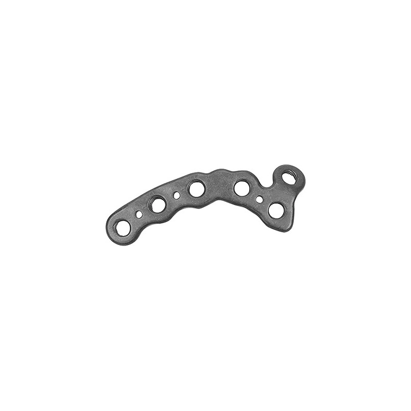
The "Perimeter" design of the calcaneal locking plate refers to its anatomical contouring and strategic placement of screw holes to provide stable fixation around the periphery of the calcaneus, encompassing key fragments and buttressing critical load-bearing structures.
Design Philosophy and Features
The Calcaneal Locking Plate (Perimeter) incorporates several innovative design elements:
- Anatomical Pre-Contouring: These plates are meticulously pre-contoured to match the complex anatomy of the lateral aspect of the calcaneus, available in left and right specific designs. This minimizes intraoperative bending, saving time and ensuring an optimal fit with a low-profile construct.
- Low-Profile Design: The plate's slim profile is crucial for minimizing soft tissue irritation, which is a common concern in the calcaneal region due to its thin soft tissue envelope.
- Perimeter Fixation Concept: The plate's shape and screw hole distribution are designed to capture and stabilize the major fracture fragments (e.g., sustentacular fragment, tuberosity fragment, posterior facet fragments) by placing screws strategically around the "perimeter" of the calcaneus, providing a strong buttress against collapse.
- Locking Screw Technology: This is the cornerstone of modern orthopedic plating. Locking screws thread into the plate, creating a fixed-angle construct. This offers:
- Enhanced Angular Stability: The construct behaves as an internal fixator, providing superior stability even in osteoporotic bone or highly comminuted fractures, where traditional screws might lose purchase.
- Reduced Plate-Bone Contact: Locking plates do not require compression between the plate and bone for stability, preserving periosteal blood supply and potentially aiding bone healing.
- Prevention of Screw Back-out: The threaded interface between the screw head and the plate prevents screws from loosening or backing out.
- Combination/Combi Holes: Many plates feature combination holes that allow for the use of both locking screws (for fixed-angle stability) and non-locking cortical screws (for compression or lag screw fixation across fracture lines). This versatility allows surgeons to customize fixation based on the fracture pattern.
- Strategic Screw Placement: Specific holes are oriented to target critical structures like the sustentaculum tali (medial column support) and the posterior facet, ensuring optimal purchase and reduction maintenance.
Materials Science
The vast majority of Calcaneal Locking Plates are manufactured from advanced medical-grade titanium alloys.
- Titanium Alloy (Ti-6Al-4V ELI): This is the gold standard material for orthopedic implants due to:
- Biocompatibility: Excellent compatibility with human tissues, minimizing adverse reactions.
- High Strength-to-Weight Ratio: Provides robust mechanical strength necessary for weight-bearing bones, while being relatively lightweight.
- Corrosion Resistance: Highly resistant to corrosion in the physiological environment, ensuring long-term implant integrity.
- MRI Compatibility: Generally considered safe for MRI scans, though patients should always inform radiologists about their implants.
- Surface Treatments: Some plates may feature specific surface treatments to enhance fatigue strength, reduce friction, or improve biocompatibility, though direct osteointegration is not typically the primary goal for plating systems.
Biomechanics of Perimeter Locking Plates
The biomechanical advantages of the Calcaneal Locking Plate (Perimeter) are profound:
- Fixed-Angle Construct: Unlike conventional plates where screws only compress the plate to the bone, locking plates create a stable, angularly fixed construct. This transforms the plate-screw interface into a rigid unit, effectively acting as an internal fixator.
- Load Sharing vs. Load Bearing: Locking plates facilitate load sharing with the bone, rather than pure load bearing. This can promote callus formation and biological healing. In highly comminuted fractures, they can bridge defects and act as a load-bearing construct until healing occurs.
- Resistance to Forces: The fixed-angle construct provides superior resistance to shear, bending, and torsional forces that are inherent in the calcaneal region during weight-bearing and movement. This stability is critical for maintaining reduction and allowing early mobilization.
- Enhanced Stability in Compromised Bone: In osteoporotic bone or highly comminuted fractures where screw pull-out is a concern with traditional screws, locking screws provide significantly improved purchase and stability.
- Maintenance of Reduction: The rigidity of the locking construct helps maintain the anatomical reduction achieved intraoperatively, preventing collapse or displacement during the healing phase.
- Potential for Earlier Mobilization: The enhanced stability often allows for a more aggressive rehabilitation protocol, potentially leading to earlier, controlled range of motion and weight-bearing, which can mitigate stiffness and improve functional recovery.
Extensive Clinical Indications & Usage
The Calcaneal Locking Plate (Perimeter) is indicated for a range of complex calcaneal fractures.
Clinical Indications
- Displaced Intra-Articular Calcaneus Fractures: Especially Sanders Type II, III, and IV fractures, which involve significant articular depression and comminution.
- Extra-Articular Calcaneus Fractures: Where stable fixation is required due to displacement or comminution, particularly those involving the calcaneal tuberosity.
- Fractures with Significant Soft Tissue Compromise: After the initial swelling subsides and the "wrinkle sign" returns, suggesting improved soft tissue viability.
- Revision Surgery: For failed previous fixation or malunion.
- Open Calcaneus Fractures: After thorough debridement and management of contamination, provided soft tissue conditions allow.
Pre-operative Planning
Meticulous pre-operative planning is crucial for successful outcomes:
- Imaging:
- X-rays: Lateral, axial (Harris view), and oblique views to assess calcaneal height, width, and tuberosity displacement.
- CT Scans: Essential for detailed fracture mapping, classification (Sanders), assessment of articular involvement, and 3D reconstruction to visualize fragment orientation.
- Fracture Classification: Accurate classification guides surgical approach and fixation strategy.
- Soft Tissue Assessment: Critical due to the tenuous soft tissue envelope. Look for blisters, abrasions, open wounds, and swelling. Delay surgery until the "wrinkle sign" returns to minimize wound complications. The Tscherne classification for closed soft tissue injuries can be applied.
- Surgical Approach Selection:
- Lateral Extensile Approach: Most common, providing wide exposure of the lateral calcaneal wall, posterior facet, and subtalar joint.
- Sinus Tarsi Approach: Less invasive, suitable for less comminuted fractures, particularly those involving the posterior facet, with potentially lower wound complication rates.
Surgical Technique (Fitting/Usage Instructions)
The surgical procedure involves precise steps to achieve optimal reduction and fixation:
- Patient Positioning: Typically lateral decubitus with the affected side up, or prone, to allow full access to the lateral aspect of the calcaneus.
- Incision & Exposure:
- Lateral Extensile Approach: An L-shaped incision is made, carefully raising a full-thickness fasciocutaneous flap to expose the lateral calcaneal wall. Meticulous care is taken to protect the sural nerve and peroneal tendons.
- Fracture Reduction:
- Indirect Reduction: Using distraction frames or Steinman pins through the talus and calcaneal tuberosity to restore length and disimpact fragments.
- Direct Reduction: Using K-wires, clamps (e.g., Verbrugge clamps), and elevators to manipulate and reduce the depressed posterior facet fragments and restore calcaneal height and width. Verification of reduction is often done with direct visualization and intraoperative fluoroscopy.
- Restoration of Böhler's angle (20-40 degrees), Gissane's angle (100-130 degrees), calcaneal height, width, and posterior facet congruity is paramount.
- Plate Application:
- The pre-contoured Calcaneal Locking Plate (Perimeter) is selected (left/right specific) and placed on the reduced calcaneus, ensuring optimal coverage of fracture fragments.
- Temporary fixation with K-wires secures the plate in position.
- Screw Placement:
- Conventional Screws: If desired, non-locking cortical screws can be inserted first through combination holes to achieve interfragmentary compression or lag screw effect across specific fracture lines.
- Locking Screws: After achieving desired compression or reduction, locking screws are inserted into the designated threaded holes. These screws provide angular stability, creating the rigid fixed-angle construct. Ensure screws are of appropriate length and do not violate the subtalar joint or other critical structures.
- Special attention is paid to placing screws into the sustentacular fragment for medial column support and into the posterior facet fragments for articular stability.
- Wound Closure: Meticulous layered closure of the fasciocutaneous flap, subcutaneous tissue, and skin. A drain is often placed to manage hematoma and reduce swelling, which can contribute to wound complications.
Post-operative Management
- Immobilization: A bulky dressing, splint, or cast is applied to protect the surgical site and provide comfort.
- Non-Weight Bearing (NWB): Typically for 6-12 weeks, depending on fracture severity and healing progress.
- Elevation: Strict elevation of the limb to reduce swelling.
- Pain Management: Appropriate analgesia is provided.
- Physical Therapy: Gradual progression from non-weight bearing range of motion exercises to partial and then full weight-bearing, guided by radiographic healing and pain tolerance.
- Follow-up: Regular clinical and radiographic follow-up to monitor healing and detect complications.
Maintenance and Sterilization Protocols (for the Implant)
Medical implants like the Calcaneal Locking Plate (Perimeter) are subject to strict regulatory and manufacturing standards to ensure patient safety and efficacy.
- Packaging: Implants are typically supplied in sterile, double-barrier packaging or in non-sterile packaging requiring sterilization prior to use. Always verify the sterility status on the product label.
- Storage: Store implants in their original, unopened packaging in a clean, dry, and cool environment, away from direct sunlight and extreme temperatures. Adhere to the manufacturer's specified storage conditions and expiry dates.
- Inspection Prior to Use: Before opening the sterile packaging, inspect it for any signs of damage, punctures, or compromise to sterility. Do not use if the package is damaged.
- Sterilization (for non-sterile implants):
- Follow the manufacturer's Instructions For Use (IFU) meticulously for sterilization parameters.
- Steam Sterilization (Autoclave): This is the most common and preferred method for titanium implants. Parameters (temperature, pressure, time) are critical and vary by device.
- Ethylene Oxide (EtO) or Gamma Irradiation: Less common for plates but may be used for certain components or if specified by the manufacturer.
- Ensure all instruments and implants are thoroughly cleaned and dried before sterilization.
- Aseptic Technique: Handle sterile implants and instruments using strict aseptic technique throughout the surgical procedure to prevent contamination.
- Traceability: Maintain records of implant lot numbers for traceability, as required by regulatory bodies.
- Disposal: Unused or contaminated implants must be disposed of according to institutional policies for medical waste. Implants that have been opened but not used should not be re-sterilized unless specifically validated and approved by the manufacturer.
Risks, Side Effects, or Contraindications
While the Calcaneal Locking Plate (Perimeter) offers significant advantages, it is essential to be aware of potential risks and contraindications.
General Surgical Risks
- Infection: Superficial or deep surgical site infection.
- Bleeding: Intraoperative or postoperative hematoma.
- Nerve Damage: Injury to the sural nerve, peroneal nerve, or other peripheral nerves, leading to numbness, weakness, or pain.
- Wound Dehiscence: Separation of wound edges, particularly common in calcaneus fractures due to the precarious soft tissue envelope.
- Deep Vein Thrombosis (DVT) / Pulmonary Embolism (PE): Blood clot formation.
- Anesthesia Risks: Adverse reactions to anesthetic agents.
Specific Risks Related to Calcaneus and Plate Fixation
- Wound Complications: The most common and challenging complication. High risk of flap necrosis, infection, and delayed wound healing due to tenuous blood supply.
- Hardware Irritation/Prominence: The plate or screws may become palpable or cause soft tissue irritation, potentially requiring hardware removal in a second surgery.
- Nonunion or Malunion: Despite stable fixation, the bone may fail to heal (nonunion) or heal in an incorrect position (malunion), leading to persistent pain and dysfunction.
- Post-traumatic Subtalar Arthritis: Even with anatomical reduction, damage to the articular cartilage can lead to arthritis, often requiring subtalar fusion in the long term.
- Nerve Entrapment: Particularly of the sural nerve in the lateral approach.
- Complex Regional Pain Syndrome (CRPS): A chronic pain condition that can develop after trauma or surgery.
- Loss of Reduction: Despite initial stable fixation, severe comminution or inadequate bone purchase can lead to a loss of the achieved reduction.
- Screw Breakage or Loosening: Though less common with locking screws, it can still occur.
Contraindications
- Active Infection: Any active infection in the surgical field or systemic infection is a absolute contraindication.
- Severe Soft Tissue Compromise: Extensive skin blistering, necrosis, or severe open fractures with gross contamination that preclude safe wound closure and increase infection risk. Surgery may be delayed until soft tissue conditions improve.
- Insufficient Bone Stock: Extremely comminuted fractures with severe osteoporosis where stable screw purchase cannot be achieved.
- Patient Inability/Unwillingness to Comply: Patients who cannot or will not adhere to post-operative non-weight bearing and rehabilitation protocols.
- Severe Comorbidities: Uncontrolled diabetes, severe peripheral vascular disease, or other medical conditions that significantly increase surgical risk and impair wound healing.
- Allergy to Implant Materials: Extremely rare for titanium, but a theoretical contraindication.
Massive FAQ Section
Q1: What is a Calcaneal Locking Plate (Perimeter)?
A1: A Calcaneal Locking Plate (Perimeter) is an advanced orthopedic implant specifically designed for the surgical fixation of complex fractures of the calcaneus (heel bone). Its "perimeter" design refers to its anatomical contouring and strategic screw placement to provide stable buttress fixation around the periphery of the calcaneus, capturing and stabilizing multiple fracture fragments. It utilizes locking screw technology for enhanced stability.
Q2: Why is the "perimeter" design important for calcaneus fractures?
A2: The perimeter design is crucial because calcaneus fractures often involve multiple displaced fragments and loss of the bone's normal height and width. This plate's pre-contoured shape and strategically placed screw holes allow for robust fixation of these fragments, particularly around the crucial articular surfaces and the calcaneal tuberosity, effectively restoring the calcaneus's anatomical shape and providing strong buttressing support against collapse.
Q3: What types of calcaneus fractures benefit most from this plate?
A3: This plate is primarily used for displaced intra-articular calcaneus fractures, especially those classified as Sanders Type II, III, and IV, which involve significant articular depression and comminution. It is also beneficial for certain extra-articular fractures requiring stable fixation and for cases of malunion or nonunion.
Q4: What material is the Calcaneal Locking Plate typically made from?
A4: Calcaneal Locking Plates are almost exclusively made from medical-grade titanium alloy (Ti-6Al-4V ELI). This material is chosen for its excellent biocompatibility, high strength-to-weight ratio, corrosion resistance, and MRI compatibility.
Q5: How long is the typical recovery period after surgery with a Calcaneal Locking Plate?
A5: Recovery is highly individual and depends on fracture severity and patient compliance. Generally, patients are non-weight bearing for 6 to 12 weeks post-surgery to allow for bone healing. Gradual weight-bearing and physical therapy then begin, with full recovery often taking 6 to 12 months, and sometimes longer for complete functional restoration.
Q6: Will I need to have the Calcaneal Locking Plate removed?
A6: Plate removal is not universally required. It is often performed if the hardware causes irritation, pain, or prominence, or if an infection develops. If the plate is not causing issues, it can often remain in place indefinitely. The decision for removal is made in consultation with your orthopedic surgeon.
Q7: What are the potential complications associated with this surgery?
A7: Potential complications include general surgical risks like infection, bleeding, and nerve damage. Specific to calcaneus fixation are high rates of wound complications (e.g., dehiscence, infection, flap necrosis) due to the tenuous soft tissue. Other specific risks include hardware irritation, nonunion/malunion, post-traumatic subtalar arthritis, and Complex Regional Pain Syndrome (CRPS).
Q8: How does locking plate technology improve patient outcomes compared to traditional plates?
A8: Locking plate technology creates a fixed-angle construct between the plate and screws, providing superior angular stability. This is particularly advantageous in comminuted fractures or osteoporotic bone, where traditional screws might lose purchase. This enhanced stability helps maintain anatomical reduction, allows for potentially earlier, controlled mobilization, and can lead to improved healing and functional outcomes by reducing the risk of loss of reduction.
Q9: Can I undergo an MRI with a Calcaneal Locking Plate implant?
A9: Yes, implants made from medical-grade titanium alloy are generally considered safe for MRI scans. However, it is crucial to always inform your radiologist and MRI technologist about your implant before undergoing any MRI procedure. The implant may cause some artifact in the images, but it typically does not pose a safety risk.
Q10: What are the alternatives to plate fixation for calcaneus fractures?
A10: Alternatives depend on the fracture type. For very mild, non-displaced fractures, non-operative management (casting, non-weight bearing) may be an option. For certain fracture patterns, minimally invasive techniques using percutaneous screws or external fixation might be considered. However, for displaced intra-articular fractures, open reduction and internal fixation with a locking plate is often the preferred method for achieving anatomical reduction and stable fixation.
Q11: How does the Calcaneal Locking Plate help restore foot anatomy?
A11: The pre-contoured design of the perimeter plate is anatomically matched to the calcaneus. During surgery, the plate acts as a template to guide the reduction of displaced fragments, helping to restore crucial anatomical parameters such as Böhler's angle, Gissane's angle, calcaneal height, and width. This anatomical restoration is vital for normal foot biomechanics and function.
Q12: Is surgery for a calcaneus fracture with a locking plate a common procedure?
A12: While calcaneus fractures are relatively common, the decision for surgical intervention depends on the fracture type and severity. For displaced intra-articular fractures, surgery with a locking plate is a well-established and frequently performed procedure in specialized orthopedic trauma centers, given its proven efficacy in restoring anatomy and function.