The Definitive Guide to Bioabsorbable Suture Anchors (3.0mm/5.5mm)
Comprehensive Introduction & Overview
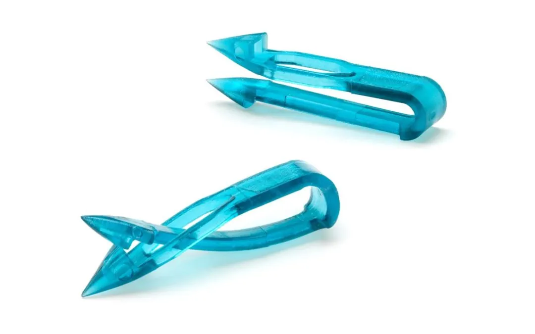
Bioabsorbable suture anchors represent a significant advancement in orthopedic surgery, offering a sophisticated solution for reattaching soft tissues to bone. Unlike traditional metallic anchors, these innovative devices are designed to gradually resorb into the body over time, eliminating the need for permanent implants and their associated potential long-term complications. This guide provides an exhaustive exploration of bioabsorbable suture anchors, specifically focusing on the widely used 3.0mm and 5.5mm sizes, detailing their design, clinical applications, biomechanics, and impact on patient outcomes.
The core principle behind bioabsorbable anchors is to provide robust initial fixation, allowing the body's natural healing processes to take hold and form a strong, biological attachment between the soft tissue and bone. As healing progresses, the anchor slowly degrades and is metabolized, transferring the mechanical load to the newly formed tissue-bone interface. This controlled degradation profile is critical, as it ensures mechanical support during the crucial early healing phase while preventing stress shielding and other issues associated with permanent foreign bodies. Orthopedic surgeons increasingly rely on these devices for a wide array of procedures, from complex rotator cuff repairs to delicate ligament reconstructions, recognizing their potential to optimize patient recovery and long-term functional success.
Deep-Dive into Technical Specifications & Mechanisms
Design & Materials Science
The efficacy of bioabsorbable suture anchors hinges on their meticulously engineered design and advanced material composition.
Anchor Body Materials:
The most common materials used for bioabsorbable anchors are biocompatible polymers, primarily from the alpha-hydroxy acid family. Each material offers distinct degradation profiles and mechanical properties:
- Poly-L-lactic Acid (PLLA): A semi-crystalline polymer known for its high initial strength and relatively slow degradation rate (typically 2-5 years). PLLA maintains mechanical integrity longer, making it suitable for applications requiring extended support.
- Polyglycolic Acid (PGA): A highly crystalline polymer with a faster degradation rate (typically 6-12 months). PGA provides excellent initial strength but loses mechanical integrity more quickly.
- Polylactic-co-glycolic Acid (PLGA): A co-polymer of PLLA and PGA, offering a tunable degradation rate depending on the ratio of lactic to glycolic acid. This allows for customized strength retention and resorption times.
- Poly-ɛ-caprolactone (PCL): A semi-crystalline polymer with a very slow degradation rate, sometimes used in specific applications requiring prolonged presence.
- Bio-composite Materials: Some anchors incorporate osteoconductive fillers like Tricalcium Phosphate (TCP) or Hydroxyapatite (HA) into a polymer matrix (e.g., PLLA-TCP). These composites are designed to promote bone ingrowth and integration, potentially enhancing the long-term biological fixation and minimizing bone tunnel enlargement.
Anchor Body Designs:
The physical design of the anchor plays a crucial role in achieving secure initial fixation:
- Threaded Anchors: Feature screw-like threads that engage directly with the bone cortex and cancellous bone, providing excellent pull-out strength, especially in denser bone.
- Barbed/Interference Fit Anchors: Utilize barbs or a press-fit mechanism to achieve fixation. These often require a precise pilot hole and may be less dependent on bone density for initial stability.
- Push-in Anchors: Designed for straightforward insertion into a pre-drilled pilot hole, relying on friction and sometimes expandable elements for fixation.
Suture Materials:
The sutures pre-loaded onto the anchors are equally vital for tissue repair. Common materials include:
- Ultra-High Molecular Weight Polyethylene (UHMWPE): Extremely strong, abrasion-resistant, and low-profile, often braided.
- Polyester (e.g., PET): Strong and durable, commonly braided.
- Polypropylene (e.g., PPL): Monofilament, smooth, and low friction.
Suture sizes typically range from USP #0 to #2, selected based on the tissue being repaired and the required strength.
Size Specifics (3.0mm vs. 5.5mm):
| Feature | 3.0mm Bioabsorbable Suture Anchor | 5.5mm Bioabsorbable Suture Anchor |
|---|---|---|
| Diameter | Smaller (e.g., 3.0mm pilot hole) | Larger (e.g., 4.5mm-5.5mm pilot hole) |
| Footprint | Minimal bone removal, ideal for smaller bone structures | Larger bone-anchor interface, higher pull-out strength |
| Bone Quality | Requires good to moderate bone quality for optimal fixation | More forgiving in slightly softer bone due to larger surface area |
| Applications | Hand, Wrist, Elbow, Foot & Ankle ligaments, smaller rotator cuff tears, labral repairs in smaller patients | Shoulder (rotator cuff, labrum), Hip labrum, larger ligament repairs, knee (MPFL) |
| Suture Capacity | Typically 1-2 sutures | Typically 2-3 sutures |
Biomechanics of Fixation
The biomechanical performance of bioabsorbable suture anchors is critical for successful surgical outcomes.
- Initial Fixation Strength: Achieved through mechanical engagement with the bone (threads, barbs, interference fit). This strength must be sufficient to withstand immediate post-operative loads and allow early healing.
- Pull-out Strength: A primary measure of anchor efficacy, influenced by:
- Anchor Design: Thread geometry, surface area, number of barbs.
- Bone Quality: Cortical thickness, cancellous bone density (higher density = higher strength).
- Insertion Technique: Proper pilot hole creation, full insertion depth, correct trajectory.
- Cyclic Loading: The ability of the anchor and suture construct to withstand repetitive stresses without failure or significant displacement. This is particularly important for joints undergoing early range of motion.
- Degradation Profile & Strength Retention: The material's degradation rate is designed to align with the biological healing cascade.
- Initially, the anchor provides maximal mechanical support.
- As the tissue heals and gains strength, the anchor gradually loses its mechanical integrity through hydrolysis.
- The goal is for the anchor to maintain sufficient strength until the tissue-bone interface is biologically strong enough to bear the full load. Premature loss of strength can lead to failure; prolonged presence can inhibit natural load transfer.
- Bone Remodeling: Bioabsorbable anchors allow for normal bone remodeling at the anchor site after resorption, potentially leaving behind a healthier bone structure compared to permanent implants. Bio-composite anchors may actively promote this remodeling.
Extensive Clinical Indications & Usage
Bioabsorbable suture anchors are versatile tools employed across various orthopedic subspecialties.
Detailed Surgical Applications
1. Shoulder:
* Rotator Cuff Repair: Both 3.0mm and 5.5mm anchors are used, depending on tear size, bone quality, and patient size. 5.5mm anchors are common for larger tears requiring robust fixation, while 3.0mm anchors may be used for marginal convergence or smaller tears. Anchors secure the torn tendon back to the humeral head.
* Labral Repair (Bankart, SLAP, Posterior Labral): Crucial for shoulder stability. 3.0mm anchors are frequently used due to the smaller bone footprint and need for precise placement in the glenoid rim.
* Capsular Shift/Plication: For shoulder instability, anchors secure the joint capsule to the glenoid.
* Biceps Tenodesis: Anchors secure the biceps tendon into the humerus, often in the bicipital groove or a suprapectoral position.
2. Knee:
* Meniscal Repair: Smaller 3.0mm anchors can be used to reattach torn meniscal fragments to the capsule, though all-inside devices are also prevalent.
* MPFL Reconstruction (Medial Patellofemoral Ligament): Anchors are used to secure the reconstructed ligament to the patella and femur for patellar stability.
* ACL/PCL Reconstruction: While not the primary fixation for the graft, anchors can be used for ancillary fixation or for repairing associated capsular/meniscal injuries.
3. Hip:
* Labral Repair/Reconstruction: 3.0mm anchors are ideal for reattaching the acetabular labrum to the rim, restoring suction seal and stability.
* Gluteus Medius/Minimus Repair: For tears of these important abductor muscles, 5.5mm anchors provide robust fixation to the greater trochanter.
4. Foot & Ankle:
* Lateral Ankle Ligament Repair (e.g., Broström-Gould): Repairing tears of the anterior talofibular ligament (ATFL) and calcaneofibular ligament (CFL) for chronic ankle instability. 3.0mm anchors are commonly used in the fibula.
* Achilles Tendon Repair: In some techniques, anchors are used to secure the Achilles tendon to the calcaneus.
* Midfoot/Forefoot Ligament Repairs: For specific instability patterns.
5. Elbow/Wrist/Hand:
* Ulnar Collateral Ligament (UCL) Repair (Elbow): For "Tommy John" injuries, anchors secure the ligament to the ulna.
* TFCC (Triangular Fibrocartilage Complex) Repair (Wrist): Smaller anchors can be used to reattach peripheral tears.
Fitting & Usage Instructions (General Principles)
The successful implantation of bioabsorbable suture anchors requires meticulous technique and adherence to specific protocols.
-
Pre-operative Planning:
- Imaging Review: X-rays, MRI, CT scans to assess bone quality, defect size, and anatomical landmarks.
- Anchor Selection: Choose the appropriate anchor size (3.0mm or 5.5mm), material, and design based on the surgical site, bone quality, and required fixation strength.
- Patient Positioning: Ensure optimal exposure and access to the surgical site.
-
Drill Hole Preparation:
- Pilot Hole Creation: A precisely sized drill bit (matching the anchor diameter or slightly smaller for press-fit designs) is used to create a pilot hole.
- Depth & Trajectory: The hole must be drilled to the correct depth and angle to ensure optimal engagement with the bone, avoiding cortical breach or neurovascular structures.
- Debridement: Clear any soft tissue or debris from the drill site.
-
Anchor Insertion:
- Loading: Ensure sutures are correctly loaded onto the anchor inserter.
- Engagement: Engage the anchor securely with the insertion tool.
- Controlled Advancement: The anchor is advanced into the pilot hole using a controlled motion.
- Self-tapping Anchors: Screwed directly into the bone.
- Non-tapping/Push-in Anchors: Tapped or pushed into the hole until fully seated.
- Confirmation of Fixation: Gently tug on the sutures to confirm solid anchor fixation in the bone. Avoid excessive force that could compromise the anchor.
-
Suture Management & Tissue Repair:
- Suture Retrieval: Retrieve the sutures from the anchor, typically through a cannula.
- Tissue Capture: Pass the sutures through the torn soft tissue using appropriate arthroscopic instruments (e.g., suture passers, shuttles).
- Tensioning & Knot Tying: Carefully tension the sutures to approximate the soft tissue to the bone without strangulating the tissue. Secure the repair with appropriate arthroscopic knot-tying techniques, ensuring sufficient knot stacks for security.
-
Post-operative Care:
- Immobilization: Prescribed period of immobilization (sling, brace) to protect the repair during initial healing.
- Rehabilitation Protocol: A structured and progressive physical therapy program tailored to the specific repair and patient needs, guiding range of motion and strengthening exercises.
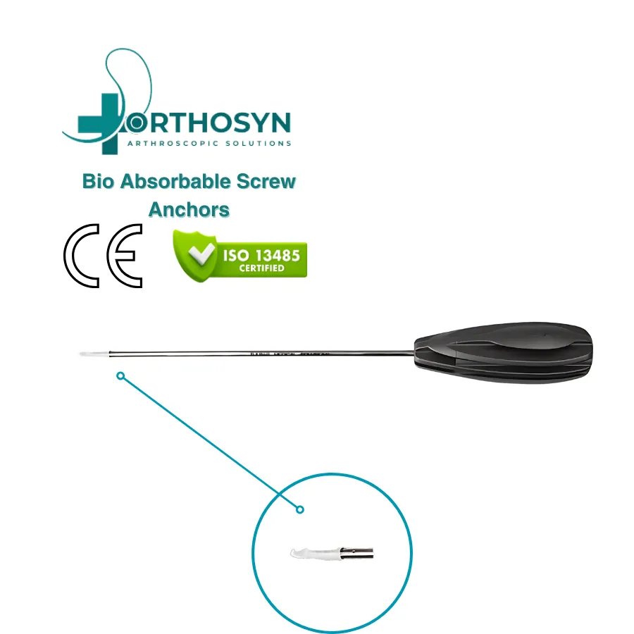
Maintenance & Sterilization Protocols
Bioabsorbable suture anchors are generally supplied as single-use, sterile devices.
- Sterilization: They are typically sterilized by gamma irradiation or ethylene oxide (ETO) gas and packaged in sterile, peel-pouch configurations.
- Single-Use: Critical to emphasize that these devices are not designed for reprocessing or re-sterilization. Re-sterilization can compromise material integrity, sterility, and mechanical performance.
- Storage: Store anchors in their original, unopened packaging in a cool, dry place, away from direct sunlight and extreme temperatures, as per manufacturer's instructions.
- Inspection Before Use: Always inspect the sterile packaging for any signs of damage, punctures, or compromise before opening. Verify the expiration date. Do not use if the package is damaged or expired.
- Disposal: Dispose of unused or expired anchors, as well as all packaging materials, according to hospital protocols for medical waste.
Risks, Side Effects, or Contraindications
While highly effective, the use of bioabsorbable suture anchors carries potential risks and contraindications.
Risks & Side Effects:
- Anchor Pull-out/Failure: Can occur due to poor bone quality, improper insertion technique, inadequate initial fixation, or excessive post-operative loading.
- Suture Breakage: Although rare with modern high-strength sutures, it can occur if sutures are improperly tied, nicked, or subjected to extreme forces.
- Infection: As with any surgical implant, there is a risk of surgical site infection, though bioabsorbable materials themselves are not typically more prone to infection than metal.
- Neurovascular Injury: Risk during drill hole preparation or anchor insertion if anatomical landmarks are not respected.
- Inflammatory Reaction/Synovitis: Rarely, some patients may experience a sterile inflammatory response or foreign body reaction to the degrading polymer material. This is usually self-limiting but can cause pain and swelling. Bio-composite anchors with high HA content have been associated with this in some older designs.
- Bone Tunnel Widening: While less common than with some permanent implants, some degree of tunnel widening can occur, especially with certain polymer degradation profiles.
- Fracture: Extremely rare, but aggressive drilling or insertion in very osteoporotic bone could theoretically lead to localized bone fracture.
Contraindications:
- Active Infection: Absolute contraindication. Implantation in an infected field significantly increases the risk of persistent infection and implant failure.
- Severely Osteoporotic Bone: A relative contraindication. While 5.5mm anchors offer better purchase than 3.0mm in softer bone, extremely poor bone quality may preclude effective fixation with any anchor. Alternative fixation methods or bone augmentation may be necessary.
- Insufficient Bone Stock: Inadequate bone volume or thickness at the intended anchor site will prevent secure fixation.
- Known Allergy to Implant Materials: Extremely rare for the common polymers used, but a theoretical contraindication.
- Compromised Healing Potential: Patients with severe systemic diseases, uncontrolled diabetes, or those on certain medications (e.g., high-dose corticosteroids) may have impaired healing, affecting the success of the repair.
Patient Outcome Improvements
The adoption of bioabsorbable suture anchors has significantly contributed to improved patient outcomes in orthopedic surgery.
- Elimination of Permanent Implant Issues:
- No Stress Shielding: Unlike metallic implants, which can bear a disproportionate amount of load, bioabsorbable anchors gradually transfer load to the healing tissue, encouraging natural bone remodeling and preventing bone weakening.
- Reduced Imaging Artifacts: Resorbing implants cause fewer artifacts on post-operative MRI or CT scans, allowing for clearer visualization of soft tissues and bone.
- No Implant-Related Pain/Irritation: Eliminates the potential for long-term irritation or pain caused by a permanent foreign body, which can sometimes necessitate a second surgery for implant removal.
- Optimized Biological Healing: The controlled degradation profile is designed to support the biological healing process by providing mechanical stability during the critical early phases, then dissolving as the body's own tissue-bone interface strengthens.
- Enhanced Functional Recovery: By providing stable initial fixation, these anchors facilitate early, controlled rehabilitation, which is crucial for restoring range of motion and strength, ultimately leading to better functional outcomes.
- Reduced Revision Rates: When appropriately selected and meticulously implanted, bioabsorbable anchors contribute to durable repairs, potentially lowering the need for revision surgeries.
- Patient Comfort and Peace of Mind: Many patients prefer the idea of an implant that will eventually disappear, offering psychological comfort and avoiding concerns about a foreign body remaining in their system indefinitely.
- Promotion of Bone Healing: Especially with bio-composite anchors, the osteoconductive components can encourage bone ingrowth and integration, further strengthening the repair site.
Massive FAQ Section
1. What is a bioabsorbable suture anchor?
A bioabsorbable suture anchor is a small, specialized implant used in orthopedic surgery to reattach soft tissues (like tendons or ligaments) to bone. Unlike metal anchors, it is made from biocompatible polymers that gradually dissolve and are absorbed by the body over time, leaving no permanent implant behind.
2. How long does it take for the anchor to absorb completely?
The absorption time varies depending on the specific polymer material used. Most bioabsorbable anchors are designed to maintain mechanical strength for several months to a year, supporting healing, and then fully absorb over 1 to 5 years. Materials like PLLA absorb more slowly than PGA or PLGA.
3. Are bioabsorbable anchors as strong as metal anchors?
Yes, modern bioabsorbable suture anchors are designed to provide comparable initial fixation strength to metallic anchors. Their strength is sufficient to withstand the forces required during the critical early healing phase. The key difference is that their strength is temporary, gradually diminishing as the body's natural healing takes over.
4. Can bioabsorbable anchors cause an allergic reaction?
Allergic reactions to the polymer materials used in bioabsorbable anchors (like PLLA, PGA) are extremely rare. These materials are highly biocompatible and have been extensively studied and used in medical devices for decades. However, a sterile inflammatory reaction to the degrading material can occasionally occur, though it is usually self-limiting.
5. What are the main differences between 3.0mm and 5.5mm bioabsorbable anchors?
The primary difference is their size and the applications they are suited for. 3.0mm anchors are smaller, require less bone removal, and are ideal for delicate repairs in smaller bones (e.g., hand, wrist, ankle, smaller labral repairs). 5.5mm anchors are larger, offer greater pull-out strength, and are used for more robust repairs in larger bones (e.g., rotator cuff, hip labrum).
6. Are these anchors visible on X-rays or MRI after surgery?
Most pure polymer bioabsorbable anchors are radiolucent (not visible on standard X-rays). However, some bio-composite anchors that contain osteoconductive fillers like tricalcium phosphate (TCP) or hydroxyapatite (HA) may be partially visible on X-rays. They typically cause minimal to no artifact on MRI scans, allowing for clearer post-operative imaging compared to metallic implants.
7. What happens to the bone after the anchor absorbs?
As the anchor degrades, the space it occupied is gradually replaced by new, healthy bone tissue through the body's natural remodeling processes. The goal is for the bone to return to a near-normal state, free of foreign material. Bio-composite anchors may even encourage this bone regeneration.
8. Is it possible for the anchor to fail or pull out?
Yes, anchor pull-out or failure is a potential risk, as with any surgical fixation device. This can be influenced by factors such as poor bone quality, improper surgical technique, inadequate initial fixation, or excessive stress on the repair during the healing period. Following post-operative rehabilitation protocols is crucial to minimize this risk.
9. What are the benefits of bioabsorbable anchors over permanent metallic implants?
Key benefits include:
* No permanent foreign body left in the patient.
* Elimination of potential long-term implant-related pain or irritation.
* Reduced risk of stress shielding in the bone.
* Fewer imaging artifacts on future MRI/CT scans.
* No need for potential future surgery to remove a symptomatic implant.
* Gradual load transfer promotes natural tissue healing and bone remodeling.
10. Who is a good candidate for bioabsorbable suture anchors?
Most patients requiring soft tissue to bone reattachment are good candidates. This includes individuals undergoing rotator cuff repair, labral repair (shoulder or hip), ligament reconstructions (e.g., ankle, knee), and other tendon repairs. Patients with severely osteoporotic bone or active infections may require careful consideration or alternative fixation methods.
11. Are there any activity restrictions after surgery with these anchors?
Yes, specific activity restrictions and a structured rehabilitation program are crucial. These protocols are designed to protect the healing repair while the anchor provides initial stability. Restrictions typically involve limiting weight-bearing, range of motion, and lifting for several weeks or months, gradually progressing as healing occurs. Your surgeon and physical therapist will provide a personalized plan.
12. Can bioabsorbable suture anchors be used in children or adolescents?
Yes, bioabsorbable anchors are often preferred in pediatric and adolescent orthopedic surgery, especially in areas where bone growth plates are present. Their temporary nature avoids potential interference with bone growth that permanent metallic implants might pose, making them a favorable choice for conditions like shoulder instability or patellar instability in younger patients.