The Arthroscopic Suture Passer: Revolutionizing Minimally Invasive Orthopedic Surgery
In the realm of modern orthopedic surgery, precision and minimal invasiveness are paramount. The arthroscopic suture passer, particularly specialized designs like the "Scorpion" and "BirdBeak," stands as a cornerstone instrument, enabling surgeons to perform complex intra-articular repairs with unparalleled accuracy through small incisions. This comprehensive guide delves into the intricate world of these essential tools, covering their design, biomechanics, clinical applications, usage protocols, maintenance, and profound impact on patient outcomes.
Arthroscopic surgery has transformed the landscape of joint repair, moving away from large open incisions to techniques that utilize small portals, a camera (arthroscope), and specialized instruments. The challenge within this confined space is the manipulation and placement of sutures – a task where traditional methods are often cumbersome or impossible. Arthroscopic suture passers were engineered to overcome this, providing the dexterity and control needed to capture, pass, and retrieve sutures within the joint, facilitating robust and anatomically correct repairs.
The Evolution of Arthroscopic Suture Management
The journey from early, basic suture manipulators to today's sophisticated passers reflects a relentless pursuit of surgical excellence. Early techniques often involved complex needle configurations or required larger portals. The advent of tools like the Scorpion and BirdBeak revolutionized this process, streamlining suture management and expanding the scope of treatable conditions via arthroscopy. These instruments are not merely tools; they are extensions of the surgeon's hands, allowing for intricate maneuvers in tight spaces, ultimately leading to better repair integrity and patient recovery.
Deep-Dive into Technical Specifications and Mechanisms
Understanding the mechanics and materials behind arthroscopic suture passers is crucial for appreciating their efficacy and reliability.
Design and Materials
Arthroscopic suture passers are engineering marvels, blending ergonomic design with robust, biocompatible materials.
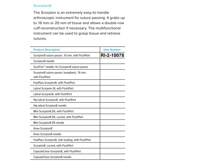
Scorpion Suture Passer
The "Scorpion" design is characterized by its unique pincer-like jaws that articulate at the tip.
- Jaws:
- Mechanism: Features a spring-loaded or trigger-activated mechanism that causes one jaw to close onto the other, capturing a suture loop or a needle.
- Needle Integration: Often incorporates a small, sharp needle within one jaw that can be advanced through tissue and then retrieved, carrying a suture strand.
- Tissue Engagement: Designed to grasp and pass through various soft tissues such as meniscus, labrum, or rotator cuff tendons with precision.
- Handle:
- Ergonomics: Ergonomically shaped to fit comfortably in the surgeon's hand, often with a pistol-grip style.
- Trigger Mechanism: A lever or button on the handle activates the jaws and/or needle, providing tactile feedback.
- Shaft:
- Length & Diameter: Available in various lengths (typically 12-20 cm) and diameters (2.7-5.0 mm) to accommodate different joint sizes and portal requirements.
- Material: Usually constructed from high-grade stainless steel or titanium alloys for strength, corrosion resistance, and biocompatibility.
- Needle:
- Fixed or Detachable: Can be fixed within the jaw or designed for single-use, detachable needles.
- Size & Curvature: Optimized for minimal tissue trauma while ensuring efficient suture passage.
BirdBeak Suture Passer
The "BirdBeak" design, as its name suggests, features a distinct curved, pointed tip resembling a bird's beak.
- Jaw/Beak Design:
- Curvature: The sharp, curved tip is designed for precise tissue penetration and navigating tight anatomical spaces.
- Suture Retrieval: Often includes a small hole or notch near the tip through which a suture can be retrieved after the beak passes through the tissue. Some designs feature a separate suture-retrieval hook or loop that deploys from the tip.
- Handle: Similar ergonomic designs to the Scorpion, often with a plunger or trigger mechanism to deploy the beak or retrieve the suture.
- Shaft: Made from similar materials (stainless steel, titanium alloys) and available in various lengths and diameters.
- Comparison: While the Scorpion excels at capturing and passing a suture loop directly through tissue, the BirdBeak is often preferred for its ability to penetrate dense tissue and retrieve a specific suture strand with its unique curved profile.
General Features & Materials
- Articulation: Some advanced models feature articulating tips, allowing for greater maneuverability and optimal angle of approach within complex joint anatomies.
- Cannula Compatibility: Designed to seamlessly integrate with standard arthroscopic cannulas, ensuring a secure and stable working portal.
- Disposable vs. Reusable: While many components of the handle and shaft are reusable, the needle or suture-engaging tip might be disposable to ensure consistent sharpness and sterility.
- Materials:
- Stainless Steel (e.g., 316L): Excellent strength, corrosion resistance, and widely used for reusable components.
- Titanium Alloys: Lighter, stronger, and more biocompatible than steel, often used for critical components or instruments requiring reduced weight.
- PEEK (Polyether Ether Ketone): Used for certain non-metallic components, offering radiolucency and strength.
Biomechanics of Suture Passage
The effective use of suture passers involves intricate biomechanical principles.
- Tissue Interaction: The design of the jaws or beak dictates how the instrument interacts with different tissue types. Sharp, thin tips minimize tissue damage while maximizing penetration efficiency.
- Suture Passage Forces: The force required to pass a suture through tissue depends on tissue density, suture diameter, and needle sharpness. Suture passers are designed to minimize this force, reducing the risk of tissue tearing or instrument fatigue.
- Needle Control & Trajectory: Precise control over the needle's trajectory is crucial to avoid iatrogenic injury to vital structures (nerves, vessels, cartilage) and ensure accurate suture placement for a strong repair.
- Load Distribution: The instrument's design ensures that forces are distributed optimally during use, preventing stress concentrations that could lead to instrument failure or suboptimal tissue engagement.
- Surgeon Ergonomics & Fatigue: The ergonomic handle design minimizes hand fatigue, allowing the surgeon to maintain fine motor control during lengthy and intricate procedures. This directly impacts the precision and quality of the repair.
Extensive Clinical Indications & Usage
Arthroscopic suture passers are indispensable across a broad spectrum of orthopedic procedures, enabling minimally invasive solutions for various joint pathologies.
Detailed Surgical Applications
| Surgical Procedure | Primary Application of Suture Passers | Specific Suture Passer Type (Commonly Used) |
|---|---|---|
| Rotator Cuff Repair | Passing sutures through torn supraspinatus, infraspinatus, subscapularis tendons for reattachment to the humerus. | Scorpion, BirdBeak |
| Meniscal Repair | Repairing meniscal tears (bucket-handle, radial, horizontal) using all-inside, inside-out, or outside-in techniques. | Scorpion, BirdBeak |
| Labral Repair (Shoulder) | Reattaching the torn glenoid labrum in cases of instability (e.g., Bankart lesions, SLAP tears). | Scorpion, BirdBeak |
| Labral Repair (Hip) | Repairing acetabular labral tears to restore hip joint stability and function. | Scorpion, BirdBeak |
| AC Joint Reconstruction | Stabilizing the acromioclavicular joint by passing sutures through ligaments or grafts. | Scorpion, BirdBeak |
| Capsular Shift/Plication | Tightening the joint capsule in cases of shoulder or hip instability. | Scorpion, BirdBeak |
| Biceps Tenodesis | Securing the biceps tendon to the humerus in certain shoulder pathologies. | Scorpion |
| Ligament Reconstruction | Less common as primary tools for ACL/PCL, but can be used for graft passage or auxiliary fixation. | BirdBeak (for specific graft handling) |
Fitting and Usage Instructions
Proper handling and technique are paramount for the safe and effective use of arthroscopic suture passers.
Pre-operative Setup
- Instrument Inspection: Before sterilization, thoroughly inspect the instrument for any signs of damage, wear, or dullness. Ensure all moving parts operate smoothly.
- Compatibility: Verify that the suture passer's shaft diameter is compatible with the chosen arthroscopic cannula.
- Suture Loading (if applicable): For systems requiring pre-loading, ensure the correct suture type and size are used and properly loaded according to manufacturer instructions.
Intra-operative Technique
- Portal Placement: Optimal portal placement is critical. The working portal should allow for direct visualization of the target tissue and provide a favorable angle for instrument insertion and manipulation.
-
Tissue Visualization: Maintain a clear arthroscopic view of the surgical field. Use fluid management and shaver debridement as necessary to optimize visibility.
-
Scorpion Technique (General Steps):
- Insertion: Insert the closed Scorpion passer through the working cannula towards the target tissue.
- Tissue Grasping: Position the open jaws of the Scorpion to encompass the desired tissue segment.
- Needle Advancement: Activate the trigger to advance the needle through the tissue, capturing the suture loop or strand within the jaw.
- Suture Retrieval: Close the jaws, securing the suture. Retract the instrument, pulling the captured suture through the tissue.
- Loop Creation: For certain repairs, the Scorpion can create a suture loop, allowing another suture limb to be passed through it.
-
BirdBeak Technique (General Steps):
- Insertion: Insert the BirdBeak passer through the working cannula.
- Tissue Penetration: Position the curved, pointed beak against the target tissue. Carefully advance the beak to penetrate the tissue.
- Suture Engagement: Once the beak has passed through the tissue, manipulate it to engage the desired suture strand or deploy the retrieval hook/loop to capture the suture.
- Suture Retrieval: Retract the BirdBeak, pulling the suture through the tissue.
Tips for Success:
- Controlled Movements: Use slow, deliberate movements to avoid accidental tissue damage or instrument breakage.
- Tissue Management: Avoid excessive tension on the tissue when passing sutures, which can lead to tearing.
- Suture Tension: Maintain appropriate suture tension throughout the repair process to ensure a secure knot and optimal tissue apposition.
- Ergonomic Positioning: Position yourself and the arthroscope monitor to minimize strain and optimize visualization, enhancing precision.
- Practice: Proficiency with these instruments comes with practice, often starting with cadaveric labs or simulation trainers.
Maintenance and Sterilization Protocols
Proper maintenance and sterilization are crucial for the longevity of arthroscopic suture passers, ensuring patient safety and instrument functionality.
Cleaning and Inspection
- Immediate Post-Use Care: Immediately after use, rinse the instrument thoroughly with distilled water to prevent blood and tissue from drying onto surfaces.
- Manual Cleaning:
- Disassemble any removable parts according to manufacturer instructions.
- Use a soft brush and enzymatic detergent to meticulously clean all surfaces, lumens, and articulations. Pay special attention to the jaws, needle mechanism, and any crevices where debris can accumulate.
- Flush lumens repeatedly until clear.
- Automated Cleaning (if applicable): If using ultrasonic cleaners or automated washer-disinfectors, follow the manufacturer's validated protocols for cycle times, temperatures, and detergent concentrations.
- Inspection: After cleaning, visually inspect the instrument under magnification for:
- Damage (bends, cracks, burrs).
- Wear (dullness of needle/beak, corrosion).
- Smooth operation of all moving parts.
- Cleanliness (absence of residual bioburden).
- Lubrication: Lubricate hinges and moving parts with a medical-grade, water-soluble lubricant as recommended by the manufacturer.
Sterilization
Arthroscopic suture passers are critical instruments and require high-level sterilization.
- Autoclave (Steam Sterilization): This is the most common and preferred method.
- Packaging: Place instruments in appropriate sterilization pouches or rigid sterilization containers, ensuring proper wrapping techniques that allow steam penetration.
- Cycle Parameters: Follow validated steam sterilization cycles:
- Gravity Displacement: 121°C (250°F) for 30 minutes, or 132°C (270°F) for 15 minutes.
- Pre-vacuum: 132°C (270°F) for 4 minutes.
- Flash Sterilization: While possible in urgent situations (e.g., 132°C for 3-10 minutes unwrapped), it should be avoided for routine sterilization due to reduced safety margins and increased risk of instrument damage.
- Ethylene Oxide (EtO) Sterilization: Less common for reusable instruments due to longer cycle times and aeration requirements, but may be used for heat-sensitive materials.
- Hydrogen Peroxide Gas Plasma: Another low-temperature sterilization option for heat- and moisture-sensitive instruments, offering shorter cycle times than EtO.
Storage
- Store sterilized instruments in a dry, temperature-controlled environment, protected from dust and physical damage, until ready for use.
- Maintain proper documentation and tracking of instrument sterilization cycles.
Patient Outcome Improvements
The adoption and refinement of arthroscopic suture passers have significantly contributed to enhancing patient outcomes in orthopedic surgery.
- Reduced Incision Size: Minimally invasive approach leads to smaller skin incisions, resulting in less tissue trauma, reduced scarring, and improved cosmesis.
- Faster Recovery and Rehabilitation: Less soft tissue disruption translates to quicker pain resolution, reduced post-operative swelling, and an accelerated return to rehabilitation protocols and daily activities.
- Lower Infection Rates: Smaller portals inherently reduce the risk of surgical site infections compared to traditional open procedures.
- Improved Surgical Precision: The ability to accurately place sutures in tight anatomical spaces leads to stronger, more anatomically correct repairs, reducing the likelihood of re-tears or repair failure.
- Reduced Post-operative Pain: Less dissection and tissue manipulation contribute to significantly less post-operative pain, often requiring fewer analgesics.
- Enhanced Patient Satisfaction: Patients generally experience a more comfortable recovery and quicker return to function, leading to higher satisfaction with the surgical outcome.
- Long-term Durability of Repair: Precise suture placement and optimal tissue apposition facilitated by these instruments contribute to a more durable and lasting repair construct.
Risks, Side Effects, or Contraindications
While highly beneficial, the use of arthroscopic suture passers is not without potential risks, side effects, or contraindications.
Instrument-Related Risks
- Breakage or Malfunction: Mechanical failure of the instrument (e.g., bent shaft, broken jaw, dull needle) can occur, potentially leading to retained foreign bodies within the joint or an incomplete repair.
- Iatrogenic Tissue Damage: Improper technique or loss of visualization can lead to inadvertent injury to articular cartilage, meniscal tissue, ligaments, or other intra-articular structures.
- Neurovascular Injury: While rare, the sharp tip or needle of the passer, if misdirected, can injure adjacent nerves or blood vessels, particularly in anatomically complex regions.
- Suture Damage: Improper handling can cause suture fraying or breakage, compromising the integrity of the repair.
Patient-Related Risks & Side Effects
- Infection: As with any surgical procedure, there is a risk of infection, though typically lower with arthroscopy.
- Bleeding/Hematoma: Post-operative bleeding or hematoma formation can occur.
- Nerve/Vessel Injury: Though minimized by visualization, direct injury or compression from swelling can lead to temporary or permanent nerve/vessel damage.
- Incomplete Repair/Re-tear: Despite precise technique, biological healing factors can lead to incomplete repair or eventual re-tear of the repaired tissue.
- Allergic Reactions: Rare reactions to instrument materials or sterilization agents.
- Anesthesia Risks: General risks associated with anesthesia.
Contraindications
- General Arthroscopic Contraindications:
- Active local or systemic infection.
- Severe underlying medical conditions that preclude safe anesthesia or surgery.
- Ankylosis (joint stiffness) that prevents adequate instrument maneuverability.
- Specific Instrument Contraindications:
- Anatomical constraints that make safe insertion and manipulation of the specific suture passer impossible or highly risky.
- Lack of surgeon familiarity or training with the particular instrument.
- Known material allergies to the instrument components (extremely rare).
- Severely osteoporotic bone or extremely friable tissue where suture passage could cause further damage rather than repair.
Massive FAQ Section
1. What is an arthroscopic suture passer?
An arthroscopic suture passer is a specialized surgical instrument used in minimally invasive joint surgery (arthroscopy) to grasp, pass, and retrieve sutures through soft tissues within the joint, facilitating repairs like rotator cuff or meniscal tears.
2. What's the main difference between a Scorpion and a BirdBeak suture passer?
The primary difference lies in their tip design and mechanism for suture handling. A "Scorpion" typically has pincer-like jaws with an integrated needle that pierces tissue and captures a suture loop directly. A "BirdBeak" features a curved, pointed tip that penetrates tissue, often with a separate mechanism (like a hook or hole) to retrieve a suture strand once the beak is through.
3. What surgical procedures commonly use these instruments?
These instruments are widely used in rotator cuff repair, meniscal repair, labral repair (shoulder and hip), capsular shifts, and certain ligament reconstructions, among other arthroscopic procedures requiring precise suture placement.
4. How do these instruments improve patient outcomes?
They enable minimally invasive surgery, leading to smaller incisions, reduced post-operative pain, faster recovery times, lower infection rates, and more precise, durable repairs compared to traditional open surgical methods.
5. Are arthroscopic suture passers reusable or disposable?
Many arthroscopic suture passers have reusable handles and shafts, while critical components like the needle or the very tip may be disposable to ensure consistent sharpness, sterility, and optimal performance for each procedure.
6. What materials are typically used in their construction?
They are commonly constructed from high-grade stainless steel or titanium alloys for strength, corrosion resistance, and biocompatibility. Some components may use advanced polymers like PEEK.
7. How are these instruments sterilized?
The most common and preferred method is steam sterilization (autoclaving). Instruments are thoroughly cleaned, inspected, and then subjected to validated high-temperature, high-pressure steam cycles.
8. What are the common challenges surgeons face when using suture passers?
Challenges include maintaining clear visualization in a confined space, achieving the optimal angle for suture passage, managing suture tension, avoiding iatrogenic tissue or neurovascular injury, and mastering the instrument's specific handling characteristics.
9. Can these instruments be used in all joint types?
Yes, they are adaptable for use in various joints, including the shoulder, knee, hip, and even smaller joints like the wrist or ankle, depending on the instrument's size and specific design. Different lengths and diameters are available for different anatomical needs.
10. What advancements are being made in suture passer technology?
Advancements include articulating tips for enhanced maneuverability, smaller profiles for less invasive access, improved ergonomic designs, integrated light sources, and more intuitive suture loading/retrieval mechanisms.
11. How do I choose the right suture passer for my procedure?
The choice depends on the specific surgical procedure, the type of tissue being repaired, the surgeon's preference, the joint size, and the desired suture passing technique. Factors like needle curvature, jaw design, and shaft length are all considered.
12. What safety precautions should be taken when using an arthroscopic suture passer?
Always ensure clear visualization, use controlled and deliberate movements, inspect the instrument for damage before use, adhere to manufacturer guidelines, and ensure proper training and proficiency with the specific instrument. Avoiding vital neurovascular structures is paramount.