The Arthroscopic Probe (Angled Hook): A Cornerstone of Minimally Invasive Joint Surgery
Welcome to this comprehensive guide on the arthroscopic probe, specifically focusing on the angled hook variant. As an essential tool in modern orthopedic surgery, understanding its function can demystify aspects of your joint health journey. This content is designed for patient information only and does not constitute medical advice. Always consult with a qualified healthcare professional for any medical concerns.
1. Comprehensive Introduction & Overview
Arthroscopy is a minimally invasive surgical procedure that allows orthopedic surgeons to visualize, diagnose, and treat problems inside a joint. Instead of making large incisions, surgeons use a small camera (arthroscope) and specialized instruments inserted through tiny keyhole incisions. Among these instruments, the arthroscopic probe, particularly the angled hook design, stands out as a fundamental tool.
The angled hook probe acts as an extension of the surgeon's hand and eye within the joint. It provides crucial tactile feedback, allowing the surgeon to "feel" the consistency and integrity of tissues like cartilage, meniscus, and ligaments that are otherwise only seen on a screen. Its precise angulation allows access to complex anatomical areas, making it indispensable for both diagnostic assessment and assisting in delicate operative procedures. It's a testament to how advanced instrumentation enhances diagnostic accuracy and surgical precision, ultimately contributing to better patient outcomes.
2. Deep-dive into Technical Specifications / Mechanisms
The efficacy of the arthroscopic probe (angled hook) lies in its meticulous design and the biomechanical principles it leverages.
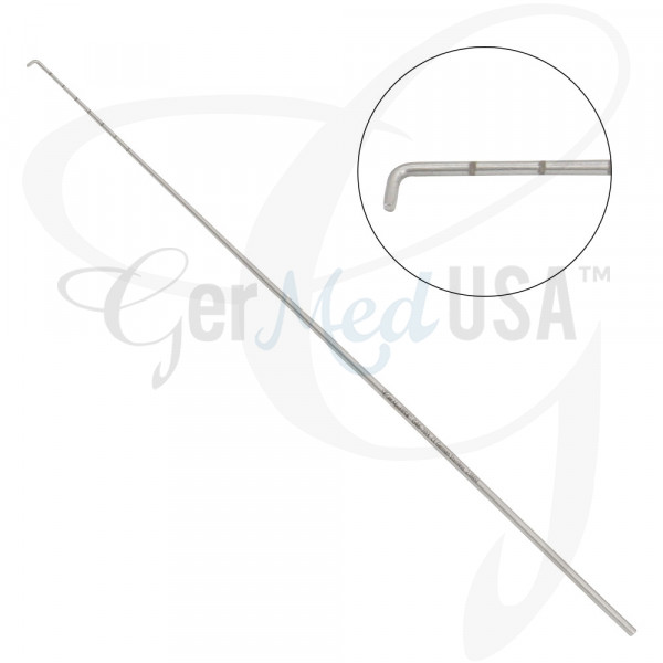
Design and Materials
Arthroscopic probes are engineered for precision, durability, and biocompatibility.
- Shaft: Typically long, slender, and rigid, made from high-grade stainless steel or titanium alloys. These materials offer:
- Strength: To withstand forces during manipulation without bending or breaking.
- Corrosion Resistance: Essential for repeated sterilization and use in a saline environment.
- Biocompatibility: Safe for use within the human body.
- Varying Lengths and Diameters: To accommodate different joint sizes and surgical approaches (e.g., knee, shoulder, hip, ankle).
- Handle: Ergonomically designed for surgeon comfort and control.
- Often made from lightweight materials like aluminum or high-grade plastics.
- Features textured grips for secure handling, even when wet.
- May be color-coded for easy identification in the operating room.
- Tip (The Angled Hook): This is the defining feature.
- Precise Angulation: Common angles include 30, 45, or 90 degrees, designed to reach specific anatomical areas and provide optimal leverage.
- Blunt or Semi-Blunt Finish: The tip is meticulously smoothed to prevent iatrogenic (surgeon-induced) damage to healthy tissue. It is designed for palpation and retraction, not cutting.
- Hook Shape: Allows for gentle lifting, probing, and retraction of tissue, providing a clear view or testing tissue stability.
- Cannula Compatibility: Designed to pass smoothly through arthroscopic cannulas (small tubes inserted into the joint to maintain access and fluid flow), minimizing friction and tissue trauma during insertion and withdrawal.
Biomechanics and Mechanisms
The angled hook probe operates on principles of leverage, tactile feedback, and precise manipulation.
- Leverage and Access: The angled tip allows the surgeon to reach around corners and under structures within the joint that would be inaccessible with a straight instrument. This leverage is crucial for:
- Retraction: Gently pulling tissue aside to improve visualization of underlying structures.
- Manipulation: Orienting tissues or fragments for better assessment or removal.
- Palpation and Tactile Feedback: This is perhaps the most critical function. The probe transmits subtle vibrations and resistance back to the surgeon's hand, allowing them to assess:
- Tissue Consistency: Differentiating between healthy, firm cartilage and soft, degenerated areas.
- Meniscal Integrity: Testing for tears by gently pushing or pulling on the meniscus to check its stability and mobility.
- Ligamentous Laxity: Assessing the stability of ligaments by applying gentle stress.
- Cartilage Delamination: Detecting areas where cartilage may be peeling away from the underlying bone.
- Visual Inspection Aid: While the arthroscope provides the primary visual, the probe helps to:
- Highlight Structures: By gently touching or moving them, making them more prominent on the monitor.
- Measure Lesions: The tip can be used as a reference point to estimate the size of cartilage defects or tears.
- Assisting Other Instruments: It often works in conjunction with other tools, guiding sutures, holding tissue during debridement, or assisting in the removal of loose bodies.
Table: Key Technical Specifications
| Feature | Description | Benefit for Patient |
|---|---|---|
| Material | High-grade Stainless Steel / Titanium | Ensures sterile, safe, and precise operation. |
| Shaft Diameter | Typically 2.5mm - 5.5mm | Minimizes incision size, less trauma, faster healing. |
| Tip Angle | 30°, 45°, 90° (Angled Hook) | Allows access to complex joint areas for thorough diagnosis. |
| Tip Finish | Smooth, Blunt/Semi-Blunt | Prevents accidental damage to healthy joint tissues. |
| Ergonomic Handle | Designed for surgeon's grip | Enhances surgeon control, leading to more precise procedures. |
3. Extensive Clinical Indications & Usage
The versatility of the arthroscopic probe (angled hook) makes it indispensable across a broad spectrum of orthopedic conditions and surgical procedures.
Diagnostic Applications
The primary role of the probe in diagnosis is to augment visual inspection with tactile information.
- Meniscal Evaluation:
- Tears: Probing helps identify the location, type (e.g., radial, horizontal, bucket-handle), and stability of meniscal tears. The surgeon can gently pull on the torn fragment to assess if it's displaced or unstable.
- Degeneration: Palpating the meniscus can reveal areas of softening or fraying.
- Articular Cartilage Assessment:
- Chondromalacia: The probe can feel for softening or roughening of the cartilage surface.
- Cartilage Defects: It helps delineate the borders and depth of focal cartilage lesions (e.g., osteochondral defects).
- Delamination: Detecting areas where cartilage may be separating from the underlying bone.
- Ligament Integrity:
- ACL/PCL: While primary assessment is often visual, the probe can gently pull on the remnants of torn ligaments to confirm instability or assess the quality of tissue for repair.
- Collateral Ligaments: Used to check for laxity or integrity near their attachments.
- Synovial Tissue Inspection:
- Inflammation: Probing can confirm the consistency of inflamed synovial tissue (synovitis).
- Plicae: Assessing the size and impingement potential of synovial folds (plicae).
- Loose Body Localization:
- The probe can gently push or trap loose cartilage or bone fragments, helping the surgeon locate them for removal.
- Capsular Assessment:
- Assessing the tightness or integrity of the joint capsule, particularly in conditions like adhesive capsulitis (frozen shoulder).
Operative Applications
Beyond diagnosis, the angled hook probe plays a crucial supportive role in various operative interventions.
- Tissue Retraction and Exposure:
- Gently holding back synovial tissue, fat pads, or meniscal fragments to provide a clearer view for other instruments (e.g., shaver, radiofrequency wand, suture passers).
- Guiding Instruments:
- Used to guide suture needles, cannulas, or other instruments to precise locations within the joint, especially in tight spaces.
- Testing Repair Stability:
- After a meniscal repair or ligament reconstruction, the probe is used to gently stress the repaired tissue to confirm its stability and integrity before closing the joint. This is a critical step to ensure the repair is sound.
- Debridement Assistance:
- While not a primary debridement tool, it can help dislodge loose fragments or gently scrape away very soft, degenerated tissue, especially in conjunction with a shaver.
- Fragment Removal:
- Assisting in the retrieval of small fragments of bone or cartilage that might be difficult to grasp with other instruments.
Joints Where It's Used
The arthroscopic probe is a universal tool adaptable to nearly all arthroscopic procedures:
- Knee: Most common, for meniscus, cartilage, ACL/PCL assessment.
- Shoulder: For rotator cuff tears, labral tears, biceps pathology, impingement.
- Hip: For labral tears, FAI (femoroacetabular impingement), loose bodies.
- Ankle: For impingement, osteochondral lesions, loose bodies.
- Wrist: For ligament tears, TFCC (triangular fibrocartilage complex) injuries.
- Elbow: For loose bodies, osteophytes, synovitis.
Fitting/Usage Instructions (Simplified for Patient Understanding)
From a patient's perspective, understanding the "usage" means knowing how the surgeon skillfully employs this tool:
- Portals: After anesthesia, small incisions (portals) are made around the joint.
- Cannula Insertion: A small tube (cannula) is inserted through a portal to maintain access.
- Arthroscope Insertion: The camera (arthroscope) is inserted through one cannula to visualize the joint.
- Probe Insertion: The angled hook probe is then inserted through another cannula.
- Exploration & Diagnosis: The surgeon systematically explores the joint, using the probe to gently touch, push, pull, and retract structures. The visual on the monitor, combined with the tactile feedback through the probe, allows for a precise diagnosis.
- Operative Assistance: If a problem is identified and requires treatment, the probe assists other specialized instruments in performing repairs, debridements, or removals.
4. Risks, Side Effects, or Contraindications
While the arthroscopic probe is a safe and highly precise instrument, it's crucial to understand that its use is part of a surgical procedure, and all surgeries carry inherent risks.
General Arthroscopy Risks (briefly mentioned)
- Infection: Risk of bacteria entering the joint (minimized by strict sterile techniques).
- Bleeding: Though minimally invasive, some bleeding can occur.
- Nerve or Blood Vessel Damage: Rare, but possible due to proximity of structures.
- Deep Vein Thrombosis (DVT): Blood clots in the leg, a general surgical risk.
- Anesthesia Risks: Related to the type of anesthesia used.
Probe-Specific Risks (extremely rare with proper technique)
- Iatrogenic Tissue Damage: The most significant, albeit rare, risk. If not handled with extreme care and precision, the probe could theoretically scratch or damage healthy cartilage or other soft tissues. This is why surgeon skill and experience are paramount.
- Instrument Breakage: Extremely rare with modern, high-quality instruments and proper handling. If a probe were to break, the fragment would need to be retrieved.
- Minor Abrasions: Very superficial scuffing of tissue can occur, usually without clinical significance.
Contraindications
There are no specific contraindications for the use of the arthroscopic probe itself. However, general contraindications for arthroscopic surgery would apply:
- Active Joint Infection: Surgery could spread the infection.
- Severe Skin Infection: Near the surgical site.
- Significant Comorbidities: Uncontrolled medical conditions that make surgery too risky (e.g., severe heart disease, uncontrolled diabetes).
- Extensive Joint Ankylosis: Severe stiffness that prevents instrument insertion.
The benefits of accurate diagnosis and targeted treatment facilitated by the probe far outweigh these minimal, well-managed risks.
5. Expert Tips from Dr. Mohammed Hutaif
"As an orthopedic surgeon specializing in joint care, I often describe the arthroscopic probe (angled hook) as my 'third eye' and 'extended finger' inside the joint," says Dr. Mohammed Hutaif. "While the arthroscope gives me the visual, it's the probe that provides the invaluable tactile feedback, allowing me to truly 'feel' the pathology."
Dr. Hutaif emphasizes several key aspects regarding this instrument:
- Diagnostic Precision: "The nuanced feel of the probe against the meniscus or cartilage allows me to confirm what I see on the screen. Is that meniscal tear truly unstable? Is this cartilage soft or firm? The probe provides answers that visual inspection alone cannot."
- Enhancing Safety and Efficacy: "By gently retracting tissues, guiding sutures, or testing the stability of a repair, the angled hook probe significantly enhances the safety and precision of my interventions. It ensures that every repair is robust and every diagnosis is accurate."
- Minimally Invasive Philosophy: "The probe is a perfect embodiment of minimally invasive surgery. It allows for highly detailed assessment and manipulation through tiny incisions, leading to less pain, faster recovery, and better cosmetic outcomes for my patients."
- Importance of Experience: "Mastering the use of the probe is an art. It requires years of experience to interpret the subtle tactile sensations and coordinate them with the visual input. This expertise is what allows for the most accurate diagnosis and effective treatment plan."
Dr. Hutaif's philosophy centers on leveraging advanced instrumentation like the angled hook probe to provide his patients with the most accurate diagnosis, the safest possible treatment, and the best path to recovery and improved joint function.
6. Massive FAQ Section
Q1: What exactly is an arthroscopic probe (angled hook)?
A1: An arthroscopic probe (angled hook) is a slender, specialized surgical instrument used in minimally invasive joint surgery (arthroscopy). It has a long shaft, an ergonomic handle, and, most distinctively, a small, blunt, angled tip designed to allow the surgeon to gently explore, palp palpate, and manipulate tissues inside a joint. It acts as an extension of the surgeon's finger, providing crucial tactile feedback not possible with just visual inspection.
Q2: Why is the tip "angled" and "hooked"?
A2: The angled and hooked design is critical for accessing complex anatomical areas within a joint. Joints have many nooks and crannies. A straight instrument might not reach behind certain structures or under edges of cartilage. The angle allows the surgeon to navigate these spaces, gently lift or retract tissues for better visualization, and get a precise feel of tissue consistency and stability, much like using a bent finger to feel around an object.
Q3: Is the angled hook probe used for cutting or repairing?
A3: No, the angled hook probe is primarily a diagnostic and manipulative tool. Its tip is blunt or semi-blunt, specifically designed not to cut tissue. While it can assist in some operative procedures by guiding other instruments or retracting tissue, its main role is to assess the integrity and stability of structures like menisci, ligaments, and cartilage through touch and gentle movement. Specialized instruments are used for cutting, shaving, or repairing.
Q4: What joints can this instrument be used in?
A4: The arthroscopic probe (angled hook) is a versatile instrument used in virtually all arthroscopic procedures. This includes, but is not limited to, the knee, shoulder, hip, ankle, wrist, and elbow joints. Its design allows for adaptation to the unique anatomy and challenges of different joints.
Q5: How does my surgeon use it to help diagnose my joint problem?
A5: Your surgeon uses the probe by inserting it through a small incision (portal) into your joint, alongside the arthroscope (camera). While viewing the inside of your joint on a monitor, the surgeon will gently touch, push, and pull on various structures like your meniscus, cartilage, and ligaments. The probe transmits tactile sensations back to the surgeon's hand, allowing them to feel if a tissue is soft, firm, unstable, or torn, confirming or refining the visual diagnosis. This detailed assessment helps in formulating the most accurate treatment plan.
Q6: Is the use of this probe safe?
A6: Yes, when used by a skilled and experienced orthopedic surgeon, the arthroscopic probe is very safe. It is designed with a blunt tip to minimize the risk of damage to healthy tissues. The primary risks associated with its use are minimal and generally fall under the broader risks of arthroscopic surgery itself (e.g., infection, bleeding), which are rare and carefully managed.
Q7: What materials is the angled hook probe made from?
A7: These probes are typically made from high-grade, biocompatible materials such as stainless steel or titanium alloys. These materials are chosen for their strength, durability, resistance to corrosion (especially important for repeated sterilization), and safety for use inside the human body.
Q8: Does the use of an arthroscopic probe prolong the surgery?
A8: No, quite the opposite. The efficient use of an arthroscopic probe often shortens surgery by facilitating a rapid and accurate diagnosis. By quickly identifying the precise nature and extent of the joint problem, the surgeon can proceed more directly to the necessary operative steps, avoiding unnecessary exploration or delays. It streamlines the diagnostic phase, making the overall procedure more efficient.
Q9: How is an arthroscopic probe cleaned and sterilized?
A9: Arthroscopic probes, like all surgical instruments, undergo rigorous cleaning and sterilization protocols after each use. They are typically:
1. Cleaned Manually: To remove visible debris.
2. Ultrasonically Cleaned: To remove microscopic particles.
3. Disinfected: Using specialized solutions.
4. Sterilized: Most commonly through steam sterilization (autoclaving) at high temperatures and pressure, which kills all microorganisms. This multi-step process ensures the instrument is completely sterile and safe for the next patient.
Q10: Will I feel anything when the probe is used during surgery?
A10: No, you will not feel anything during the surgery itself. Arthroscopic procedures are performed under anesthesia (either general, regional, or local with sedation), ensuring you are comfortable and pain-free throughout the entire process, including the use of the probe and other instruments.
Q11: How does the angled hook probe contribute to improved patient outcomes?
A11: The angled hook probe significantly improves patient outcomes by enabling:
* More Accurate Diagnosis: Leading to a precise understanding of the joint problem.
* Targeted Treatment: Ensuring that the correct surgical intervention is performed.
* Minimally Invasive Approach: Less tissue trauma, smaller scars, reduced post-operative pain.
* Faster Recovery: Patients often experience quicker rehabilitation and return to function.
* Reduced Complications: By enhancing surgical precision and safety.
Q12: Can the probe detect early signs of arthritis?
A12: Yes, the arthroscopic probe is highly effective in detecting early signs of articular cartilage degeneration, which is a hallmark of arthritis. By palpating the cartilage, the surgeon can feel subtle changes in its texture, softening, or early fraying that might not be immediately obvious visually. This allows for earlier diagnosis and potential intervention to slow the progression of arthritis.
Disclaimer: This information is intended for educational purposes only and should not be considered medical advice. Always consult with a qualified healthcare professional for diagnosis and treatment.