The Arthroscopic Knot Pusher: A Cornerstone of Minimally Invasive Joint Repair
1. Comprehensive Introduction & Overview
In the realm of modern orthopedics, arthroscopy has revolutionized the way joint conditions are diagnosed and treated. This minimally invasive surgical technique involves small incisions, a tiny camera (arthroscope), and specialized instruments to visualize and repair damage inside a joint. One such indispensable instrument, often overlooked by patients but critical to surgical success, is the arthroscopic knot pusher.
At its core, the arthroscopic knot pusher is a precision tool designed to facilitate the secure tying and advancement of surgical knots deep within the joint space, through small portals. In arthroscopic procedures, surgeons often need to repair torn ligaments, tendons, or cartilage by suturing them back into place. Tying a strong, secure knot in the confined, fluid-filled environment of a joint, using long instruments and limited visibility, is a complex task. The knot pusher simplifies this, allowing surgeons to precisely place and tighten knots, ensuring the stability and longevity of the repair.
This guide will provide a comprehensive overview of the arthroscopic knot pusher, delving into its design, various applications, biomechanical principles, and the profound impact it has on patient outcomes. Our goal is to demystify this critical instrument, offering patients a deeper understanding of the advanced techniques employed in their orthopedic care, all while emphasizing that this information is for educational purposes only and not medical advice.
2. Deep-dive into Technical Specifications / Mechanisms
The efficacy of an arthroscopic knot pusher lies in its ingenious design and the precision of its mechanism. These instruments are engineered to meet the demanding requirements of arthroscopic surgery.
2.1. Design and Materials
Arthroscopic knot pushers are typically slender, elongated instruments, designed to pass through small cannulas (tubes) into the joint.
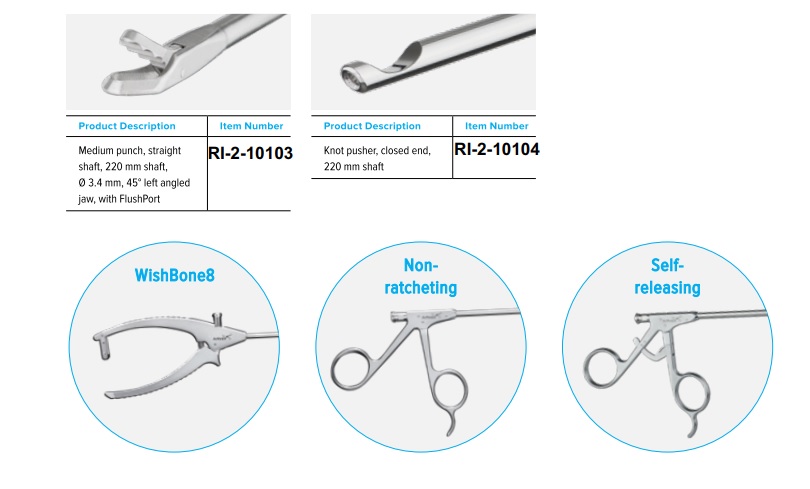
- Shaft: The shaft is usually made from high-grade medical stainless steel or titanium, offering rigidity, durability, and corrosion resistance. It comes in various lengths (e.g., 15 cm to 25 cm) and diameters (e.g., 2.7 mm, 3.5 mm, 4.0 mm) to accommodate different joint sizes and surgical approaches. Many shafts are cannulated, meaning they have a central lumen (channel) through which the suture can pass, allowing for precise control.
- Tip Configuration: This is the most crucial part, designed to engage and push the knot. Tips vary widely:
- Open-ended Cannula: A simple tube that slides over one limb of the suture, pushing the knot down.
- Slotted Tip: Features a small slot or notch to capture the suture loop or knot, offering more control.
- Closed-ended/Blunt Tip: Used for pushing knots, often with a small indent or hook to engage the suture.
- Specialized Jaw Designs: Some advanced pushers incorporate small jaws or prongs that can grasp and manipulate the suture more actively.
- Angled Tips: Available in various angles (e.g., 30°, 45°, 90°) to allow access to difficult-to-reach anatomical areas within the joint.
- Handle: The handle is ergonomically designed for surgeon comfort and control. It can be a simple rod with finger rings, a pistol grip, or a T-handle. Materials often include lightweight, high-grade polymers or stainless steel. Color-coding might be used to differentiate sizes or types.
- Materials: Primarily medical-grade stainless steel (e.g., 17-4 PH, 316L) for its strength and biocompatibility. Titanium is also used for lighter weight and enhanced MRI compatibility. Some components might utilize high-performance polymers for insulation or specific tactile properties.
2.2. Mechanism of Action
The fundamental principle of an arthroscopic knot pusher is to deliver a securely tied knot from outside the joint to its intended position on the tissue inside the joint.
- Suture Loop Formation: After passing a suture through the target tissue (e.g., a torn meniscus), the surgeon creates an initial knot (often a surgeon's throw or a half-hitch) outside the body, at the portal site.
- Suture Engagement: One limb of the suture (the "post" or "static" limb) is threaded through the cannulated shaft of the knot pusher, or the pusher's tip is engaged with the knot or the "running" limb of the suture.
- Knot Delivery: The surgeon then advances the knot pusher down the suture, guiding the pre-formed knot through the cannula and into the joint space.
- Knot Seating and Tightening: Once the knot pusher reaches the target tissue, the surgeon applies controlled pressure, "pushing" the knot down firmly against the tissue. Simultaneously, tension is maintained on the suture limbs to ensure the knot seats snugly and securely, without loosening.
- Sequential Knot Tying: This process is repeated for subsequent throws of the knot (e.g., square knots, sliding knots, half-hitches) until a secure, multi-throw knot complex is formed, providing stable fixation of the repaired tissue.
2.3. Types of Knot Pushers
Beyond general design variations, specific types cater to different surgical needs:
- Cannulated Knot Pushers: Most common, allowing the suture to pass through the instrument's center, offering excellent control.
- Non-Cannulated Knot Pushers: Used to push knots formed by other instruments or to apply counter-pressure.
- Suture-Grabbing Knot Pushers: Feature small, retractable jaws or hooks to actively grasp and pull the suture, useful in complex situations.
- Loaded Knot Pushers: Some systems integrate a pre-tied knot or an anchor delivery mechanism with the pusher.
3. Extensive Clinical Indications & Usage
The arthroscopic knot pusher is an essential tool across a wide spectrum of arthroscopic procedures, facilitating repair and reconstruction in various joints.
3.1. General Arthroscopic Applications
- Shoulder Arthroscopy:
- Rotator Cuff Repair: Reattaching torn supraspinatus, infraspinatus, subscapularis, or teres minor tendons to the humerus. Secure knot tying is paramount for healing.
- Labral Repair (SLAP, Bankart): Repairing tears in the glenoid labrum, often using suture anchors and precise knot placement.
- Capsular Plication: Tightening the joint capsule for instability, requiring multiple secure sutures.
- Knee Arthroscopy:
- Meniscus Repair: Suturing torn meniscus tissue to promote healing and preserve knee function. Various repair techniques (inside-out, outside-in, all-inside) often rely on knot pushers.
- ACL/PCL Reconstruction: Securing grafts (e.g., hamstring, patellar tendon) within bone tunnels using suspensory fixation or interference screws, often augmented with sutures requiring knot tying.
- MPFL Reconstruction: Reconstructing the medial patellofemoral ligament for patellar instability.
- Hip Arthroscopy:
- Labral Repair: Repairing tears in the acetabular labrum, crucial for hip joint stability and pain relief.
- Capsular Closure: Closing the hip capsule after capsulotomy for FAI (femoroacetabular impingement) or instability.
- Ankle Arthroscopy:
- Lateral Ankle Ligament Repair: Repairing torn ligaments after sprains or instability.
- Elbow Arthroscopy:
- Ligament Repair/Reconstruction: Addressing instability or tears in collateral ligaments.
3.2. Detailed Usage Instructions (Simplified for Patients)
While the surgeon performs these steps, understanding the sequence provides insight into the procedure:
- Portal Creation & Visualization: Small incisions (portals) are made, and the arthroscope is inserted to visualize the joint.
- Suture Passage: A specialized instrument passes a suture through the torn tissue and the bone (if an anchor is used), creating a loop or two free ends.
- Initial Knot Formation: The surgeon manipulates the suture ends outside the body to form the first "throw" of a surgical knot.
- Engaging the Knot Pusher: One limb of the suture is threaded through the cannulated knot pusher, or the pusher's tip is carefully engaged with the knot or the running suture limb.
- Controlled Advancement: The knot pusher is gently advanced through the portal, guiding the knot down towards the tissue requiring repair.
- Knot Seating & Tensioning: Once the knot reaches the target, the surgeon uses the knot pusher to apply precise, controlled pressure, seating the knot firmly against the tissue. Simultaneously, tension is applied to the suture ends to tighten the knot without overtightening or damaging the tissue.
- Sequential Throws: This process is repeated several times, creating multiple throws (e.g., 3 to 6 throws) to build a strong, secure, and permanent knot complex.
- Suture Cutting: Once the knot is secure, another arthroscopic instrument is used to cut the excess suture tails close to the knot.
3.3. Biomechanics
The biomechanical role of the arthroscopic knot pusher is critical for the success and durability of the repair.
- Precise Knot Seating: The pusher ensures the knot is seated firmly against the tissue, minimizing gaps and maximizing compression. This is vital for tendon-to-bone or ligament-to-ligament healing.
- Controlled Tension: It allows the surgeon to apply optimal tension to the suture. Too little tension can lead to a loose repair, while too much can cause tissue strangulation or suture breakage. The tactile feedback through the pusher is essential.
- Reproducible Knot Strength: By facilitating consistent knot formation and seating, the pusher helps achieve reproducible knot strength, which is directly correlated with the overall strength of the repair construct.
- Minimizing Knot Bulk: A well-seated knot is often a low-profile knot, reducing potential impingement or irritation of surrounding soft tissues within the joint.
- Reduced Friction & Trauma: The smooth passage of the knot pusher minimizes friction on the suture and reduces potential trauma to the portal edges or surrounding soft tissues compared to trying to manipulate knots with less specialized instruments.
3.4. Patient Outcome Improvements
The use of an arthroscopic knot pusher significantly contributes to enhanced patient outcomes in several ways:
- Minimally Invasive Benefits: As part of arthroscopic surgery, it supports smaller incisions, less post-operative pain, reduced blood loss, and lower infection rates compared to open surgery.
- Stronger, More Reliable Repairs: The ability to tie secure, reproducible knots directly translates to a more robust repair, reducing the risk of re-tear or failure.
- Faster Rehabilitation: A stable repair allows for earlier initiation of rehabilitation protocols, leading to faster recovery of joint function and return to daily activities.
- Improved Long-Term Function: The durability of the repair contributes to better long-term functional results, reduced chronic pain, and a higher quality of life for the patient.
- Reduced Surgical Time: Efficient knot tying saves valuable operating time, which can contribute to better patient safety.
4. Risks, Side Effects, or Contraindications
While the arthroscopic knot pusher itself is a safe and inert instrument when used correctly, it is part of a surgical procedure, and as such, general risks associated with arthroscopic surgery apply.
- General Surgical Risks:
- Infection: Any surgical procedure carries a risk of infection, although rates are low in arthroscopy due to sterile techniques and prophylactic antibiotics.
- Bleeding/Hematoma: Minor bleeding is common; significant bleeding or hematoma is rare.
- Nerve or Vessel Damage: Accidental damage to nerves or blood vessels, though uncommon, is a potential risk of any invasive procedure.
- Stiffness/Arthrofibrosis: Some patients may develop stiffness in the joint post-surgery, requiring additional therapy or intervention.
- Deep Vein Thrombosis (DVT): Blood clots, though rare, can occur.
- Knot-Specific Risks (Rare with proper technique):
- Knot Slippage/Failure: While the goal is a secure knot, in rare instances, a knot might slip or fail, leading to repair failure. This is often mitigated by using multiple throws and specific knot configurations.
- Suture Breakage: Excessive tension during knot tying can cause the suture to break, requiring re-suturing.
- Tissue Damage: Incorrect application of the knot pusher could theoretically cause localized tissue damage, but this is extremely rare in the hands of an experienced surgeon.
- Contraindications:
- General Contraindications for Arthroscopy: These include active joint infection, severe uncontrolled bleeding disorders, or medical conditions that make general anesthesia unsafe for the patient. The knot pusher itself has no specific contraindications beyond those for the overall procedure.
5. Expert Tips from Dr. Mohammed Hutaif
As an orthopedic specialist, Dr. Mohammed Hutaif emphasizes that while technology provides advanced tools, the surgeon's skill and experience remain paramount. Here are his expert tips regarding the arthroscopic knot pusher:
- "Choosing the Right Tool for the Job: Not all knot pushers are created equal. I meticulously select the specific type and tip configuration based on the suture material, the anatomical location, and the nature of the repair. An angled pusher for a posterior shoulder labral repair, for instance, is vastly different from a straight pusher for a meniscal repair."
- "Mastering Tactile Feedback: Arthroscopic surgery often relies on visual cues, but the knot pusher demands a keen sense of touch. I train to feel the knot seating against the tissue, ensuring optimal tension without overtightening. It's an art to apply just enough pressure for a secure, low-profile knot."
- "The Importance of Initial Knot Formation: The quality of the final knot begins with the first throw. A well-formed initial knot outside the joint provides a stable foundation for the pusher to work with, minimizing slippage during advancement."
- "Controlled Tension is Key: One of the most common pitfalls is either too little or too much tension. Too little, and the repair is loose; too much, and you risk cutting through tissue or breaking the suture. The knot pusher allows for controlled, incremental tensioning, which is crucial for the biomechanical integrity of the repair."
- "Sequential Security: I always advocate for multiple throws (typically 4-6) to ensure maximal knot security. Each throw adds to the overall strength and reduces the chance of slippage, especially with modern, low-friction suture materials."
- "Patient Education and Compliance: While the knot pusher ensures a strong repair, the patient's role in post-operative care, including adherence to rehabilitation protocols, is equally vital for successful healing and long-term outcomes. A perfectly tied knot needs time and proper care to integrate with healing tissue."
6. Massive FAQ Section
Q1: What is an arthroscopic knot pusher?
A1: An arthroscopic knot pusher is a specialized surgical instrument used by orthopedic surgeons during minimally invasive (arthroscopic) joint surgery. Its primary function is to help tie and tighten surgical knots deep inside a joint, through small incisions, to repair damaged ligaments, tendons, or cartilage.
Q2: Why is this instrument important for my surgery?
A2: It's crucial because it allows the surgeon to create strong, secure knots in a very confined space without needing large open incisions. This precision is vital for the stability and success of repairs like rotator cuff tears, meniscus repairs, or ACL reconstructions, leading to better healing and long-term results.
Q3: Is the arthroscopic knot pusher left inside my body?
A3: No, absolutely not. The arthroscopic knot pusher is a temporary surgical tool used by the surgeon during the operation. Once the surgical knots are tied and secured, the instrument is removed from your body. Only the sutures (which are usually absorbable or inert non-absorbable materials) remain to hold the repaired tissues.
Q4: What types of surgeries commonly use an arthroscopic knot pusher?
A4: It's commonly used in almost all types of arthroscopic joint surgeries where tissue needs to be sutured. This includes repairs in the shoulder (e.g., rotator cuff, labrum), knee (e.g., meniscus, ACL), hip (e.g., labrum), ankle, and elbow.
Q5: How does this tool improve patient outcomes?
A5: By enabling precise and secure knot tying, it contributes to:
* Stronger Repairs: Reducing the risk of the repair failing.
* Minimally Invasive Benefits: Less pain, smaller scars, and faster recovery compared to open surgery.
* Improved Long-Term Function: Better joint stability and reduced chronic pain.
* Faster Rehabilitation: A secure repair allows you to start your physical therapy sooner.
Q6: How is the knot pusher cleaned and sterilized?
A6: After each use, the knot pusher undergoes a rigorous cleaning process, typically involving immediate rinsing, manual cleaning with enzymatic solutions, and ultrasonic cleaning. It is then thoroughly inspected, and finally sterilized using validated methods, most commonly steam sterilization (autoclave), to eliminate all microorganisms and ensure it is completely safe for the next patient.
Q7: Can the knots tied with this instrument fail?
A7: While extremely rare with proper surgical technique, no surgical repair is 100% immune to failure. Surgeons use specific knot types and multiple "throws" (repetitions) to maximize knot security. Factors like patient compliance with post-operative restrictions and the body's healing capacity also play a significant role in the overall success of the repair.
Q8: What should I expect immediately after surgery involving this tool?
A8: Post-surgery, you can expect some pain and swelling, managed with medication. The joint will likely be immobilized or protected in a brace or sling, and you'll receive specific instructions on weight-bearing, movement, and wound care. Physical therapy usually begins soon after to restore strength and range of motion.
Q9: Does the use of a knot pusher make the surgery more painful?
A9: No, the knot pusher itself does not increase pain. It is used while you are under anesthesia and is part of a minimally invasive procedure designed to reduce post-operative pain compared to traditional open surgery. Any pain you experience after surgery is related to the healing process of the repaired tissues.
Q10: How does Dr. Hutaif ensure the knots are secure during my surgery?
A10: Dr. Hutaif employs a combination of advanced techniques and experience. He carefully selects the appropriate knot pusher and suture material, uses proven knot configurations (e.g., surgeon's knot, sliding knots with locking throws), and applies precise, controlled tension to each throw. He also relies on tactile feedback to confirm the knot is seated firmly against the tissue, ensuring maximal security for your repair.
Q11: Are there different types of knot pushers, and does it matter which one is used?
A11: Yes, there are various types with different tip designs (e.g., open-ended, slotted, angled, cannulated) and shaft diameters. The choice of knot pusher is crucial and depends on the specific surgical procedure, the type of suture being used, and the anatomical location within the joint. Dr. Hutaif selects the instrument that offers the best access and control for optimal knot tying in each unique case.
Q12: What is the benefit of a "low-profile" knot created with this instrument?
A12: A "low-profile" knot means the knot is compact and lies flat against the tissue, rather than being bulky. This is beneficial because it reduces the chance of the knot impinging on or irritating surrounding healthy tissues within the joint, potentially leading to less post-operative discomfort and better long-term function. The knot pusher helps achieve this precise seating.
Disclaimer: This information is intended for patient education only and does not constitute medical advice. Please consult with a qualified healthcare professional for any health concerns or before making any decisions related to your health or treatment.