Understanding the Arthroscopic Cannula System (5.0mm, 6.0mm, 8.0mm): A Patient's Guide
Welcome to our comprehensive guide on the Arthroscopic Cannula System, an indispensable tool in modern orthopedic surgery. As an expert medical SEO copywriter and orthopedic specialist, our goal is to provide you with authoritative, easy-to-understand information about this crucial instrument, empowering you with knowledge about your potential treatment options. This content is for patient information only and is not medical advice. Always consult with a licensed medical professional for diagnosis and treatment.
1. Comprehensive Introduction & Overview
Arthroscopy is a minimally invasive surgical procedure used to visualize, diagnose, and treat problems inside a joint. Instead of making large incisions, surgeons use a tiny camera called an arthroscope and specialized instruments inserted through small "portals" in the skin. The Arthroscopic Cannula System is at the heart of this technique, acting as a protective sleeve and conduit.
What is an Arthroscopic Cannula System?
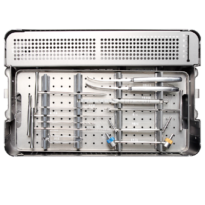
An arthroscopic cannula system consists of a hollow tube (the cannula) and an obturator (a pointed or blunt rod inserted into the cannula to facilitate its passage through tissue). Once the cannula is in place, the obturator is removed, leaving a clear channel through which the arthroscope and other surgical instruments can be inserted and manipulated.
Why are Different Sizes Important?
The specific sizes – 5.0mm, 6.0mm, and 8.0mm – refer to the inner diameter of the cannula. The choice of size is critical and depends on several factors:
* Joint Size: Larger joints (e.g., shoulder, knee, hip) often require larger cannulas to accommodate bigger instruments or multiple instruments simultaneously.
* Surgical Instruments: The size and type of instruments needed for a specific procedure dictate the cannula's diameter.
* Surgical Access: Different sizes allow for varying degrees of surgical maneuverability and visualization.
* Tissue Protection: Selecting the appropriate size minimizes trauma to surrounding soft tissues while providing stable access.
The use of these systems greatly enhances the precision and safety of arthroscopic procedures, contributing to the overall success of minimally invasive surgery and improved patient outcomes.
2. Deep-dive into Technical Specifications / Mechanisms
The design and material science behind arthroscopic cannula systems are crucial for their performance and patient safety.
Design and Materials
Arthroscopic cannulas are engineered for optimal performance, combining strength, biocompatibility, and user-friendliness.
- Materials:
- Polycarbonate/PEEK (Polyether Ether Ketone): Common for disposable cannulas due to their transparency, lightweight nature, and biocompatibility. They allow for clear visualization of instruments passing through.
- Stainless Steel/Titanium: Used for reusable cannulas, offering superior durability and resistance to repeated sterilization cycles.
- Hydrophilic Coatings: Some cannulas feature coatings to reduce friction during insertion and instrument manipulation.
- Cannula Types:
- Smooth-Walled: Provide unrestricted passage but may be prone to dislodgement.
- Threaded: Feature external threads that grip soft tissues, offering enhanced stability and reducing the risk of dislodgement during vigorous instrument manipulation. This is particularly useful in joints with thick capsules or during complex procedures.
- Fenestrated (Windowed): Designed with openings to allow for suture retrieval or tissue manipulation outside the primary lumen.
- Valved Systems: Incorporate a valve (e.g., trumpet valve, duckbill valve) at the proximal end to prevent fluid leakage and maintain joint distension when instruments are removed. This is vital for continuous clear visualization.
- Non-Valved Systems: Simpler designs without a valve, often used for inflow/outflow or when continuous instrument presence is expected.
- Obturators:
- Blunt Obturators: Used after initial portal creation to dilate the tissue safely without sharp edges, minimizing nerve and vessel injury.
- Trocar (Sharp) Obturators: Employed for initial penetration through skin and subcutaneous tissue, typically with extreme care to avoid damage to underlying structures.
- Dilating Obturators: Gradually expand the portal, often used in conjunction with a series of progressively larger cannulas.
Biomechanics
The biomechanical principles behind cannula use are focused on minimizing tissue trauma and optimizing the surgical environment.
- Tissue Protection: The smooth, rounded edges of the cannula protect the soft tissues and articular cartilage from repeated friction and potential damage caused by the entry and exit of instruments.
- Maintaining Joint Distension: Inflow and outflow cannulas are strategically placed to ensure a continuous flow of sterile saline solution, which distends the joint capsule. This distension creates a working space and washes away debris, providing a clear field of view for the surgeon.
- Minimizing Friction: The smooth internal surface of the cannula reduces friction as instruments are passed through, allowing for precise and controlled movements. Threaded designs, while providing stability, can also distribute pressure more evenly across the tissue.
- Portal Stability: Threaded cannulas, in particular, engage with the soft tissues, creating a stable working portal that resists unwanted movement, crucial for delicate surgical maneuvers.
Mechanism of Action
The cannula's primary mechanism is to establish and maintain a stable, protected gateway into the joint space.
1. Portal Creation: After localizing the anatomical landmarks, a small skin incision is made.
2. Insertion: The cannula, with an obturator inserted, is carefully advanced through the tissue layers into the joint capsule. The obturator guides the cannula and creates the initial pathway.
3. Obturator Removal: Once the cannula is securely positioned within the joint, the obturator is withdrawn.
4. Instrument Access: The hollow cannula then serves as a channel for the arthroscope, grasping instruments, shavers, suture passers, and other specialized tools.
5. Fluid Management: Valved cannulas prevent fluid leakage, while dedicated inflow/outflow cannulas manage the constant irrigation and suction necessary for clear visualization.
3. Extensive Clinical Indications & Usage
Arthroscopic cannula systems are integral to a wide range of orthopedic procedures across various joints.
Detailed Surgical Applications
The choice of cannula size and type is tailored to the specific joint and procedure.
- Knee Arthroscopy:
- 5.0mm/6.0mm: Often used for diagnostic arthroscopy, meniscectomy (trimming damaged meniscus), chondroplasty (cartilage smoothing), and removal of loose bodies. Smaller instruments for precise work.
- 8.0mm: Crucial for more extensive procedures like ACL/PCL reconstruction, meniscal repair (allowing passage of suture devices), and complex chondral defect repair, where larger instruments or multiple instruments are simultaneously required.
- Shoulder Arthroscopy:
- 5.0mm/6.0mm: Common for diagnostic scopes, subacromial decompression, biceps tenodesis, and minor labral debridement.
- 8.0mm: Essential for rotator cuff repair (to pass suture anchors and sutures), labral repair (Bankart, SLAP), capsular release, and instability procedures, providing stable portals for complex suturing and anchor placement.
- Hip Arthroscopy:
- 8.0mm (and sometimes larger, 8.5mm, 10mm): Hip arthroscopy often requires larger cannulas due to the depth of the joint and the need for robust instruments for procedures like labral repair, femoroacetabular impingement (FAI) osteoplasty, and removal of loose bodies. The 8.0mm size offers excellent stability and instrument passage.
- Ankle Arthroscopy:
- 5.0mm/6.0mm: Typically used for anterior ankle impingement, osteochondral defect debridement, and removal of loose bodies. Smaller joints usually require smaller cannulas.
- Elbow/Wrist Arthroscopy:
- 5.0mm: Often the preferred size due to the smaller joint spaces and delicate neurovascular structures. Used for loose body removal, synovectomy, and treatment of osteochondral lesions.
Fitting and Usage Instructions (Simplified for Patients)
While surgeons undergo extensive training, understanding the basic steps can demystify the process for patients.
- Patient Positioning & Anesthesia: The patient is positioned appropriately, and regional or general anesthesia is administered.
- Sterile Field: The surgical area is prepped and draped to maintain sterility.
- Portal Marking: The surgeon identifies and marks the precise locations for the small incisions (portals) based on anatomical landmarks and the planned procedure.
- Incision: A small skin incision (typically 3-10mm) is made at each portal site.
- Cannula Insertion: The chosen cannula, with its obturator, is carefully inserted through the incision and guided into the joint space. The obturator is then removed.
- Arthroscope Insertion: The arthroscope is inserted through one cannula to visualize the joint interior.
- Instrument Insertion: Other instruments are passed through additional cannulas to perform the surgical tasks.
- Fluid Management: Inflow and outflow cannulas are connected to an irrigation system to maintain joint distension and clear the field.
- Procedure Completion: Once the surgical objectives are met, instruments and cannulas are carefully removed.
- Closure: The small incisions are closed with sutures or sterile strips, and a dressing is applied.
4. Risks, Side Effects, or Contraindications
While arthroscopy is generally safe, it's important to be aware of potential risks associated with any surgical procedure, including those related to cannula use.
Potential Risks and Side Effects
- Infection: Although rare, infection at the portal site or within the joint is a possibility. Strict sterile technique minimizes this risk.
- Nerve or Vessel Injury: As cannulas are inserted, there's a small risk of injuring nearby nerves or blood vessels, especially in areas with complex anatomy. Careful anatomical knowledge and technique are paramount.
- Extravasation: Fluid used to distend the joint can sometimes leak into surrounding soft tissues, causing swelling and discomfort. This is usually temporary.
- Hematoma/Bleeding: Minor bleeding can occur at the portal sites, leading to bruising or a small collection of blood (hematoma).
- Cartilage Damage: Though cannulas protect joint surfaces, improper insertion or excessive movement can potentially cause superficial damage to articular cartilage.
- Loss of Joint Distension: If cannulas are dislodged or valves fail, fluid can leak, making visualization difficult and prolonging surgery.
- Instrument Breakage: Although rare, instruments can sometimes break within the joint, requiring careful retrieval.
Contraindications
Certain conditions may make arthroscopic surgery, and thus cannula use, less advisable:
* Active Joint Infection: Existing infection typically requires treatment before arthroscopy.
* Severe Skin Conditions/Infection at Portal Sites: Increases the risk of introducing infection into the joint.
* Severe Osteoarthritis: In some cases, extensive joint degeneration may limit the benefits of arthroscopy, making open surgery or joint replacement a more appropriate option.
* Extensive Scarring/Adhesions: May make portal creation difficult and increase the risk of complications.
* Poor General Health/Coagulopathy: Patients with significant medical comorbidities or bleeding disorders may have increased surgical risks.
5. Expert Tips from Dr. Mohammed Hutaif
"As an orthopedic surgeon, I view the arthroscopic cannula system as an extension of my hands and eyes within the joint. Its proper selection and meticulous use are fundamental to successful minimally invasive surgery. Here are my key insights for patients to understand its importance:
- Precision and Protection: The primary role of the cannula is to create a safe, stable, and protected pathway. This allows me to work with extreme precision, minimizing trauma to surrounding healthy tissues. The choice of a threaded cannula, for instance, provides exceptional stability, which is critical during complex tasks like intricate suture repairs in the shoulder or knee.
- Optimal Visualization: Maintaining clear visualization is paramount. Valved cannulas are invaluable for preventing fluid leakage, ensuring the joint remains adequately distended and free of debris. This uninterrupted view allows for accurate diagnosis and precise execution of repairs.
- Tailored Approach: We don't use a 'one-size-fits-all' approach. The specific size (5.0mm, 6.0mm, or 8.0mm) and type of cannula are carefully chosen based on the joint, the planned procedure, and the instruments required. For example, an 8.0mm cannula might be essential for passing robust suture anchors in a rotator cuff repair, while a 5.0mm might suffice for a diagnostic knee scope. This customized approach directly contributes to a smoother surgery and better functional outcomes.
- Minimizing Patient Impact: The beauty of arthroscopy, facilitated by these cannula systems, is the significantly reduced invasiveness compared to traditional open surgery. Smaller incisions mean less post-operative pain, a lower risk of infection, and a faster return to daily activities and rehabilitation. My focus is always on achieving the best possible surgical repair with the least possible impact on the patient's body.
- Rigorous Sterilization: For reusable systems, the sterilization protocols are incredibly stringent. Every instrument, including cannulas, undergoes meticulous cleaning and sterilization to eliminate any risk of infection. Patient safety is our highest priority."
6. Massive FAQ Section
Here are some frequently asked questions about arthroscopic cannula systems:
Q1: What exactly is an arthroscopic cannula and why is it used?
A1: An arthroscopic cannula is a hollow tube, often made of plastic or metal, inserted through a small incision into a joint during arthroscopic surgery. It acts as a protective sleeve, creating a stable portal for the surgeon to insert the arthroscope (camera) and various surgical instruments, while also managing fluid flow to keep the joint distended and clear.
Q2: Why are there different sizes like 5.0mm, 6.0mm, and 8.0mm?
A2: Different sizes are used to accommodate the specific needs of the surgery. Smaller joints (like the ankle or elbow) or less complex procedures may use 5.0mm or 6.0mm cannulas for smaller instruments. Larger joints (like the knee or shoulder) or more complex procedures requiring larger instruments (e.g., suture anchors, shavers) or multiple instruments simultaneously, often necessitate 8.0mm cannulas. The size chosen optimizes surgical access and instrument maneuverability.
Q3: What materials are arthroscopic cannulas typically made from?
A3: Disposable cannulas are often made from medical-grade polycarbonate or PEEK (Polyether Ether Ketone) for their transparency, lightweight, and biocompatibility. Reusable cannulas are typically made from durable materials like stainless steel or titanium, designed to withstand repeated sterilization cycles.
Q4: Are arthroscopic cannulas reusable or single-use?
A4: Both single-use (disposable) and reusable cannula systems exist. Single-use cannulas are discarded after one procedure, ensuring sterility. Reusable cannulas are meticulously cleaned, inspected, and sterilized after each use according to strict protocols to ensure patient safety.
Q5: How do cannulas improve the outcome of my surgery?
A5: Cannulas significantly improve surgical outcomes by facilitating a minimally invasive approach. They allow surgeons to work with greater precision, protect surrounding tissues from instrument friction, maintain a clear surgical field, and ultimately lead to smaller incisions, less post-operative pain, reduced risk of infection, and faster recovery times for patients.
Q6: What are the main risks associated with using an arthroscopic cannula?
A6: While generally safe, potential risks include infection at the portal site, injury to nearby nerves or blood vessels, extravasation (fluid leaking into surrounding tissues causing swelling), minor bleeding/hematoma, or, rarely, superficial damage to joint cartilage if not used properly. These risks are minimized by expert surgical technique.
Q7: How are reusable cannulas sterilized?
A7: Reusable cannulas undergo a rigorous sterilization process. First, they are thoroughly cleaned to remove all organic matter. Then, they are sterilized using validated methods such as steam sterilization (autoclaving) or ethylene oxide (ETO) gas sterilization, depending on the material. Strict adherence to these protocols is essential to prevent surgical site infections.
Q8: Can I feel the cannula during surgery?
A8: No, you will not feel the cannula during surgery. Arthroscopic procedures are performed under anesthesia (general, regional, or local with sedation), ensuring you are comfortable and pain-free throughout the procedure.
Q9: How long does the cannula stay in during surgery?
A9: The cannulas remain in place only for the duration of the arthroscopic procedure, providing continuous access for instruments and visualization. Once the surgical tasks are completed, the instruments and cannulas are carefully removed before the small incisions are closed.
Q10: What is the difference between a valved and non-valved cannula?
A10: A valved cannula has a mechanism (like a trumpet valve or duckbill valve) that automatically closes when an instrument is removed, preventing the irrigation fluid from leaking out and helping maintain joint distension and clear visualization. A non-valved cannula is an open tube, allowing fluid to flow freely, and is often used for continuous inflow or outflow of fluid, or when an instrument will remain in place for an extended period.
Q11: How does the use of a cannula affect my recovery time?
A11: The use of cannulas is integral to minimally invasive arthroscopic surgery, which generally leads to faster recovery times compared to traditional open surgery. Smaller incisions mean less tissue trauma, reduced pain, lower risk of complications, and quicker healing, allowing patients to start rehabilitation sooner and return to their activities more rapidly.
Q12: Are there alternatives to using cannulas for joint surgery?
A12: Yes, the primary alternative to arthroscopy (which uses cannulas) is traditional open surgery. Open surgery involves making a larger incision to directly visualize and access the joint. While necessary for certain complex conditions, open surgery typically results in more pain, larger scars, longer recovery times, and a higher risk of complications compared to arthroscopic procedures facilitated by cannula systems.
Disclaimer: This information is for educational purposes only and does not constitute medical advice. Please consult with a qualified healthcare professional for any medical concerns or before making any decisions related to your health or treatment.