The Arthroscopic Burr (Round / Oval): Precision in Minimally Invasive Orthopedic Surgery
1. Comprehensive Introduction & Overview
In the realm of modern orthopedic surgery, precision and minimal invasiveness are paramount to optimizing patient recovery and outcomes. Among the sophisticated tools that enable these advancements, the arthroscopic burr stands out as a fundamental instrument. This guide delves into the specifics of arthroscopic burrs, particularly the round and oval variations, explaining their critical role in diagnosing and treating a wide array of joint conditions.
An arthroscopic burr is a specialized, high-speed rotating surgical instrument used in conjunction with an arthroscope – a small camera inserted into a joint – to meticulously remove, shape, or debride bone, cartilage, and certain soft tissues. Its design allows surgeons to perform intricate procedures through tiny incisions, significantly reducing the trauma associated with traditional open surgery. By providing controlled tissue removal, these burrs enable surgeons to address joint impingement, prepare bone surfaces for repair, smooth rough cartilage, and remove osteophytes (bone spurs) or other problematic structures that impede joint function.
The ability to precisely contour bone and cartilage arthroscopically has revolutionized the treatment of many musculoskeletal conditions, from knee and shoulder pathologies to hip and ankle issues. This guide will explore the intricate design and materials of these instruments, their extensive clinical applications, the crucial protocols for their maintenance, the biomechanical principles governing their use, and ultimately, how they contribute to superior patient outcomes.
2. Deep-dive into Technical Specifications & Mechanisms
Understanding the technical aspects of arthroscopic burrs reveals the engineering marvel behind their surgical efficacy.
Design and Materials
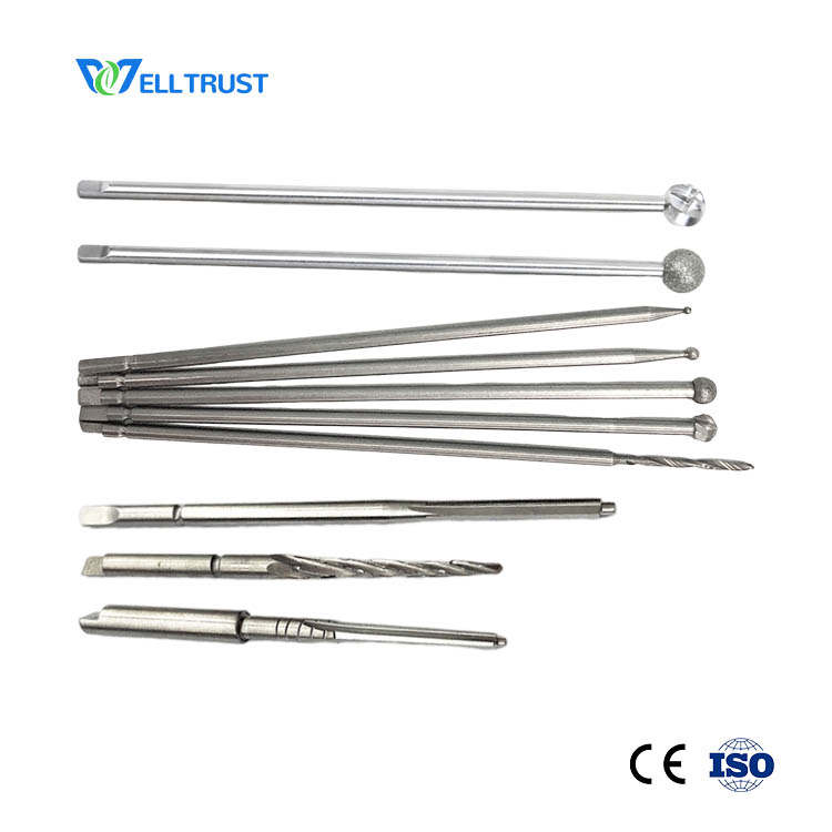
Arthroscopic burrs are engineered for durability, precision, and biocompatibility. They typically consist of a long, slender shaft connected to a cutting head, designed to attach to a motorized handpiece.
- Shaft:
- Material: Usually high-grade stainless steel or titanium alloys, chosen for their strength, corrosion resistance, and biocompatibility. The length varies depending on the joint being accessed.
- Flexibility/Rigidity: Designed to be rigid enough for controlled force transmission but slender enough to navigate through tight joint spaces.
- Head (Burr): This is the active cutting component and comes in various shapes and sizes, with round and oval being two of the most common and versatile.
- Round Burr: Features a spherical cutting surface.
- Uses: Ideal for general bone and soft tissue debulking, creating smooth contours, and preparing surfaces for procedures like microfracture or graft fixation. Its even cutting surface provides consistent material removal.
- Oval Burr: Characterized by an elongated, elliptical cutting surface.
- Uses: Particularly useful for broader, flatter surfaces, resecting osteophytes, shaping larger bone defects, or performing acromioplasty in the shoulder. The oval shape allows for more efficient material removal over a wider area while still maintaining control.
- Cutting Surfaces:
- Fluted/Toothed: Designed for aggressive bone removal, often made from tungsten carbide for superior hardness and sharpness.
- Abrasive/Diamond-tipped: Used for finer sculpting, smoothing, and cartilage debridement, where precision and minimal tissue trauma are critical. Diamond tips offer exceptional control and a very fine finish.
- Size Range: Burrs are available in diameters ranging from very small (e.g., 2.0 mm) for delicate work to larger sizes (e.g., 5.5 mm) for more extensive resections.
- Round Burr: Features a spherical cutting surface.
- Connection Mechanism: The proximal end of the burr shaft securely connects to a specialized motorized handpiece, which provides the rotational power. This connection must be robust and allow for quick, secure attachment and detachment.
- Irrigation Ports: Many burr designs incorporate internal or external channels for continuous irrigation fluid flow. This serves multiple purposes:
- Debris Removal: Flushes away bone fragments and tissue, maintaining clear visualization.
- Cooling: Prevents heat buildup at the cutting site, minimizing the risk of thermal necrosis (tissue damage due to heat).
Mechanism of Action
The arthroscopic burr operates on the principle of high-speed rotational cutting or abrasion.
1. Motorized Handpiece: The burr is attached to a powered handpiece, which can rotate at speeds ranging from thousands to tens of thousands of revolutions per minute.
2. Controlled Resection: The surgeon guides the rotating burr under direct arthroscopic visualization. The cutting head's sharp flutes or abrasive surface precisely removes targeted bone or tissue.
3. Suction: Simultaneously, a suction mechanism integrated into the handpiece or via a separate cannula removes the irrigation fluid and tissue debris, maintaining a clear surgical field.
4. Precision: The combination of high rotational speed, sharp cutting surfaces, and constant visualization allows for extremely precise and controlled tissue removal, critical in the confined spaces of a joint.
Biomechanics
The biomechanical implications of using arthroscopic burrs are significant for joint health and function.
* Bone Contouring: Burrs allow for precise shaping of bone, correcting anatomical deformities (e.g., in FAI of the hip) that cause impingement. This restores normal joint kinematics and reduces abnormal stresses.
* Cartilage Preparation: When preparing for microfracture or other cartilage repair techniques, burrs create a clean, stable base for new cartilage growth. The ability to create smooth, non-jagged edges is crucial for optimal healing.
* Surface Finish: The type of burr (e.g., diamond-tipped) and the surgeon's technique can create a smooth bone or cartilage surface, minimizing friction within the joint and potentially delaying degenerative changes.
* Minimizing Thermal Necrosis: The continuous irrigation and high speed of rotation are crucial in dissipating heat generated during cutting, thereby protecting surrounding vital tissues, including bone and cartilage, from thermal damage.
3. Extensive Clinical Indications & Usage
Arthroscopic burrs are indispensable across various orthopedic subspecialties, facilitating precise tissue management in numerous joint pathologies.
General Principle
The primary application of arthroscopic burrs is the minimally invasive removal, shaping, or debridement of bone, articular cartilage, or specific soft tissues that are diseased, damaged, or causing mechanical symptoms within a joint.
Specific Joint Applications
Knee Arthroscopy
- Meniscus Repair Preparation: Debridement of unstable meniscal edges to create a stable, vascularized bed for repair.
- Chondroplasty: Smoothing of frayed or rough articular cartilage defects to reduce pain and mechanical irritation.
- Osteophyte Removal: Resection of bone spurs (e.g., in the patellofemoral joint or around the intercondylar notch) that can cause impingement or limit range of motion.
- ACL/PCL Reconstruction: Precision shaping of femoral and tibial tunnels for graft placement, ensuring optimal graft fit and positioning.
- Microfracture Preparation: Decorticating subchondral bone to expose cancellous bone, stimulating a healing response for cartilage defects.
Shoulder Arthroscopy
- Acromioplasty (Subacromial Decompression): Resection of bone from the undersurface of the acromion to relieve impingement on the rotator cuff tendons, often using an oval burr.
- Rotator Cuff Repair Preparation: Debridement of the greater tuberosity to create a fresh, bleeding bone bed for tendon reattachment.
- Glenoid Preparation: Preparing the glenoid rim for labral repair or stabilization procedures.
- Removal of Loose Bodies: Grinding down or removing free fragments of bone or cartilage within the joint.
Hip Arthroscopy
- Femoroacetabular Impingement (FAI) Correction:
- Cam Lesion Resection: Shaping the femoral head-neck junction to remove the bony bump causing impingement.
- Pincer Lesion Resection: Trimming excess bone from the acetabular rim.
- Labral Debridement: Smoothing frayed portions of the labrum.
- Osteophyte Removal: Addressing bone spurs around the hip capsule.
Ankle Arthroscopy
- Ankle Impingement: Resection of anterior or posterior osteophytes causing pain and limited dorsiflexion/plantarflexion.
- Osteochondral Lesion Debridement: Preparing the bed of an osteochondral defect for repair or microfracture.
Wrist and Elbow Arthroscopy
- While less common than in larger joints, burrs can be used for specific debridement, loose body removal, or osteophyte resection in the wrist (e.g., distal ulna head shaping) or elbow (e.g., radial head shaping, olecranon fossa debridement).
Fitting/Usage Instructions (Patient-Friendly Explanation)
The use of an arthroscopic burr is a highly skilled procedure performed by an orthopedic surgeon.
1. Pre-operative Planning: Before surgery, detailed imaging (X-rays, MRI) is reviewed to identify the exact pathology and plan the surgical approach.
2. Incision & Portals: Small incisions (typically less than 1 cm) called portals are made around the joint. One portal is for the arthroscope, which projects images onto a monitor. Other portals are for instruments like the burr.
3. Visualization: The surgeon uses the arthroscope to gain a clear, magnified view of the joint's interior on a high-definition screen.
4. Burr Insertion: The appropriate burr, selected based on the specific surgical need (e.g., round for smoothing, oval for broader resection), is inserted through a separate portal.
5. Controlled Resection: Under constant visual guidance, the surgeon activates the motorized burr. With precise, controlled movements, the burr removes the targeted bone or tissue. The surgeon's experience and tactile feedback are crucial for accurate tissue removal.
6. Continuous Irrigation & Suction: Sterile fluid is continuously pumped into the joint to keep it distended, clear away debris, and cool the instrument. A suction mechanism simultaneously removes this fluid and the excised tissue fragments.
7. Post-Resection Inspection: After the desired shaping or debridement is complete, the joint is thoroughly flushed and inspected via the arthroscope to ensure the surgical goals have been met and no debris remains.
4. Risks, Side Effects, or Contraindications
While arthroscopic procedures using burrs are generally safe and effective, like any surgical intervention, they carry potential risks.
General Arthroscopy Risks
- Infection: Though rare, any surgical procedure carries a risk of infection.
- Bleeding/Hematoma: Accumulation of blood within the joint.
- Nerve or Vessel Damage: Injury to surrounding nerves or blood vessels, though minimized by careful portal placement.
- Deep Vein Thrombosis (DVT) / Pulmonary Embolism (PE): Blood clot formation, particularly in lower extremity surgery.
- Anesthesia Risks: Adverse reactions to general or regional anesthesia.
- Joint Stiffness: Post-operative stiffness, often managed with physical therapy.
Burr-Specific Risks
- Excessive Tissue Removal: Inadvertent removal of too much healthy bone or cartilage, potentially altering joint mechanics or stability.
- Thermal Injury: Despite irrigation, localized heat buildup can occur, potentially damaging surrounding tissues.
- Instrument Breakage: Although rare with quality instruments, a burr can potentially break within the joint.
- Incomplete Debridement: Failure to remove all problematic tissue, leading to persistent symptoms.
- Damage to Healthy Cartilage/Bone: Accidental contact with non-targeted healthy structures.
Contraindications
- Active Joint Infection: Surgery should be delayed until the infection is resolved.
- Severe Osteoporosis: In some cases, extremely brittle bone may be less suitable for burr use due to fracture risk (relative contraindication).
- Severe Arthritis: In advanced cases where joint surfaces are extensively damaged, arthroscopy may offer limited benefit, and joint replacement might be a more appropriate solution.
- Inability to Tolerate Anesthesia: Patients with severe medical comorbidities that preclude safe anesthesia administration.
- Unrealistic Patient Expectations: Patients must understand the potential outcomes and limitations of the procedure.
5. Expert Tips from Dr. Mohammed Hutaif
Dr. Mohammed Hutaif, a leading orthopedic specialist, shares his insights on optimizing the use of arthroscopic burrs for superior patient care:
"The arthroscopic burr is an incredibly powerful tool in our surgical armamentarium, but its effectiveness lies in meticulous planning, precise execution, and a deep understanding of joint biomechanics. My approach emphasizes several key principles:
- Careful Patient Selection: Not every patient or every pathology is suitable for burr-based arthroscopic intervention. A thorough evaluation, including a detailed history, physical examination, and advanced imaging, is crucial to determine if a patient will truly benefit.
- Advanced Imaging Interpretation: High-resolution MRI and CT scans are invaluable. They allow us to precisely map the pathology, understand the bone and cartilage architecture, and pre-plan the exact extent of resection required.
- Surgeon Experience and Skill: The control of an arthroscopic burr demands significant training and experience. The ability to differentiate between healthy and pathological tissue, coupled with fine motor control, is paramount to achieving optimal results while minimizing complications.
- Choosing the Right Burr: Selecting the appropriate burr type (round vs. oval), size, and cutting surface (fluted vs. diamond-tipped) is critical for each specific surgical task. A round burr might be perfect for general contouring, while an oval burr might be more efficient for broader bone resection, and a diamond burr for delicate cartilage smoothing.
- Meticulous Irrigation and Suction: Maintaining a continuously clear surgical field through efficient irrigation and suction is non-negotiable. This not only ensures optimal visualization but also protects tissues from thermal injury and prevents the accumulation of debris.
- Continuous Arthroscopic Visualization: Never operate the burr without a clear, unobstructed view of the cutting site and surrounding structures. This vigilance prevents inadvertent damage to healthy tissues.
- Patient Education and Realistic Expectations: It's essential to have an open dialogue with patients about what the procedure entails, the expected recovery, and potential outcomes. Managing expectations is key to patient satisfaction.
- Post-operative Rehabilitation: The surgical intervention is only one part of the journey. A well-structured, personalized post-operative rehabilitation program is vital for restoring strength, flexibility, and function, maximizing the long-term benefits of the surgery."
6. Massive FAQ Section
Q1: What is an arthroscopic burr primarily used for?
A1: An arthroscopic burr is a specialized surgical instrument used in minimally invasive joint surgery (arthroscopy) to precisely remove, shape, or smooth bone, cartilage, and certain soft tissues. Its main purpose is to correct anatomical deformities, debride damaged tissues, or prepare bone surfaces for other reconstructive procedures within the joint.
Q2: Are arthroscopic burrs reusable or disposable?
A2: The majority of arthroscopic burrs used today are single-use, disposable instruments. This ensures maximum sharpness, sterility, and reduces the risk of cross-contamination or instrument fatigue. Some components of the motorized handpiece they attach to are reusable and undergo rigorous sterilization protocols.
Q3: What's the difference between a round and an oval arthroscopic burr, and when are they used?
A3: The main difference lies in their cutting head shape and application.
* Round Burrs: Have a spherical cutting head. They are excellent for general bone and soft tissue debulking, creating smooth, concave contours, and preparing small, specific areas (e.g., for microfracture or tunnel creation).
* Oval Burrs: Have an elongated, elliptical cutting head. They are more effective for resecting broader, flatter surfaces, removing larger osteophytes, or shaping wider areas of bone (e.g., in acromioplasty or FAI correction). The oval shape allows for more efficient material removal over a larger surface area.
Q4: Is arthroscopic surgery involving a burr painful?
A4: Arthroscopic surgery is performed under anesthesia (general, regional, or local with sedation), so you will not feel pain during the procedure. Post-operatively, you will experience some pain and discomfort, which is managed with pain medication. The minimally invasive nature of arthroscopy generally results in less post-operative pain and a faster recovery compared to open surgery.
Q5: How long is the recovery from surgery involving an arthroscopic burr?
A5: Recovery time varies significantly depending on the joint involved, the extent of the pathology, the specific procedure performed, and individual patient factors. It can range from a few weeks for minor debridement to several months for more complex reconstructive procedures requiring significant bone shaping and subsequent rehabilitation (e.g., FAI correction). Your surgeon will provide a personalized recovery plan.
Q6: Can an arthroscopic burr damage healthy tissue?
A6: While rare, there is a potential risk of inadvertent damage to healthy bone, cartilage, nerves, or blood vessels if the burr is not used with extreme precision. This risk is minimized by the surgeon's skill, continuous arthroscopic visualization, and meticulous technique, along with constant irrigation to keep the field clear and cool.
Q7: What materials are arthroscopic burrs typically made of?
A7: The shafts are usually made of high-grade stainless steel or titanium alloys for strength and biocompatibility. The cutting heads often feature tungsten carbide for aggressive cutting or diamond tips for finer abrasion and smoothing. These materials are chosen for their hardness, durability, and resistance to corrosion.
Q8: How does the surgeon see what they are doing inside the joint?
A8: The surgeon uses an arthroscope, which is a thin fiber-optic camera inserted into the joint through a small incision. The arthroscope is connected to a video monitor, providing a magnified, high-definition view of the joint's interior. This allows the surgeon to precisely visualize the burr's action and the surrounding anatomy.
Q9: Is this procedure suitable for all joint problems?
A9: No, arthroscopic surgery with a burr is not suitable for all joint problems. It is most effective for conditions requiring precise removal or shaping of bone or cartilage, debridement of damaged tissues, or preparation for repairs. For very advanced arthritis with extensive joint destruction, joint replacement might be a more appropriate treatment. Your orthopedic surgeon will determine if it's the right option for your specific condition.
Q10: What are the main benefits of using an arthroscopic burr in surgery?
A10: The primary benefits include:
* Precision: Allows for highly accurate removal and shaping of tissue.
* Minimally Invasive: Smaller incisions lead to less pain, reduced scarring, and faster recovery compared to open surgery.
* Improved Outcomes: Contributes to better joint function, reduced pain, and potentially delays or prevents the need for more extensive surgeries like joint replacement.
* Enhanced Visualization: Direct, magnified view of the joint interior for targeted treatment.
Q11: How is the burr controlled during surgery to ensure accuracy?
A11: The burr is controlled by the surgeon's hand, which is attached to a motorized handpiece. The surgeon uses delicate movements, guided by continuous visualization on the arthroscopic monitor, along with their tactile sense, to precisely remove the targeted tissue. The speed of rotation can also be adjusted for different tasks, further enhancing control.
Q12: What happens to the tissue that is removed by the burr?
A12: As the burr removes bone or tissue, the fragments are immediately flushed away by continuous irrigation fluid and simultaneously removed from the joint through a suction mechanism integrated into the surgical system. This ensures a clear surgical field and prevents debris from remaining within the joint.