The Advanced 4.0mm, 30-Degree Lens, HD Arthroscope: A Comprehensive Orthopedic Guide
Introduction & Overview
In the realm of modern orthopedics, the arthroscope stands as a cornerstone instrument, revolutionizing how joint pathologies are diagnosed and treated. Far removed from the era of extensive open surgeries, arthroscopy offers a minimally invasive pathway, translating to reduced patient morbidity, faster recovery times, and superior cosmetic outcomes. At the forefront of this technological advancement is the 4.0mm, 30-Degree Lens, High-Definition (HD) Arthroscope, a sophisticated tool that has become an indispensable asset for orthopedic surgeons worldwide.
This guide delves deeply into the intricacies of this specific arthroscope, exploring its design, technical specifications, diverse clinical applications, meticulous maintenance protocols, underlying biomechanical principles, and the profound impact it has on patient outcomes. As expert medical SEO copywriters and orthopedic specialists, our aim is to provide an exhaustive, authoritative resource for medical professionals, procurement specialists, and anyone seeking a profound understanding of this critical orthopedic instrument.
Deep-Dive into Technical Specifications & Mechanisms
The efficacy of the 4.0mm, 30-degree lens, HD arthroscope is rooted in its precision engineering and advanced optical capabilities. Understanding these elements is crucial for appreciating its clinical value.
Design & Materials
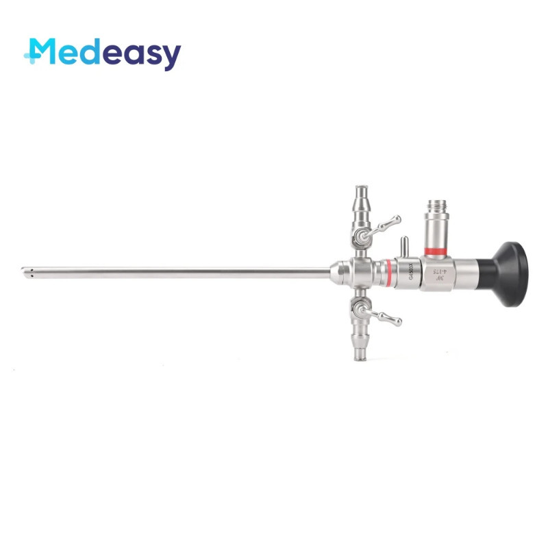
The construction of a high-quality arthroscope is a testament to sophisticated material science and optical engineering.
* Outer Sheath: Typically crafted from medical-grade stainless steel or titanium alloys, the outer sheath provides structural integrity, biocompatibility, and resistance to corrosion and repeated sterilization cycles. Its smooth finish minimizes tissue trauma during insertion.
* Optical System: The heart of the arthroscope is its rod lens system, often based on the Hopkins rod lens design. This system uses a series of glass rods instead of conventional air spaces, allowing for brighter images, wider fields of view, and superior light transmission.
* Lens: The distal lens, which directly interacts with the joint environment, is frequently made from sapphire glass. Sapphire offers exceptional hardness, making it highly resistant to scratches and abrasions, crucial for maintaining optical clarity in a dynamic surgical field.
* Fiber Optic Illumination: Integrated fiber optic bundles run alongside the rod lens system, delivering intense cold light from an external light source to illuminate the joint cavity. This prevents heat generation within the joint while providing optimal visualization.
* Sealing Mechanisms: Meticulous sealing ensures the arthroscope is watertight, preventing fluid ingress into the optical system during surgery and safeguarding against contamination during sterilization.
* Ergonomics: The proximal end features a comfortable handle for surgeon control and a standardized connector for attaching the light guide cable and camera head.
Specifics of 4.0mm Diameter
The 4.0mm diameter represents a critical balance in arthroscopic instrumentation.
* Invasiveness vs. Visualization: It offers a generous field of view and excellent light transmission, suitable for a wide range of procedures, while remaining minimally invasive enough to reduce tissue disruption.
* Joint Versatility: This size is highly versatile, making it the workhorse for larger joints like the knee and shoulder, and often suitable for smaller joints such as the ankle or hip, depending on the patient's anatomy and specific pathology.
Specifics of 30-Degree Lens Angle
The 30-degree angle is a defining feature that significantly enhances surgical capability compared to a 0-degree (straight ahead) scope.
* Panoramic View: It allows the surgeon to "look around corners," providing an oblique view that can visualize structures not directly in front of the scope.
* Access to Hidden Areas: This angle is invaluable for inspecting posterior aspects of menisci, superior labrum in the shoulder, or specific areas within the hip joint that would otherwise require additional portals or more aggressive manipulation.
* Enhanced Maneuverability: By rotating the scope, the 30-degree angle provides a 360-degree panoramic sweep of the joint from a single portal, reducing the need for multiple portal placements and minimizing tissue damage.
HD (High Definition) Capability
The "HD" designation is no mere marketing term; it signifies a profound leap in visual quality.
* Resolution & Clarity: HD optics, when paired with compatible HD camera systems and monitors, deliver images with significantly higher pixel density. This translates to sharper, clearer images, enabling surgeons to distinguish subtle tissue textures, vascular patterns, and minute pathological changes with unprecedented detail.
* Improved Diagnostic Accuracy: The enhanced visualization directly improves diagnostic accuracy, allowing for precise identification of lesions, tears, and degenerative changes that might be missed with standard definition systems.
* Surgical Precision: For interventional procedures, HD clarity allows for more precise instrument manipulation, better tissue debridement, and more accurate repair placements, ultimately leading to better surgical outcomes.
* Reduced Surgeon Fatigue: Sharper images reduce the cognitive load on the surgeon, potentially leading to less eye strain and fatigue during lengthy procedures.
Mechanism of Action
The arthroscope functions as a sophisticated internal camera and light source.
1. Light Transmission: A powerful external light source (e.g., Xenon, LED) connects via a fiber optic cable to the arthroscope. The fiber optic bundles within the scope transmit this cold light into the joint cavity.
2. Image Capture & Transmission: Light reflected from the joint's internal structures passes back through the objective lens, through the rod lens system, to the eyepiece (ocular) at the proximal end. An HD camera head attaches to this ocular, converting the optical image into an electronic signal.
3. Visualization: This electronic signal is then transmitted to a high-definition monitor, displaying real-time, magnified images of the joint interior for the surgical team.
4. Fluid Management: While not directly part of the scope's optical mechanism, fluid management (inflow and outflow through separate cannulas or integrated channels) is crucial. Sterile saline solution distends the joint, washes away debris, and maintains a clear visual field, preventing scope fogging and improving visibility.
Extensive Clinical Indications & Usage
The 4.0mm, 30-degree HD arthroscope is central to a vast array of orthopedic procedures, embodying the principles of minimally invasive surgery.
General Arthroscopy Principles
- Minimally Invasive: Smaller incisions (portals) mean less tissue disruption, reduced pain, and faster healing.
- Reduced Recovery Time: Patients typically experience quicker mobilization and return to activities.
- Less Scarring: Cosmetic benefits due to smaller incisions.
- Diagnostic & Therapeutic: Can be used solely for diagnosis or to perform surgical repairs simultaneously.
Specific Joint Applications
| Joint | Common Clinical Indications |
|---|---|
| Knee | Meniscus tears (repair/resection), ACL/PCL reconstruction, chondroplasty, loose body removal, synovial biopsy, patellofemoral disorders, plica excision. |
| Shoulder | Rotator cuff repair, labral repair (SLAP, Bankart), subacromial decompression, biceps tenodesis/tenotomy, capsular release for adhesive capsulitis, AC joint debridement. |
| Hip | Labral tears, femoroacetabular impingement (FAI) correction (cam/pincer osteoplasty), loose body removal, capsular plication, treatment of gluteal tendinopathy. |
| Ankle | Osteochondral lesions of the talus (OCLT), impingement syndromes (anterior/posterior), loose body removal, synovectomy, ligament repair/reconstruction. |
| Elbow | Loose body removal, capsular release, synovectomy for synovitis, osteophyte removal. |
| Wrist | TFCC repair, ganglion excision, carpal instability workup, synovectomy. |
Pre-operative Considerations
Before arthroscopy, meticulous planning is essential:
* Patient Selection: Appropriateness of arthroscopy based on symptoms, physical examination, and patient expectations.
* Imaging: Comprehensive imaging studies (X-rays, MRI, CT scans) are vital for diagnosis, surgical planning, and identifying anatomical variants.
* Informed Consent: Detailed discussion with the patient regarding the procedure, potential risks, benefits, and alternatives.
Intra-operative Usage (Fitting/Usage Instructions)
The precise and sterile handling of the arthroscope is paramount for successful outcomes.
* Preparation:
* Ensure the arthroscope has undergone proper sterilization.
* Connect the fiber optic light cable to the scope and the external light source.
* Attach the HD camera head to the scope's ocular.
* Connect the camera system to the video monitor.
* Prepare the fluid management system (pump, inflow/outflow tubing, sterile saline).
* Perform white balance and focus adjustments on the camera system.
* Insertion:
* After surgical prep and draping, a small incision (portal) is made.
* A trocar (sharp obturator) is inserted through a cannula (working sleeve) into the joint.
* Once the joint capsule is breached, the trocar is removed, leaving the cannula in place.
* The arthroscope is gently inserted through the cannula.
* Visualization & Maneuverability:
* The joint is distended with sterile saline via the fluid management system to create a working space and clear visibility.
* The surgeon systematically inspects the joint, using the 30-degree angle and scope rotation to visualize all anatomical structures.
* Additional portals may be created for the insertion of other surgical instruments (probes, shavers, graspers, suture passers) to perform therapeutic interventions.
* Maintaining a clear visual field is critical, often requiring adjustment of fluid flow and occasional debridement of debris.
Biomechanics Considerations
The use of an arthroscope inherently involves biomechanical principles:
* Portal Placement: Optimal portal placement minimizes soft tissue damage, avoids neurovascular structures, and provides the best triangulation for scope and instrument manipulation.
* Joint Distension: The fluid pressure used to distend the joint must be carefully managed to maintain adequate visualization without causing excessive extravasation or compromising joint integrity.
* Scope Manipulation: Gentle, controlled movements of the scope prevent iatrogenic cartilage scuffing or damage to intra-articular structures. The 30-degree angle allows for less physical manipulation of the scope itself to achieve a wider view.
* Surgeon Ergonomics: The overall setup, including monitor placement and instrument handling, impacts surgeon posture and fatigue, influencing precision and safety.
Patient Outcome Improvements
The benefits of arthroscopy with a 4.0mm, 30-degree HD arthroscope are directly reflected in superior patient outcomes:
* Reduced Post-operative Pain: Smaller incisions and less tissue trauma contribute to significantly less pain compared to open surgery.
* Faster Recovery & Rehabilitation: Patients can often begin rehabilitation sooner and return to daily activities and sports more quickly.
* Improved Cosmesis: Minimal scarring is a significant aesthetic advantage.
* Lower Infection Risk: While not entirely eliminated, the smaller incisions and controlled environment generally reduce the risk of infection.
* Enhanced Diagnostic Accuracy: HD visualization leads to more precise diagnoses, ensuring targeted and effective treatment plans.
* Better Functional Outcomes: Precise repairs and debridements, guided by high-definition imaging, often result in improved long-term joint function and reduced recurrence rates.
Maintenance & Sterilization Protocols
The longevity, safety, and optimal performance of the 4.0mm, 30-degree HD arthroscope depend heavily on strict adherence to maintenance and sterilization protocols.
Immediate Post-Procedure Care
- Gross Contaminant Removal: Immediately after use, visible blood and tissue debris should be wiped off the exterior of the scope.
- Flushing: The internal channels (if applicable, though less common for standard scopes) and external surfaces should be thoroughly flushed with water to prevent drying of organic material.
Disassembly & Cleaning
- Disassembly: Separate the arthroscope from the camera head, light cable, and any attached cannulas.
- Manual Cleaning:
- Submerge the scope in an enzymatic detergent solution designed for medical instruments.
- Use soft brushes (specific for arthroscopes) to clean the outer sheath, light post, and particularly the distal lens.
- Ensure all surfaces are meticulously scrubbed to remove biofilm and organic residues.
- Rinse thoroughly with sterile or deionized water to remove all detergent residue.
- Inspection: After cleaning, carefully inspect the arthroscope under magnification for:
- Scratches or cracks on the distal lens.
- Bends, dents, or damage to the outer sheath.
- Dark spots or broken fibers in the light guide (indicating reduced light transmission).
- Fluid leaks or fogging within the optical system (a sign of seal compromise).
- Any signs of corrosion. Damaged scopes must be sent for repair.
Sterilization Methods
Sterilization is critical to prevent surgical site infections.
* Steam Sterilization (Autoclaving):
* Principle: Uses high-temperature steam under pressure to kill microorganisms.
* Advantages: Widely available, cost-effective, rapid, non-toxic.
* Considerations: Arthroscopes are heat-sensitive. Specific low-temperature or "flash" cycles designed for delicate instruments must be used, following manufacturer guidelines precisely. Over-exposure to high temperatures can degrade optical cement and seals.
* Low-Temperature Sterilization:
* Ethylene Oxide (EtO):
* Principle: Alkylating agent that denatures proteins and nucleic acids.
* Advantages: Effective for heat-sensitive instruments.
* Considerations: Requires aeration time, toxic, environmental concerns.
* Hydrogen Peroxide Plasma (e.g., Sterrad):
* Principle: Uses hydrogen peroxide vapor and plasma to generate free radicals that inactivate microorganisms.
* Advantages: Rapid, non-toxic byproducts (water, oxygen), effective for heat-sensitive instruments.
* Considerations: Not suitable for instruments with lumens less than 1mm or certain materials.
* Manufacturer Guidelines: Always consult the instrument manufacturer's instructions for use (IFU) for specific cleaning agents, sterilization parameters, and compatible methods. Deviating from these guidelines can void warranties, damage the instrument, and compromise patient safety.
Storage
Sterilized arthroscopes should be stored in a clean, dry, and protected environment, ideally in sterile pouches or containers, until ready for use. This prevents recontamination and physical damage.
Risks, Side Effects, or Contraindications
While arthroscopy with a 4.0mm, 30-degree HD arthroscope is a safe and effective procedure, it is not without potential risks, side effects, or contraindications.
General Arthroscopy Risks
- Infection: Although rare with sterile technique, any surgical procedure carries a risk of surgical site infection.
- Bleeding/Hematoma: Minor bleeding is common; significant hematoma formation is less frequent.
- Nerve or Vessel Damage: Accidental injury to nearby nerves or blood vessels, though uncommon, can occur during portal placement or instrument manipulation.
- Deep Vein Thrombosis (DVT) / Pulmonary Embolism (PE): As with any surgery, there is a small risk of blood clot formation.
- Anesthesia Risks: All risks associated with general or regional anesthesia apply.
- Joint Stiffness (Arthrofibrosis): Post-operative stiffness can occur, requiring physiotherapy or further intervention.
- Persistent Pain: Some patients may experience ongoing pain despite successful surgery.
Scope-Specific Risks
- Scope Breakage/Damage: While rare with proper handling, the scope itself can break within the joint. This is a serious complication requiring removal of fragments.
- Cartilage Scuffing: Inadvertent contact of the scope or instruments with articular cartilage can cause damage.
- Fluid Extravasation: Excessive fluid pressure or prolonged surgery can lead to fluid leaking into surrounding soft tissues, causing swelling, compartment syndrome (rare), or impaired visualization.
- Thermal Injury: Though cold light is used, prolonged contact of the light post with tissue or excessive use of electrocautery near the scope can cause thermal injury.
Contraindications
- Active Joint Infection: Arthroscopy is generally contraindicated in the presence of an active joint infection due to the risk of spreading the infection.
- Severe Arthritis: In cases of end-stage osteoarthritis, arthroscopy may offer minimal benefit and could potentially exacerbate symptoms. Joint replacement is often a more appropriate treatment.
- Ankylosed Joint: A completely stiff or fused joint may not provide adequate space for arthroscope insertion or manipulation.
- Severe Coagulopathy: Uncontrolled bleeding disorders significantly increase the risk of hemorrhage.
- Poor General Health/Anesthesia Risks: Patients with severe systemic diseases or those deemed high-risk for anesthesia may not be candidates for elective arthroscopy.
- Skin Lesions/Infection at Portal Sites: Active skin infections or lesions in the planned portal areas are contraindications.
Massive FAQ Section
Q1: What is the primary advantage of a 30-degree arthroscope over a 0-degree scope?
A1: The 30-degree lens provides an oblique, panoramic view, allowing the surgeon to "look around corners" and visualize structures that are not directly in front of the scope. This enhances diagnostic capabilities and surgical access to difficult-to-reach areas within the joint, often reducing the need for additional portals compared to a 0-degree (straight-ahead) scope.
Q2: Why is HD (High Definition) crucial for modern arthroscopy?
A2: HD capability provides significantly higher resolution and clarity of images. This allows surgeons to distinguish subtle tissue textures, identify minute pathological changes, and perform precise instrument manipulation with greater confidence, leading to improved diagnostic accuracy and surgical precision.
Q3: Is the 4.0mm arthroscope suitable for all joint sizes?
A3: The 4.0mm arthroscope is highly versatile and considered the standard for medium to large joints like the knee and shoulder. It is also frequently used in the hip and ankle. For very small joints like the wrist or specific pediatric applications, smaller diameter scopes (e.g., 2.7mm or 1.9mm) might be preferred, but the 4.0mm offers a good balance of invasiveness and visualization for most common procedures.
Q4: How often should an arthroscope be serviced or inspected?
A4: Arthroscopes should be thoroughly inspected after every use, particularly during the cleaning and sterilization process, for any signs of damage (scratches, bends, broken fibers, fluid ingress). Regular preventive maintenance by qualified technicians, typically annually or semi-annually, is also recommended to ensure optimal performance and extend the instrument's lifespan.
Q5: What are the common signs of damage to an arthroscope that warrant repair?
A5: Common signs of damage include:
* Scratches, cracks, or chips on the distal lens, affecting image clarity.
* Bends or dents in the outer sheath, which can impede insertion or damage tissue.
* Black spots, shadows, or significantly reduced brightness in the image, indicating broken fiber optics or internal lens damage.
* Fogging or fluid ingress within the scope, suggesting a compromised seal.
* Corrosion on any part of the instrument.
Q6: Can arthroscopes be reused, and how are they made safe for subsequent procedures?
A6: Yes, most arthroscopes are designed as reusable instruments. To ensure safety, they undergo a rigorous process of meticulous cleaning, disinfection (if applicable), and high-level sterilization (typically steam sterilization or low-temperature plasma sterilization) after each use, strictly following manufacturer guidelines and institutional protocols.
Q7: What is the role of fluid management during arthroscopy?
A7: Fluid management is critical. Sterile saline solution is continuously pumped into the joint to:
1. Distend the joint capsule: Creating a working space for visualization and instrument manipulation.
2. Wash away debris: Clearing blood, tissue fragments, and particulate matter for a clear visual field.
3. Prevent fogging: Maintaining a constant flow over the lens helps prevent condensation.
Proper fluid pressure is maintained to avoid extravasation or inadequate distension.
Q8: How does arthroscopy improve patient outcomes compared to traditional open surgery?
A8: Arthroscopy generally leads to:
* Reduced post-operative pain due to smaller incisions and less tissue trauma.
* Faster recovery and rehabilitation times.
* Lower risk of infection in many cases.
* Improved cosmetic results with minimal scarring.
* More precise diagnosis and treatment thanks to magnified, high-definition visualization.
Q9: What materials are arthroscopes typically made from?
A9: High-quality arthroscopes commonly feature:
* Outer Sheath: Medical-grade stainless steel or titanium alloys for durability and biocompatibility.
* Lenses: Sapphire glass for the distal lens (scratch resistance) and specialized optical glass for the internal rod lens system.
* Light Guides: Fiber optic bundles made of glass.
* Seals: Biocompatible polymers for watertight integrity.
Q10: Are there any non-surgical alternatives to arthroscopy for joint diagnosis?
A10: Yes, non-surgical diagnostic alternatives include:
* Physical Examination: Clinical assessment of joint stability, range of motion, and pain.
* Imaging Studies: X-rays (for bony structures), MRI (gold standard for soft tissues like ligaments, tendons, cartilage), CT scans (detailed bone imaging), and ultrasound (for superficial soft tissues).
* Diagnostic Injections: Injecting local anesthetic into the joint to localize pain sources.
While these can provide significant information, arthroscopy offers direct visual confirmation and the ability to perform biopsies or therapeutic interventions concurrently.
Q11: What is the typical lifespan of a well-maintained arthroscope?
A11: The lifespan of an arthroscope can vary significantly depending on usage frequency, handling care, and adherence to maintenance/sterilization protocols. With proper care, a high-quality arthroscope can last several years, often 5-10 years or even longer, though components like the distal lens or fiber optics may require occasional repair or replacement.
Q12: How is the light source connected to the arthroscope, and why is it important?
A12: The light source is connected to the arthroscope via a flexible fiber optic light guide cable. This cable transmits intense "cold" light from an external light generator into the arthroscope, illuminating the joint cavity without generating heat within the patient. A strong, consistent light source is crucial for optimal visualization and image quality, especially with HD systems.