Chronic Unreduced Dislocations of the Lower Extremity
Key Takeaway
Chronic unreduced dislocations of the lower extremity present complex reconstructive challenges. In the hip, neglected anterior dislocations often require subcapital displacement osteotomies or modified Girdlestone arthroplasties to restore biomechanics while preserving bone stock for future arthroplasty. Similarly, chronic patellar dislocations demand meticulous extensor mechanism realignment, capsular balancing, and potential tibial tuberosity transfer. This guide details the indications, surgical approaches, and postoperative protocols for managing these rare but debilitating orthopedic presentations.
INTRODUCTION TO CHRONIC UNREDUCED DISLOCATIONS
Chronic unreduced (old) dislocations of the lower extremity represent a formidable challenge to the orthopedic surgeon. While acute dislocations are orthopedic emergencies managed with urgent reduction, chronic or neglected dislocations are relatively uncommon in modern practice, particularly in developed nations. When they do occur, they are typically the sequelae of high-energy motor vehicle collisions accompanied by severe polytrauma. Concomitant traumatic brain injuries, prolonged intensive care admissions, ipsilateral femoral fractures, or contralateral pelvic ring disruptions frequently draw clinical attention away from the dislocated joint, leading to a delayed diagnosis. In developing countries, however, neglected traumatic dislocations present with higher frequency due to delayed access to specialized orthopedic care.
The management of chronic unreduced dislocations requires a paradigm shift from acute trauma principles to complex reconstructive and salvage strategies. Attempting a delayed closed reduction is universally contraindicated due to severe soft tissue contractures, capsular fibrosis, and the unacceptably high risk of iatrogenic fracture or neurovascular compromise. This masterclass details the evidence-based surgical management of two distinct but highly challenging clinical entities: the chronic unreduced anterior dislocation of the hip, and the chronic unreduced lateral dislocation of the patella.
PART I: CHRONIC UNREDUCED ANTERIOR DISLOCATIONS OF THE HIP
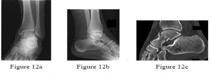
Traumatic anterior dislocation of the hip is a comparatively rare injury, accounting for only 10% to 15% of all traumatic hip dislocations. Consequently, the literature regarding old, unreduced anterior dislocations is sparse. The femoral head is typically displaced into either the obturator foramen (inferior) or the pubic/iliac region (superior). Over time, the acetabulum fills with fibrofatty tissue, the capsule heavily scars, and the articular cartilage of the femoral head undergoes irreversible degeneration or avascular necrosis (AVN).
Preoperative Evaluation and Pathoanatomy
Patients typically present with a fixed deformity. In an obturator dislocation, the hip is locked in flexion, abduction, and external rotation. In a pubic dislocation, the hip is extended, abducted, and externally rotated.
Surgical Warning: In chronic anterior dislocations, the femoral neurovascular bundle (femoral artery, vein, and nerve) is frequently draped directly over the displaced femoral head or stretched across the anterior capsule. Meticulous preoperative imaging, including CT angiography, is highly recommended to map the distorted vascular anatomy prior to any surgical intervention.
Treatment Modalities and Salvage Operations
The treatment algorithm for a neglected anterior hip dislocation depends on the duration of the dislocation, the patient's age, functional demands, and the status of the articular cartilage.
- Open Reduction: Rarely successful if the dislocation is older than 3 to 4 weeks. The incidence of AVN and severe post-traumatic osteoarthritis approaches 100%, often resulting in a stiff, painful joint.
- Primary Total Hip Arthroplasty (THA): Technically demanding due to severe anatomic distortion, massive soft tissue contractures, and the high risk of neurovascular injury. Furthermore, the acetabulum is often dysplastic or filled with mature scar tissue, and the proximal femur is osteopenic.
- Trochanteric Osteotomy: Historically reported to correct the mechanical axis and improve body mechanics and balance. While a trochanteric osteotomy may yield a stable hip in the short term, the long-term functional results are unpredictable. Furthermore, subsequent salvage operations, such as THA, become significantly more difficult due to the distorted proximal femoral anatomy.
- Modified Girdlestone Arthroplasty (The Nagi Procedure): A highly effective salvage procedure that restores a functional range of motion, corrects the fixed deformity, and critically preserves the femoral neck bone stock to facilitate a future, less complex THA.
The Nagi Procedure: Modified Subcapital Displacement Osteotomy
Nagi et al. pioneered a modified Girdlestone resection arthroplasty specifically designed for patients with unreduced anterior hip dislocations. The primary objective is to bypass the incarcerated, necrotic femoral head by performing a subcapital osteotomy, thereby allowing the preserved femoral neck to be relocated into the true acetabulum.
Indications
- Neglected anterior hip dislocations (>4 weeks old).
- Severe articular degeneration or established AVN of the femoral head.
- Patients requiring restoration of ambulatory alignment but in whom primary THA is contraindicated or technically prohibitive.
Surgical Approach
The femoral neck is exposed through either an anterior or anterolateral approach, depending on the exact position of the dislocated head.
- Anterior (Smith-Petersen) Approach: Utilizes the internervous plane between the sartorius (femoral nerve) and the tensor fasciae latae (superior gluteal nerve) superficially, and the rectus femoris (femoral nerve) and gluteus medius (superior gluteal nerve) deeply. This provides excellent exposure of the anterior capsule and the pubic eminence.
- Anterolateral (Watson-Jones) Approach: Utilizes the interval between the tensor fasciae latae and the gluteus medius. This is often preferred if the head is displaced superiorly and laterally.
Step-by-Step Surgical Technique
- Patient Positioning: Place the patient supine on a radiolucent operating table. Ensure the entire hemipelvis and lower extremity are prepped and draped free to allow for intraoperative manipulation.
- Incision and Dissection: Develop the chosen surgical interval. Carefully identify and retract the neurovascular structures, which may be displaced laterally or stretched over the prominent femoral head.
- Capsulotomy: Perform a generous T-shaped or H-shaped anterior capsulotomy. Excise the thickened, fibrotic capsule to mobilize the proximal femur.
- Subcapital Osteotomy: Identify the junction of the femoral head and neck. Using an oscillating saw, perform a subcapital osteotomy.
- Crucial Step: Attempt to leave as much of the femoral neck as possible attached to the distal fragment. This preserved neck length is vital for maintaining leg length and providing a robust bony bed for a future femoral stem during subsequent THA.
- Management of the Femoral Head: Depending on the degree of integration with the surrounding pelvis, the necrotic femoral head may be excised or, if heavily fused and asymptomatic, left in situ to avoid catastrophic bleeding from the obturator vessels.
- Acetabular Preparation: Clear the true acetabulum of fibrofatty tissue and the pulvinar to create a receptive space for the femoral neck.
- Displacement and Relocation: By applying longitudinal traction and manipulating the leg (internal rotation and adduction), displace the cut surface of the femoral neck upward and medially into the prepared true acetabulum.
- Closure: Insert deep surgical drains. Close the deep fascia, subcutaneous tissue, and skin in layers.
Clinical Pearl: The preservation of the femoral neck is the cornerstone of the Nagi procedure. It acts as a biologic spacer, preventing severe proximal migration of the femur, and makes subsequent conversion to a total hip arthroplasty significantly easier by preserving the proximal femoral metaphysis.
Postoperative Protocol
- Skeletal Traction: Postoperative skeletal traction (via a proximal tibial pin) with 5 kg of weight is maintained continuously for 6 weeks. This prevents proximal migration of the femur while the soft tissues heal and a pseudoarthrosis forms.
- Early Mobilization: Gentle, active-assisted hip flexion is initiated 10 days after surgery while in traction.
- Weight Bearing: At 6 weeks, traction is removed, and the patient begins non–weight-bearing ambulation with crutches. Gradual, progressive weight-bearing is initiated at 3 months, advancing as tolerated.
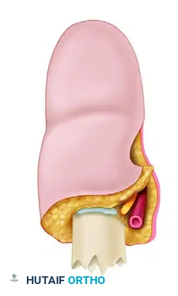
PART II: CHRONIC UNREDUCED DISLOCATIONS OF THE PATELLA
While the hip represents a major weight-bearing ball-and-socket joint, chronic unreduced dislocations of the patellofemoral joint present an entirely different biomechanical challenge. Old, unreduced lateral dislocations of the patella are relatively uncommon in adults but may arise from neglected acute trauma, severe untreated congenital dysplasia, or progressive neuromuscular disorders.
Pathoanatomy and Biomechanics
In a chronic lateral patellar dislocation, the extensor mechanism undergoes profound adaptive changes:
* Lateral Structures: The lateral retinaculum, vastus lateralis, and iliotibial band become severely contracted and fibrotic, tethering the patella to the lateral femoral condyle.
* Medial Structures: The medial patellofemoral ligament (MPFL) and the vastus medialis obliquus (VMO) are stretched, attenuated, and mechanically incompetent.
* Articular Cartilage: Prolonged abnormal tracking leads to severe chondromalacia or complete eburnation of the patellar and trochlear articular surfaces.
* Bony Alignment: The tibial tuberosity is often laterally translated, resulting in an abnormally high Q-angle.
To restore knee kinematics, a comprehensive realignment of the extensor mechanism is mandatory. The following technique (historically referenced as Technique 61-3) details the extensive soft-tissue and bony reconstruction required.
Comprehensive Extensor Mechanism Realignment
Indications
- Chronic, fixed lateral dislocation of the patella.
- Failure of conservative management in recurrent subluxators with fixed lateral contractures.
- Severe patellofemoral maltracking requiring both proximal soft-tissue and distal bony realignment.
Surgical Pitfall: Attempting to reduce a chronic patellar dislocation with a medial plication alone, without an adequate lateral release and distal realignment, will inevitably result in catastrophic failure, suture pull-out, and immediate redislocation.
Step-by-Step Surgical Technique
- Incision and Exposure:
- Make a 7.5-cm to 10-cm longitudinal midline incision over the anterior knee, extending from the superior pole of the patella to the tibial tuberosity. A midline incision is highly versatile, allowing extensive access to both the medial and lateral aspects of the extensor mechanism without creating large skin flaps that are prone to necrosis.
-
Dissect laterally, deep to the subcutaneous tissue, to expose the lateral retinaculum and the vastus lateralis insertion.
-
Lateral Release and Mobilization:
- Incise the capsule and synovium parallel with the lateral border of the quadriceps tendon, the patella, and the patellar tendon. This release must be extensive, often extending proximally into the vastus lateralis fascia to fully mobilize the extensor mechanism.
- Free the deep surfaces of the quadriceps tendon and the patella from any intra-articular adhesions to the lateral femoral condyle.
-
Manually reduce the patella into the trochlear groove. It should sit in the groove without requiring manual pressure. If it springs back laterally, further proximal lateral release is required.
-
Medial Capsular Management:
- Once the patella is centralized, the medial capsule will be grossly redundant.
- Excise the redundant, attenuated portion of the capsule from the medial side of the knee.
-
Close the capsule on the medial side using heavy, non-absorbable sutures (e.g., #2 FiberWire or Ethibond) in a pants-over-vest or imbricated fashion to provide a robust medial restraint.
-
Vastus Medialis Obliquus (VMO) Advancement:
- It is critical that the general alignment of the extensor mechanism be biomechanically optimized at the completion of the procedure to prevent lateral redislocation.
- The fibers of the vastus medialis muscle must be appropriately oriented (approximately 50 to 55 degrees relative to the femoral shaft) to exert a dynamic medial pull on the patella.
-
This frequently requires the sharp detachment, distal advancement, and reattachment of the VMO to the superomedial pole of the patella and the adductor tubercle of the femur.
-
Distal Realignment (Tibial Tuberosity Transfer):
- If the Q-angle remains excessive (>20 degrees) after proximal soft-tissue balancing, a distal bony realignment is necessary.
- Perform an osteotomy of the tibial tuberosity (e.g., Elmslie-Trillat or Fulkerson osteotomy).
- Transfer the tibial tuberosity medially (and anteriorly if patellofemoral offloading is desired) to centralize the distal portion of the extensor mechanism.
-
Rigidly fix the transferred bony block with two cortical lag screws.
-
Management of the Degenerated Patella (Patellaplasty vs. Patellectomy):
- Inspect the articular surface of the patella. In chronic dislocations, the cartilage is often completely destroyed.
- Patellaplasty: If the cartilage damage is partial, perform a chondroplasty, removing loose flaps and contouring the remaining bone.
- Patellectomy: If the articular surface is entirely degenerated and the subchondral bone is exposed and sclerotic, a partial or total patellectomy may be necessary to relieve intractable anterior knee pain.
- Crucial Biomechanical Note: Realignment of the extensor mechanism is just as important after a patellectomy as it is after an open reduction of the patella. Even without the patella, the quadriceps tendon will subluxate laterally over the femoral condyle if the lateral release and medial imbrication are not meticulously performed.
Postoperative Protocol
- Immobilization: The knee is immobilized in a hinged knee brace locked in full extension for the first 2 to 4 weeks to protect the medial soft-tissue repair and the tibial tuberosity osteotomy.
- Weight Bearing: Touch-down weight bearing is permitted immediately, advancing to partial and full weight bearing over 6 weeks as radiographic evidence of tuberosity healing is observed.
- Rehabilitation: Passive and active-assisted range of motion (0 to 90 degrees) is initiated early (usually within the first 2 weeks) to prevent arthrofibrosis. Isometric quadriceps strengthening is begun immediately, with progressive resistance exercises introduced after 6 weeks.
CONCLUSION
The surgical management of chronic unreduced dislocations requires a profound understanding of altered pathoanatomy and joint biomechanics. In the hip, the Nagi modified subcapital displacement osteotomy provides an elegant salvage solution that restores ambulatory capacity while strategically preserving the femoral neck for future arthroplasty. In the knee, chronic patellar dislocations demand a meticulous, multi-level reconstruction of the extensor mechanism, combining extensive lateral releases, medial imbrications, and distal bony transfers. By adhering to these strict, evidence-based surgical principles, the orthopedic surgeon can successfully salvage these complex, neglected injuries and restore meaningful function to the patient.
You Might Also Like