Acute Hand Injuries: Principles of Management and Soft Tissue Coverage
Key Takeaway
Acute hand injuries require a systematic approach prioritizing meticulous debridement, skeletal stabilization, and adequate soft tissue coverage. While primary nerve and tendon repairs are valuable, they remain secondary to preventing infection and achieving a closed, stable wound. This guide details the critical order of tissue repair, indications for skin grafts, and advanced flap techniques essential for restoring optimal hand function and anatomy in the acute trauma setting.
Introduction to Acute Hand Injuries
The hand and fingers are the most frequently injured body parts in both occupational and domestic settings. In the United States alone, more than one million emergency department visits annually are attributed to work-related hand trauma. The ultimate goal in the management of the acutely injured hand is the maximal restoration of function.
To achieve this, the surgeon must adhere to strict foundational principles: prevention of infection, salvage of viable tissues, and the promotion of primary healing. While the primary repair of nerves and tendons is highly desirable, these procedures are strictly secondary in importance to thorough cleansing, meticulous surgical debridement, rigid stabilization of fractures and dislocations, and the achievement of definitive wound closure or coverage.
Through exhaustive history taking and careful physical examination, the orthopedic surgeon must personally appraise the injury to determine which primary procedures can be executed safely and which reconstructive efforts must be delayed for secondary procedures.
Initial Assessment and Preparation
History and Physical Examination
A detailed history is paramount. The mechanism of injury (crush, avulsion, sharp laceration, high-pressure injection) dictates the zone of injury and the potential for tissue necrosis. The surgeon must document the exact time of injury, hand dominance, patient occupation, and any relevant comorbidities (e.g., diabetes, smoking) that may compromise microvascular perfusion.
Physical examination must be systematic, evaluating the hand's resting cascade, vascular perfusion (capillary refill, Allen test), and neurologic status (two-point discrimination) prior to the administration of any local anesthetic.
Clinical Pearl: Always assess and document two-point discrimination and motor function before injecting local anesthesia. Once a digital block is administered, accurate neurologic assessment becomes impossible.
Anesthesia and Tourniquet Application
Optimal surgical management requires a painless, bloodless field. Depending on the extent of the injury, anesthesia may range from local digital blocks to regional brachial plexus blocks or general anesthesia.
A pneumatic tourniquet is indispensable for identifying vital structures and preventing iatrogenic injury during exploration. The arm is exsanguinated using an Esmarch bandage (unless contraindicated by severe infection or malignancy), and the tourniquet is typically inflated to 250 mm Hg, or 100 mm Hg above the patient's systolic blood pressure.
Surgical Warning: Tourniquet time should generally not exceed 120 minutes. If prolonged surgery is anticipated, the tourniquet must be deflated for 15 to 20 minutes to allow for reperfusion before re-inflation.
Cleansing, Draping, and Debridement
The foundation of acute hand trauma surgery is debridement. The hand is prepped with a broad-spectrum antiseptic, and the wound is irrigated copiously with sterile saline. All devitalized tissue, foreign bodies, and severely contaminated structures must be excised. Skin edges should be sharply debrided to healthy, bleeding dermis.
Surgical Principles in Acute Hand Trauma
Order of Tissue Repair
In complex, multi-tissue hand injuries, a standardized sequence of repair minimizes the disruption of previously repaired structures and ensures a stable foundation. The universally accepted order of tissue repair is:
- Bone and Joint Stabilization: Rigid skeletal fixation (using K-wires, plates, or external fixators) restores the anatomical framework.
- Extensor Tendons: Repaired next as they are dorsal and less prone to disruption during subsequent volar repairs.
- Flexor Tendons: Repaired using core and epitendinous sutures.
- Arteries: Microvascular anastomosis to restore perfusion.
- Nerves: Epinurial or group fascicular repair without tension.
- Veins: Repaired to ensure adequate venous outflow and prevent congestion.
- Skin Coverage: Direct closure, grafting, or flap coverage.
Considerations for Amputation
Not all injured digits are salvageable. The decision to amputate versus replant or salvage depends on the level of injury, the mechanism (crush/avulsion vs. sharp), ischemic time, and patient factors. Absolute indications for completion amputation include irreversible ischemia with prolonged warm ischemia time (>12 hours for digits), severe multilevel crush injuries, and life-threatening systemic instability.
Arterial Injuries
Arterial injuries in the hand require prompt recognition. If the superficial palmar arch or common digital arteries are lacerated, primary microvascular repair with 8-0 or 9-0 nylon is indicated to prevent cold intolerance and claudication, even if the digit appears viable via collateral flow. If a tension-free repair is impossible, reversed interposition vein grafts (typically harvested from the distal forearm or foot) are required.
Soft Tissue Coverage: Principles and Techniques
Considerations for Skin Closure
The primary objective of skin closure is to achieve a healed wound without tension, thereby preventing infection, protecting underlying neurovascular and tendinous structures, and allowing early mobilization.
Methods and Indications for Skin Closure
- Direct Suture: Indicated for clean, linear lacerations where skin edges can be approximated without tension.
- Skin Grafts: Indicated for superficial defects where the wound bed is well-vascularized (muscle, fascia, or healthy granulation tissue). Grafts will not survive over bare bone without periosteum, bare tendon without paratenon, or bare nerve.
- Skin Flaps: Indicated for deep defects exposing bare bone, joints, tendons, or neurovascular bundles. Flaps bring their own blood supply, facilitating healing in avascular beds.
Free Skin Grafts
Split-Thickness Skin Grafts (STSG)
STSGs include the epidermis and a variable portion of the dermis. They are highly reliable and have a lower metabolic demand, allowing them to survive in less-than-ideal wound beds.
Obtaining Skin Grafts with a Dermatome:
STSGs are typically harvested from the anterior or lateral thigh using an air or electric dermatome. The thickness is usually set between 0.012 and 0.015 inches. The graft may be meshed (e.g., 1.5:1 ratio) to allow for the egress of hematoma/seroma and to expand the graft, though unmeshed (sheet) grafts provide superior cosmetic results on the dorsum of the hand.
Pitfall: Failure to achieve absolute hemostasis in the recipient bed is the most common cause of skin graft failure, as a hematoma prevents plasmatic imbibition and subsequent revascularization.
Free Full-Thickness Grafts (FTSG)
FTSGs contain the epidermis and the entire dermis. They contract less than STSGs, provide better durability, and offer superior cosmetic matches for the palmar surface of the hand.
Donor Sites:
For palmar defects, glabrous skin from the hypothenar eminence or the plantar instep is ideal. For dorsal defects, the groin crease, lower abdomen, or medial arm provides excellent donor sites that can be closed primarily. FTSGs must be meticulously defatted prior to application to ensure graft take.
Skin Flaps for Hand Coverage
When the wound bed cannot support a free graft, flap coverage is mandatory. Flaps are categorized by their blood supply (random vs. axial) and their proximity to the defect (local, regional, distant).
Local Flaps
Local flaps utilize adjacent tissue, providing an excellent color and texture match.
Local Flaps in Fingers:
* V-Y Advancement Flap (Atasoy): Ideal for transverse or dorsal oblique fingertip amputations. A V-shaped volar flap is advanced distally and closed in a Y-configuration.
* Moberg Volar Advancement Flap: Used primarily for the thumb. The entire volar skin is elevated with both neurovascular bundles and advanced distally to cover tip defects up to 1.5 cm.
Cross Finger Flaps:
Indicated for volar defects of the digits exposing bare flexor tendon or bone. A random-pattern flap is raised from the dorsum of the adjacent intact finger (cleaving above the paratenon of the extensor mechanism) and hinged laterally to cover the volar defect of the injured finger. The donor site is covered with an STSG. The flap is divided and inset at 2 to 3 weeks.
Forearm Flaps for Hand Coverage
Regional flaps from the forearm provide robust, single-stage coverage for large hand defects.
Radial Forearm Flap:
A highly versatile fasciocutaneous flap based on the radial artery and its venae comitantes. It can be raised as a pedicled reverse-flow flap to cover massive dorsal or volar hand defects.
* Prerequisite: A normal Allen test confirming that the hand will remain adequately perfused by the ulnar artery once the radial artery is sacrificed.
* Surgical Technique: The flap is designed on the volar forearm. The radial artery and venae comitantes are ligated proximally and elevated with the fascia. The superficial radial nerve must be carefully identified and preserved. The donor site usually requires STSG coverage.
Posterior Interosseous Flap:
A reverse-flow fasciocutaneous flap based on the posterior interosseous artery (PIA), anastomosing with the anterior interosseous artery distally. It is ideal for covering defects on the dorsum of the hand and the first web space without sacrificing a major artery to the hand.
Distant Flaps: Abdominal and Groin Flaps
When local or regional options are exhausted or contraindicated, distant flaps are utilized.
Random Pattern Abdominal Pedicle Flaps:
Historically used for large hand defects, these flaps rely on the subdermal plexus. They require a broad base to ensure viability and necessitate the hand being attached to the abdomen for 3 to 4 weeks, leading to significant joint stiffness.
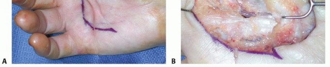
Axial Pattern Flaps (The Groin Flap):
The groin flap is an axial pattern flap based on the superficial circumflex iliac artery (SCIA). It provides a massive amount of skin and subcutaneous tissue.
* Indications: Extensive degloving injuries of the hand or multiple digit amputations requiring soft tissue banking.
* Technique: The flap is centered over the SCIA, originating just below the inguinal ligament and extending toward the anterior superior iliac spine. The flap is tubed, and the hand is inset. Division and final inset are performed at 3 weeks.
Filleted Grafts (Spare Parts Concept)
In severe crush injuries where a digit is rendered non-salvageable (e.g., irreparable neurovascular damage) but its skin envelope remains viable, the digit can be "filleted." The bone and tendons are excised, and the remaining vascularized skin flap is unfolded to cover adjacent defects. This "spare parts" approach preserves valuable, highly specialized glabrous skin.
Management of Scars and Granulating Areas
Granulating Areas
Wounds left to heal by secondary intention or those with failed primary coverage will develop granulation tissue. While healthy granulation tissue can accept a skin graft, chronic granulating wounds often harbor high bacterial loads and become fibrotic. These areas must be sharply excised down to healthy tissue before definitive coverage with grafts or flaps is attempted.
Scars
Post-traumatic scars in the hand can lead to severe functional impairment, particularly if they cross flexion creases, resulting in burn-like contractures.
Methods of Correcting Linear Scars:
Linear scars crossing joint creases are prone to longitudinal contracture. The Z-plasty is the workhorse technique for correcting this. By transposing two triangular flaps, the Z-plasty achieves two goals: it lengthens the contracted scar and reorients the scar line so it no longer crosses the crease perpendicularly.
Methods of Correcting Area Scars:
Broad, flat area scars (often resulting from avulsion injuries or burns) restrict the gliding of underlying tendons and limit joint mobility. These scars must be completely excised. If the underlying bed is well-vascularized, a thick FTSG is applied. If tendons or joints are exposed following scar excision, flap coverage (e.g., a radial forearm flap or groin flap) is required to restore a supple, gliding soft tissue envelope.
Postoperative Protocols and Rehabilitation
The success of acute hand trauma surgery relies heavily on postoperative rehabilitation.
* Immobilization: The hand is initially immobilized in the "intrinsic plus" or "safe" position (wrist extended 20-30 degrees, MCP joints flexed 70-90 degrees, IP joints fully extended) to prevent collateral ligament contracture.
* Elevation: Strict elevation above the level of the heart is mandatory to minimize edema, which can compromise microvascular perfusion and lead to stiffness.
* Therapy: Early, controlled mobilization under the guidance of a certified hand therapist is initiated as soon as the skeletal and soft tissue repairs allow. Tendon gliding exercises and edema control are critical to achieving the ultimate goal: the restoration of a functional, painless hand.
You Might Also Like