DEFINITION
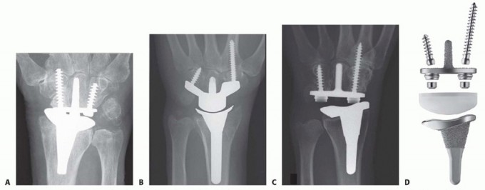
The wrist is a common site for end-stage joint degeneration, particularly in patients with rheumatoid disease. Osteoarthritis and posttraumatic arthritis following distal radius fractures, scaphoid nonunion advanced collapse (SNAC), and scapholunate advanced collapse (SLAC) are other common causes for advanced arthritis.The gold standard of treatment for severe wrist arthritis has historically been complete wrist arthrodesis. Although arthrodesis provides good pain relief and durability, it is associated with substantial functional loss, especially if both wrists have arthritis.1,9,13Total wrist arthroplasty is a motion-preserving alternative to arthrodesis that provides excellent pain relief. Sufficient motion and strength is retained for activities of daily living.Preservation of wrist joint motion is of particular importance for patients who are debilitated by arthritis affecting multiple joints and those with specific joint motion requirements.9Similar to arthroplasty in other joints, early wrist arthroplasty implants had poor long-term survivorship.2,3,7Wrist arthroplasty has continually improved since the introduction of articulated implants more than 40 years ago. Advancements in design include distal component fixation being primarily within the carpus and not the metacarpal; intercarpal fusion to provide broad, solid support for the component; screw augmentation for carpal component fixation; minimal bone resection; preservation of the wrist capsule; cementless fixation; a broad semiconstrained ellipsoid articulation; and an option to preserve the distal radioulnar joint (DRUJ).FIG 1•A-D.Total wrist arthroplasty implants (fromlefttoright): Re-Motion (Small Bone Innovations), Maestro (Biomet), Universal 2 (Integra LifeSciences), and Freedom (Integra LifeSciences).Through improved materials, designs, and fixation techniques, total wrist arthroplasty has emerged as aviable option for selected patients with end-stage wrist arthritis.Regardless of the desire for arthroplasty, patients must commit to a lifetime of restricted activities to obtain a durable outcome.There are currently three total wrist implant systems in the United States: Re-Motion (Small Bone Innovations, Inc., Morrisville, PA), Maestro (Biomet, Warsaw, IN), and most recently Freedom (Integra LifeSciences, Plainsboro, NJ) (FIG 1). The Freedom wrist system evolved from its predecessor, the Universal 2 (Integra LifeSciences).The Re-Motion wrist offers a mobile bearing attached to the carpal component that theoretically improves motion and load transfer, thus reducing stresses known to contribute to loosening.The Maestro allows complete resection of the proximal carpal row and has a polyethylene surface proximally. The system is also approved for hemiarthroplasty using the distal component alone.The Freedom wrist is the newest design, with a more anatomic articulation that provides physiologic wrist motion, improved bone fixation, and precise instrumentation.
ANATOMY
The wrist joint consists of the distal radial articular surface, distal ulna, triangular fibrocartilage complex (TFCC), eight carpal bones arranged in two rows (proximal and distal), and five metacarpal bases.There are four major wrist articulations: radiocarpal (and ulnocarpal), midcarpal, carpometacarpal (CMC), and distal radioulnar joints.P.999In addition to the wrist capsule, multiple interosseous, intrinsic and extrinsic ligaments provide joint stability, with intrinsic ligaments referring to those between carpal bones and located primarily within the joint and extrinsic ligaments located within the joint capsule.Normal radiographic parameters of the distal articular surface of the radius include 11 degrees of volar tilt, 22 degrees of radial inclination, and 11 mm of radial height.Ulnar variance refers to the length of ulna relative to the radius, with positive variance indicating the ulna is longer. Approximately 70% of the population is ulnar neutral.The sigmoid notch of the distal radius provides the radial articulation for the DRUJ. The DRUJ is stabilized by dorsal and volar radioulnar ligaments. Current implant systems are designed to preserve both the joint surfaces and ligaments of the DRUJ.The center of wrist motion is located near the center of the head of the capitate.
PATHOGENESIS
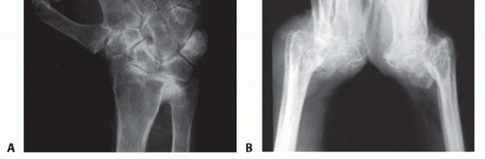
FIG 2 • A. PA radiograph of a rheumatoid wrist showing ulnocarpal translocation and radial deviation deformity. B. Lateral view of a rheumatoid wrist showing severe arthritis with volar subluxation of the carpus. Posttraumatic arthritis may develop years after an intraarticular distal radius fracture or fracture-dislocation of the carpus. In regard to implant arthroplasty, a malunion of the radius presents additional surgical challenges, but with proper planning and technique, a successful outcome is possible.SNAC and SLAC wrist conditions are the most common causes of nonrheumatoid wrist arthritis, often with predictable degenerative patterns but only modest deformity.
NATURAL HISTORY
End-stage wrist arthritis, no matter the cause, is a painful condition resulting in progressive stiffness and diminished function.In addition to pain and functional loss, deformity may be a cosmetic concern for patients.Inflammatory arthritis may cause severe deformity and bone loss, precluding wrist implant arthroplasty.
PATIENT HISTORY AND PHYSICAL FINDINGS
The history should confirm the presence of substantial pain that indicates the need for wrist arthroplasty.Age, activity desires, hand dominance, presence of contralateral wrist arthritis, use of walking aids, and occupation are important factors in the preoperative assessment.In rheumatoid patients, disease activity should be optimally controlled medically prior to surgical treatment because highly active disease reduces the durability of an arthroplasty.12Lower limb surgery, such as total hip or knee arthroplasty, should be done prior to wrist replacement surgery to avoid weight bearing through the wrist implant during rehabilitation.The ideal candidate for total wrist arthroplasty is an elderly patient with a low-demand lifestyle who desires pain reliefP.1000and can accept modest motion and strength and be willing to avoid stressful use.Younger patients may qualify for wrist arthroplasty if activities can be modified, particularly when the nondominant wrist is involved.
IMAGING AND OTHER DIAGNOSTIC STUDIES
Standard posteroanterior (PA), lateral, and oblique views of the wrist are adequate to assess the extent of disease, alignment, and bone stock.In patients with rheumatoid arthritis, cervical spine radiographs are indicated to assess for instability.
DIFFERENTIAL DIAGNOSIS
Rheumatoid arthritis Posttraumatic arthritisOsteoarthritis, including SLAC and SNAC wrists Avascular necrosis (eg, Kienböck disease) Other inflammatory arthritis (eg, psoriatic)
NONOPERATIVE MANAGEMENT
Conservative treatment for severe wrist arthritis includes activity modification, bracing, nonsteroidal anti-inflammatory medications, and corticosteroid injections.Failure of pain relief by conservative treatment or progression of deformity are indications for surgical treatment.
SURGICAL MANAGEMENT
Relative contraindications include poor bone stock, lack of wrist motor control, and wrist instability due to severely damaged tendons or ligaments,Active infection locally or systemically is an absolute contraindication to total wrist arthroplasty. Inflammatory arthritis should be well controlled medically prior to proceeding with surgical treatment.
PREOPERATIVE PLANNING
If severe bone loss or erosive disease is suspected but not confirmed by preoperative imaging, then the surgical permit should include other options such as arthrodesis. Proper instruments and devices should be available.The proper implant size is estimated using PA and lateral wrist x-rays; however, final sizing is determined intraoperatively.The DRUJ is assessed preoperatively for arthritis and instability.A partial or complete resection of the distal ulna is planned when needed. If the DRUJ is unaffected, then the DRUJ is not exposed.
POSITIONING
The patient is positioned supine with the affected limb resting on a hand table. A padded tourniquet is placed on the upper arm.Prophylactic antibiotics are administered intravenously.
TECHNIQUES
FREEDOM TOTAL WRIST PROSTHESIS
DORSAL APPROACH

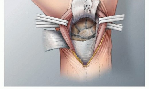
TECH FIG 1 • To expose the joint, the extensor retinaculum is elevated as a radially based flap and the joint capsule is raised as a distally based flap.
PREPARATION OF THE CARPUS
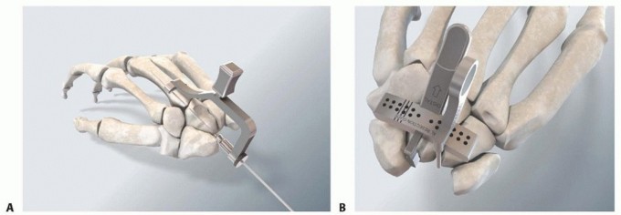
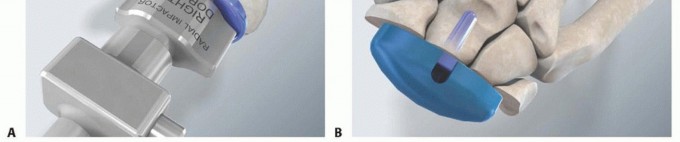
TECH FIG 2 • A. The lunate is excised to expose the capitate. The modular drill guide is applied with the barrel pressed against the center of the capitate head and the saddle on the third metacarpal. A guidewire is inserted through the center of the capitate into the third metacarpal. B. Insert the carpal guide bar into the capitate and mount the resection guide with the hamate feeler just touching the proximal pole of the hamate.(continued)

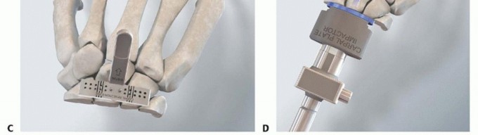
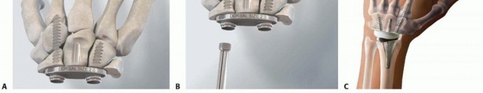
TECH FIG 2 •(continued)C. Secure the resection guide to the carpus and ensure the osteotomy will pass through the proximal pole of the hamate, a small portion of the capitate head, and approximately half of the scaphoid and triquetrum (avoid excessive carpal resection). D. Impact the carpal plate trial into the capitate and confirm that it seats properly at the osteotomy site.
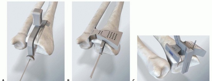
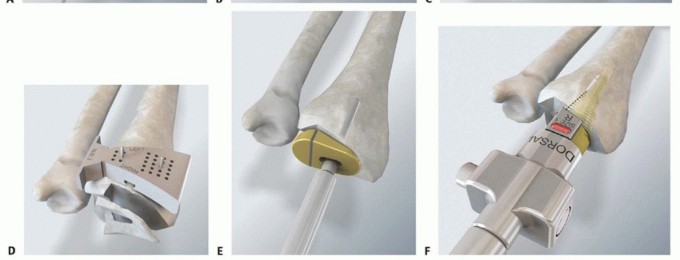
TECH FIG 3 • A. Using the modular drill guide, a guidewire is inserted through the articular surface of the radius and down the center of the radius canal (confirm correct position with fluoroscopy). Drill the hole for the guide rod using the cannulated drill bit. B. Apply the radius resection guide in a position that will resect only the articular surface of the distal radius (a laser mark on the guide arm corresponds to the level where the barrel is pressed against the articular surface). C. Use the radius score guide to make an initial vertical cut, which will mark the ulnar extent of the horizontal cut to protect the DRUJ. D. Complete the distal radius articular surface resection. E. After reinserting the guide rod, use the radius drill guide followed by the box punch to remove the remaining hard subchondral bone of the distal radius. F. Perform sequential broaching to the appropriate size.
TRIAL REDUCTION

TECH FIG 4 • A. Insert the radius trial component into the prepared radius. B. Insert the carpal component and apply the trial poly bearing.
IMPLANTATION

TECH FIG 5 • A. Impact the carpal component and insert the fixation screws, beginning with the radial screw using the modular drill guide, guidewire, and cannulated drill for preparation. B. Apply the locking caps over radial and ulnar carpal screws. C. Impact the radial component, apply the appropriate poly bearing, reduce the joint, and assess range of motion and joint stability. Drill to the proper depth with a cannulated drill bit and insert a 4.5-mm screw of the measured depth.Using a similar technique for the ulnar screw, position the modular drill guide barrel in the ulnar hole of the carpal component and its saddle on the fourth metacarpal shaft. Insert a guidewire, measure, anddrill. Insert a screw of proper length. Do not penetrate the fourth CMC joint (TECH FIG 5A). Apply locking caps over the two carpal screw heads (TECH FIG 5B).Confirm appropriate carpal poly thickness using carpal poly trials.Apply the carpal poly implant and impact it into place, ensuring there is no impinging soft tissue. Reduce the joint and make a final assessment of the balance and stability (TECH FIG 5C).7. Bone Graft and ClosureP.1005Perform an intercarpal fusion by carefully resecting portions of the joint surfaces and then inserting cancellous bone chips from the previously resected bone into the prepared fusion sites.Reattach the dorsal capsule to the dorsal rim of the radius using the previously placed sutures. The medial and lateral edges of the capsule are also closed.If the capsule is insufficient for closure with the wrist flexed 30 degrees, it is supplemented with a portion of the extensor retinaculum.A suction drain is placed, skin is closed, and a bulky dressing with a below-elbow plaster splint is applied.Indications1. Wrist degeneration resulting from all causes of arthritis serves as an indicationfor this surgery; however, patients should be of appropriate age and have fewer activity demands.2. Severe loss of bone stock and active and aggressive inflammatory arthritis are contraindications.Surgicaltechnique1. Adequate exposure is mandatory to achieve accurate component implantation.2. Intercarpal arthrodesis is a key factor in obtaining long-term fixation of the implants.DRUJ ▪ Preservation of the DRUJ including the TFCC improves stability of the wrist andmanagement overall functional outcome.1. When treatment is needed, expose the DRUJ through the same skin incision but through a separate capsulotomy.
PEARLS AND PITFALLS
POSTOPERATIVE CARE
Strict elevation and early active digital range of motion are encouraged.At 2 weeks postoperatively, the sutures are removed. Implant reduction is confirmed by x-ray and gentle wrist exercises are begun, including active flexion, extension, radial and ulnar deviation, and pronation and supination. A removable splint is applied.The splint is weaned at 4 weeks postoperatively, and strengthening is added to the rehabilitation. The patient is advised against impact loading of the wrist and repetitive forceful use of the hand.
OUTCOMES
Long-term outcomes of modern generation implants have been reported with the first generation Universal implant by Ward et al.12Carpal component loosening was found to be the most common reason for revision surgery. Survival of stable implants was found to be 75% at 5 years and 60% at 7 years.Early failures in this series of patients all had highly active inflammatory arthritis and severe wrist laxity, demonstrating the importance of medical control of rheumatoid arthritis.Cooney et al2 performed a retrospective review comparing the 16 Biaxial resurfacing implants to a series of 30 anatomic implants (Re-Motion and Universal 2). At an average of 6 years follow-up, they found that 50% of the Biaxial implants had failed, whereas 1 out of 30 of the new generation implants had failed indicating significant improvements in implant design.The Universal 2 implant has been reported in several midterm follow-up series with positive results.Ferreres et al,4 Morapudi et al,8 and van Winterswijk11 reported three separate series with follow-up between 3 and 5 years.All studies demonstrated high levels of patient satisfaction and improved postoperative standardized outcome scores (Disability of the Arm, Shoulder, and Hand [DASH] and Patient-Rated Wrist Evaluation [PRWE]).There was only 1 revision in the combined total of 57 wrists in these series. Average arc of motion ranged from 52 to 68 degrees; average motion was improved postoperatively in each study.P.1006The Maestro prosthesis has shown promising results in short follow-up.Nydick et al10 retrospectively reviewed 23 total wrist arthroplasties at mean 28-month follow-up and found pain scores improved from 8.0 to 2.2. A 30% complication rate was reported in this series. There was only one failure due to infection.Results of the Re-Motion prosthesis reported by Herzberg5 showed good to excellent outcomes in 16 of 20 wrists at an average 32-month follow-up.These results were part of a larger multicenter web-based database which included 215 wrists. At average follow-up of 4 years, they reported a 96% survival rate in patients with rheumatoid arthritis and a 92% survival in nonrheumatoid arthritis patients. Average postoperative motion was 60.5degrees, and there were significant improvements in postoperative pain scores.6The Freedom total wrist has yet to be reported in clinical follow-up; however, it has been designed based on the successful concepts of the Universal 2 implant. Improvements in design aim to decrease rates of loosening and increase range of motion.
COMPLICATIONS
Superficial or deep infection Stiffness or contracture Joint imbalance or instabilityImplant loosening
REFERENCES
- Adey L, Ring D, Jupiter JB. Health status after total wrist arthrodesis for posttraumatic arthritis. J Hand Surg Am 2005;30(5):932-936.
- Cooney W, Manuel J, Froelich J, et al. Total wrist replacement: a retrospective comparative study. J Wrist Surg 2012;1(2):165-172.
- Dennis DA, Ferlic DC, Clayton ML. Volz total wrist arthroplasty in rheumatoid arthritis: a long-term review. J Hand Surg Am 1986;11(4):483-490.
- Ferreres A, Lluch A, Del Valle M. Universal total wrist arthroplasty: midterm follow-up study. J Hand Surg Am 2011;36(6):967-973.
- Herzberg G. Prospective study of a new total wrist arthroplasty: short term results. Chir Main 2011;30(1):20-25.
- Herzberg G, Boeckstyns M, Sorensen AI, et al. “Remotion” total wrist arthroplasty: preliminary results of a prospective international multicenter study of 215 cases. J Wrist Surg 2012;1(1):17-22.
- Kistler U, Weiss AP, Simmen BR, et al. Long-term results of silicone wrist arthroplasty in patients with rheumatoid arthritis. J Hand Surg Am 2005;30(6):1282-1287.
- Morapudi SP, Marlow WJ, Withers D, et al. Total wrist arthroplasty using the universal 2 prosthesis. J Orthop Surg 2012;20(3): 365-368.
- Murphy DM, Khoury JG, Imbriglia JE, et al. Comparison of arthroplasty and arthrodesis for the rheumatoid wrist. J Hand Surg Am 2003;28(4):570-576.
- Nydick JA, Greenberg SM, Stone JD, et al. Clinical outcomes of total wrist arthroplasty. J Hand Surg Am 2012;37(8):1580-1584.
- van Winterswijk PJ, Bakx PA. Promising clinical results of the universal total wrist prosthesis in rheumatoid arthritis. Open Orthop J 2010;4:67-70.
- Ward CM, Kuhl T, Adams BD. Five to ten-year outcomes of the universal total wrist arthroplasty in patients with rheumatoid arthritis. J Bone Joint Surg Am 2011;93(10):914-919.
- Weiss AC, Wiedeman G Jr, Quenzer D, et al. Upper extremity function after wrist arthrodesis. J Hand Surg Am 1995;20(5):813-817.
