 .
.
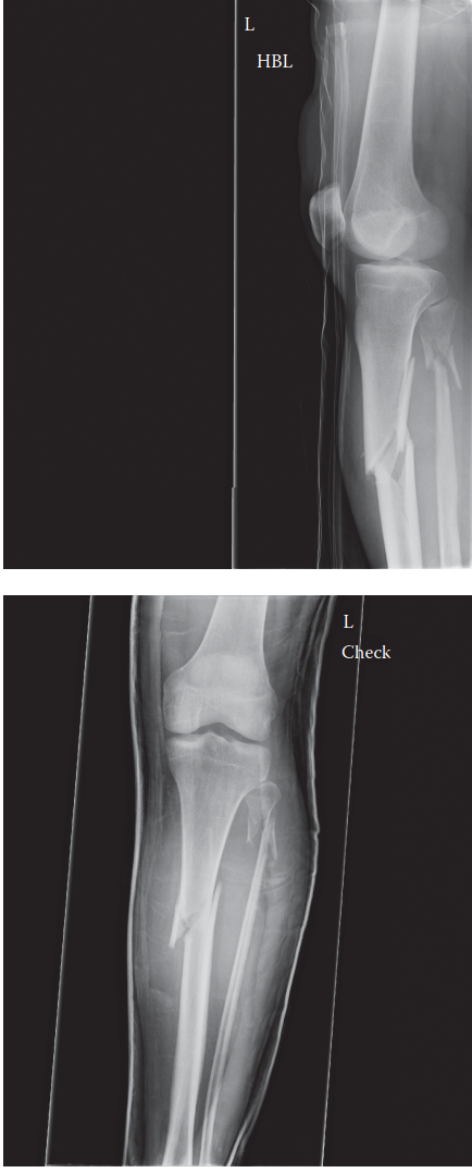
Can you describe the radiographs?
These are AP and lateral radiographs of the left tibia and fibula showing a proximal tibial diaphyseal fracture. The fracture is in valgus and procurvatum (apex anterior).
What is responsible for the deformities seen here?
The procurvatum is due to the unopposed pull of the patellar tendon, whereas the valgus deformity is due to the pull of the pes anserinus attached to the proximal fragment, and the bulky anterior compartment musculature preventing the fracture from displacing into a varus configuration. If you were to treat this fracture with an IM nail, what techniques could you use to counteract these deformities?
There are several techniques one could utilise to prevent this valgus and procurva- tum deformity. Since the nail is not in contact with the cortical bone at the level of the fracture, the fragment can displace until the posterior or lateral cortex lies in contact with the nail:
Poller
*
blocking screws: These are bicortical screws inserted before reaming and nailing. Alternatives include thick K-wires or 3/16-inch smooth Steinmann Pins. The screw/wire/pin blocks the incorrect path of the nail and channels the nail along the correct path preventing a mal-union. In the case of the fracture pictured above, a poller screw placed posteriorly will prevent the procurvatum deformity and a lateral screw will prevent the valgus deformity.
Unicortical plate: A mini-open approach can be utilised to reduce the fracture and maintain fixation using a small fragment dynamic compression plate with unicortical fixation.
Lateral starting point: A lateral parapatellar approach and lateral proximal starting point will allow the nail to abut the lateral cortex of the proximal frag- ment and prevent a valgus deformity.
Semi-extended nailing: Nailing with the knee in flexion can exaggerate any procur- vatum deformity. Most companies can now supply instrumentation to allow for nail- ing of tibial fractures in a semi-extended position, preventing fracture malalignment.
L
 *
Poller
is German for bollard.
*
Poller
is German for bollard.
Tibial Fracture: Proximal Deformity Answers for Surgeons
Updated: Feb 2026
84 Views
Key Medical Takeaway
Learn more about Tibial Fracture: Proximal Deformity Answers for Surgeons and how to manage it. A fracture proximal tibia often presents with valgus and procurvatum deformities due to specific muscle pulls. The treatment answe to counteract these during IM nailing involves techniques like Poller blocking screws, unicortical plates, lateral starting points, or semi-extended nailing, ensuring proper alignment.

Keywords