DEFINITION
Primary osteoarthritis (OA) of the elbow is a relatively rare condition that has an idiopathic etiology, although it is frequently associated with heavy use of the arm.Unlike OA of other large joints, elbow OA is characterized by relatively preserved joint space and articular cartilage but with hypertrophic osteophyte formation and capsular contracture.Primary OA at the elbow is characterized by pain and lost motion, strength, and, ultimately, function.
ANATOMY
The elbow is a modified hinge joint composed of the ulnotrochlear, radiocapitellar, and proximal radioulnar articulations.The elbow has two planes of motion: flexion and extension in the sagittal plane and pronosupination in the axial plane.Flexion-extension comes from motion at the ulnotrochlear articulation and has normal range of motion of 0 degree of extension to 145 degrees of flexion.Pronosupination is a result of motion at the radiocapitellar and proximal radioulnar articulation and has a normal range of motion of 80 degrees of pronation and 80 degrees of supination.Collateral ligamentous complexes are present on the medial and lateral sides of the elbow that—along with the inherent bony geometry—confer static stabilization to the elbow.Lateral collateral ligamentous complex functions as a lateral stabilizer preventing posterolateral rotatory instability (PLRI) and is described as a Y-shaped confluence of three components.Radial collateral ligament—extends from the lateral epicondyle of the humerus to the annular ligamentLateral ulnar collateral ligament—extends from the lateral epicondyle of the humerus to the crista supinatoris of the ulnaAnnular ligament—forms a ring around the radial neck, extending from the anterior sigmoid notch of the ulna to the crista supinatorisMedial collateral ligament functions as the most important stabilizer to valgus stress and is composed of three components.Anterior oblique ligament—extends from the medial epicondyle of the humerus to the sublime tubercle of the anteromedial facet of the coronoidPosterior oblique ligament—extends from the medial epicondyle of the humerus and fans out to insert along the sigmoid notchTransverse ligament—this thin band extends transversely from the posterior to anterior margin of the sigmoid notch
PATHOGENESIS
The cause of primary OA is likely multifactorial with hand dominance, history of heavy use, race, and other factors contributing.2
NATURAL HISTORY
Primary OA is a relatively rare condition that commonly affects the dominant arm of middle-aged males who have a history of heavy use due to sports or heavy equipment use.The severity of the presentation is variable and dependent on disease progression.Early OA is characterized by pain at terminal extension and flexion with patients frequently complaining of pain with carrying a heavy object with the elbow extended.Radiographically, early disease is characterized by relatively preserved joint space and impinging osteophytes on the margins of anterior and posterior ulnotrochlear articulation as well as radiocapitellar osteophyte formation.Late-stage OA presents with pain throughout the arc of motion with loss of articular cartilage and joint space narrowing.
PATIENT HISTORY AND PHYSICAL FINDINGS
Primary OA of the elbow is unique amongst OA of other joints, in that the disease severity and anatomic source of pathology allow for a variety of successful treatment options ranging from simple débridement to arthroplasty.
HISTORY
The classic presentation is a middle-aged male with a history of heavy use who presents with symptoms on his dominant side.Symptoms may include pain, loss of motion, mechanical symptoms of catching or locking, or weakness.When pain is the primary complaint, it is important to understand the degree of disability and impact on the patient's function; identify the anatomic source of pain whether ulnotrochlear, radiocapitellar, or extra-articular; and identify whether pain occurs at the extremes of flexion and/or extension or throughout the arc of motion.Patients with predominantly mechanical symptoms may need only arthroscopic débridement.In patients who primarily complain of lost motion, it is important to understand the degree to which this affects the patient and whether the lost motion is in flexion, extension, or both.Further history that is necessary to evaluate when considering arthroplasty include prior surgeries, history of trauma, history of infection, and the patient's demand level and posttreatment expectations.P.934
PHYSICAL EXAMINATION
The physical examination begins with inspection of the skin for prior surgical scars, other wounds, or evidence of infection.Flexion and extension as well as pronosupination range of motion should be assessed.It is essential to document at what point in the arc of motion pain is experienced as well as any catching, locking, or other mechanical symptoms.Rotation through the radiocapitellar joint may not generate significant pain or stiffness despite a degenerative appearance radiographically.Neurovascular examination specifically focused on evaluation for ulnar nerve compression at the elbow is essential to document.
IMAGING AND OTHER DIAGNOSTIC STUDIES
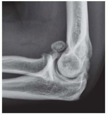
Anteroposterior (AP), lateral, and oblique radiographic views are adequate to make the diagnosis of OA.Characteristic findings on plain films include osteophyte and/or loose body formation emanating from the olecranon and coronoid processes and extending into their fossae (FIG 1).The radiocapitellar joint may also be affected and demonstrate osteophytes around the radial head.Typically, radiographic changes in the ulnotrochlear joint precede those in the radiocapitellar or proximal radioulnar joints.Computed tomography (CT) scan with three-dimensional reconstructions may be performed and is especially helpful for localizing osteophytes not well visualized on plain films.Impinging shelf osteophytes within the olecranon, radial, and coronoid fossae are best appreciated on CT and may be missed on plain films.Similarly, CT is helpful for identifying osteophytes in the medial gutter within close proximity to the ulnar nerve.
DIFFERENTIAL DIAGNOSIS
Posttraumatic arthritis of the elbow Rheumatoid or other inflammatory arthropathy Chronic septic arthritisCrystalline arthropathy Haemophilic arthropathy FIG 1 • Lateral x-ray demonstrating osteophyte formation, a loose body, and relatively preserved joint space.
NONOPERATIVE MANAGEMENT
Nonoperative management is appropriate for the early stages of the disease, when the patient reports mild pain and motion loss of less than 15 degrees.Nonoperative treatments include activity modification, nonsteroidal anti-inflammatory drugs, intra-articular corticosteroid injections, and physical therapy.Physical therapy should focus on pain control, antiinflammatory modalities, and preserving range of motion and strength.Intra-articular injections provide transient relief that is suitable for maintenance therapy.
SURGICAL MANAGEMENT
Total elbow arthroplasty (TEA) is considered in patients with the following:Disabling elbow OA in patients older than 65 yearsMid-arc pain with activity resulting from cartilage loss of the ulnotrochlear joint Willing to comply with low activity levels with their operative extremityProstheses come in several basic designs:Linked prosthesesLinked devices have mechanically linked ulnar and humeral components that function as a hinge. Contemporary designs are semiconstrained implants, which about 7 degrees of varus/valgus and rotational laxity at the articulation.Unlinked prosthesesUnlinked prostheses have no mechanical connection between the ulnar and humeral components and rely on the ulnar and humeral components' congruence and the capsuloligamentous structures for stability. They have the theoretical benefit of lower bone-cement interface stress leading to less loosening, although clinical data has not yet demonstrated this.Convertible prosthesesThese implants allow for use in either a linked or unlinked fashion.
PREOPERATIVE PLANNING
Full-length x-rays of the arm and forearm must be obtained and scrutinized for deformity, prior hardware, or pathologic lesions.Preoperative templates are available by many manufacturers and may be used as a guide for intraoperative component selection.The condition of the soft tissue must be assessed preoperatively, including prior surgical or traumatic scars. We avoid creating skin bridges of less than 1 cm when there is a prior scar. When any doubt exists about the quality of closure, arrangements should be made preoperatively to have a wound vacuum-assisted closure device available.Regional anesthetic infusion through an interscalene catheter is used throughout the perioperative period.
POSITIONING
The patient is positioned supine on a standard operating room table.A nonsterile tourniquet is applied below the drapes as proximal as possible on the arm.P.935The extremity distal to the tourniquet is prepped circumferentially; exposed skin is isolated with the use of an impervious stockinet and an Ioban (3M Health Care, St. Paul, MN) antimicrobial drape.The arm is brought over the patient's chest. A padded Mayo stand is brought into the operative field from the patient's contralateral side, and the arm may rest on the Mayo stand during surgery.
APPROACH
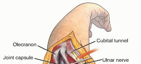
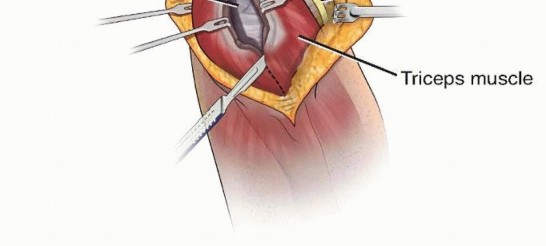
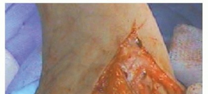
FIG 2 • Triceps-splitting approach: The triceps is split sharply in the midline to the olecranon process where it may be partially detached medially and laterally.
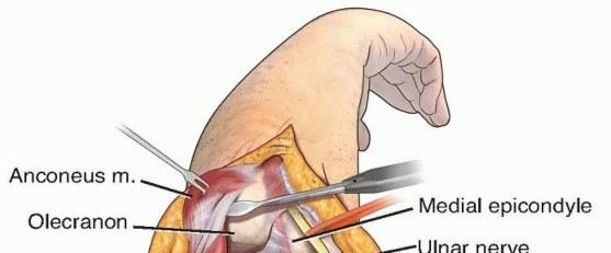
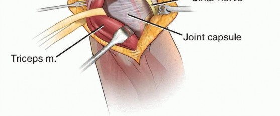
FIG 3 • Triceps-reflecting approach: The triceps in continuity with the antebrachial fascia, proximal ulnar periosteum, and anconeus are elevated and retracted laterally.
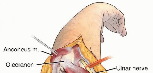
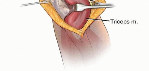
FIG 4 • Triceps-sparing approach: The triceps is elevated from the intermuscular septa and humerus medially and laterally and the anconeus is elevated from the humerus. Regardless of how the triceps is handled, the entire distal humerus must be visualized necessitating the elevation of the collateral ligaments and flexor/pronator and extensor masses from their origins on the medial and lateral epicondyles.The elbow may then be dislocated allowing access for bony preparation and prosthesis insertion.
TECHNIQUES
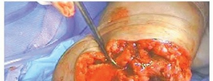
PREPARATION OF THE HUMERUS
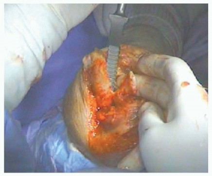
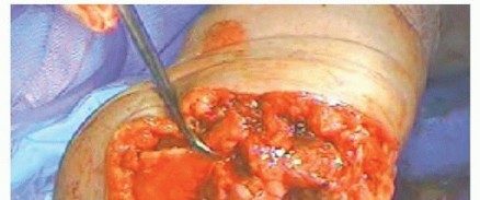
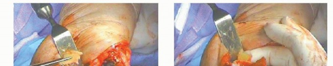
TECH FIG 1 • After skeletonizing, the distal humerus entry into the medullary canal is gained with a rongeur, removing bone from the articular surface in line with the midpoint of the olecranon fossa.
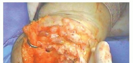
TECH FIG 2 • The medullary canal of the humerus is prepared with rasps of increasing size.
PREPARATION OF THE ULNA
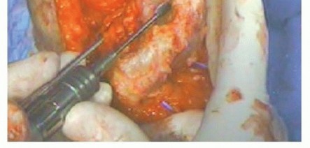
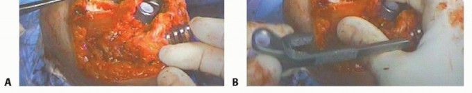
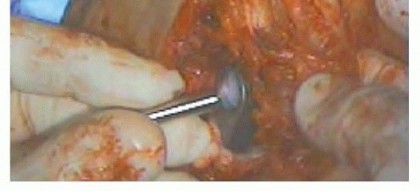
TECH FIG 3 • Entry into the medullary canal of the ulna is gained with a high-speed burr hole through the distal articular surface of the sigmoid notch.
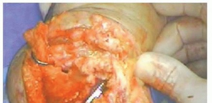
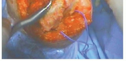
TECH FIG 4 • The medullary canal of the ulna is prepared with rasps of increasing size.
TRIAL THE COMPONENTS
COMPONENT INSERTION
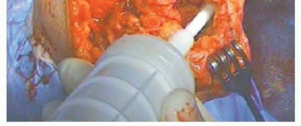
TECH FIG 5 • A long-stemmed cement gun is used to ensure an adequate cement mantle in the ulna and humerus.
TECH FIG 6 • The ulnar component is seated with gentle mallet blows on its inserter.
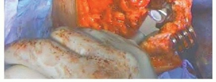
TECH FIG 7 • A. A wafer of bone graft from the resected portions of the humerus is contoured to fit along the anterior cortex of the humerus. B. The anterior flange of this humeral component seats around the bone graft's position along the anterior cortex.
TECH FIG 8 • The ulnar and humeral components are coupled before final seating of the humeral component.
Closure
Depending on the approach technique, repair the triceps to the olecranon process with nonabsorbable suture.
We routinely perform an anterior subcutaneous transposition of the ulnar nerve.
A well-padded anterior splint is placed to maintain the elbow in terminal extension, and instructions are given to elevate the arm as much as possible for the first 1 to 2 weeks. This is thought to minimize skin tension along the incision line, allow for epithelialization to occur, and postoperative edema to subside. The splint should be abundantly padded to prevent pressure necrosis.
Wound complications:
dehiscence, hematoma formation, drainage, wound infection- We emphasize careful and meticulous soft tissue handling.
- The wound is closed in layers over a drain.
- The elbow is splinted in a well-padded dressing, in a position of extension to minimize wound tension and allow for epithelialization and edema control.
Infection - Antibiotic cement is routinely used.
Ulnar nerve complication - Routine transposition of the nerve after it has been carefully
isolated and released proximally along the intermuscular septum and distally to the deep fascia of the flexor carpi ulnaris
Aseptic loosening of the
prostheses over time - Patients should be counseled on permanent activity restrictions.
CLOSURE
PEARLS AND PITFALLS
P.939
POSTOPERATIVE CARE
Sutures are removed 2 weeks following surgery.No formal range of motion instructions are imposed following splint removal if the patient had a triceps-sparing approach.If the patient had a triceps-reflecting or triceps-splitting approach, then no active extension against resistance is allowed for 8 weeks postoperatively.We recommend a lifetime weight-bearing restriction of 5 pounds for the operative extremity.
OUTCOMES
Results from the Scottish Arthroplasty Project registry identified 1146 primary TEAs of which 108 were performed for primary OA.The 10-year implant survivorship was 85% for TEA for primary OA.4Early implant-specific complications rates for all primary TEAs were reported as follows: Infection: 1.9%Dislocation: 0.7%Periprosthetic fracture: 3.1%Naqui et al5 reported on 11 patients with primary OA older than 65 years of age who underwent TEA with the Acclaim (DePuy, Warsaw, IN) convertible prosthesis at an average of 57.6 months.Ten were placed in linked mode and 1 in unlinked.Average arc of motion improved from an average of 70 to 110 degrees, statistically significantlyimproved American Shoulder and Elbow Surgeons (ASES) scores, and all patients were pain free at rest.There were no cases of loosening, although 4 patients had 1 mm radiolucencies.Complications included 1 patient with an intraoperative periprosthetic fracture and 1 patient with transient ulnar neuritis.
COMPLICATIONS
Ulnar neuropathy, in the form of paresthesias, is common after this surgery. This condition is usually self-limited and resolves within the first 6 months postoperatively. Careful handling of the ulnar nerve and protecting it from inadvertent stretching or trauma during surgery may minimize these symptoms.Infection is one of the most devastating complications after this surgery. The elbow is more prone to infection and wound complications due to its relatively thin soft tissue envelope. Prevention of wound dehiscence and meticulous soft tissue handling are important factors to remember in order to minimize the chance of developing a postoperative infection.Aseptic loosening is of greater concern in patients with primary OA than with rheumatoid arthritis given that primary OA patients generally impose higher physical demands on their elbows.Strict adherence to postoperative limitations as well as proper implant selection may help limit loosening and early failure. Evolution in implant designs in the future may improve long-term outcomes in this challenging patient population.
REFERENCES
- Bryan RS, Morrey BF. Extensive posterior exposure of the elbow. A triceps-sparing approach. Clin Orthop Relat Res 1982;166:188-192.
- Cheung EV, Adams R, Morrey BF. Primary osteoarthritis of the elbow: current treatment options. J Am Acad Orthop Surg 2008;16:77-87.
- Choo A, Ramsey ML. Total elbow arthroplasty: current options. J Am Acad Orthop Surg 2013;21:427-437.
- Jenkins PJ, Watts AC, Norwood T, et al. Total elbow replacement: outcome of 1,146 arthroplasties from the Scottish Arthroplasty Project. Acta Orthop 2013;84:119-123.
- Naqui SZ, Rajpura A, Nuttall D, et al. Early results of the Acclaim total elbow replacement in patients with primary osteoarthritis. J Bone Joint Surg Br 2010;92:668-671.
